Brief Report
234 Ann Dermatol
Received May 27, 2016, Revised March 22, 2017, Accepted for publication March 30, 2017
*These authors have equally contributed to the article.
Corresponding author: Mi Woo Lee, Department of Dermatology, Asan Medical Center, University of Ulsan College of Medicine, 88 Olympic-ro 43-gil, Songpa-gu, Seoul 05505, Korea. Tel: 82-2-3010-3460, Fax: 82-2-3010-3460, E-mail: miumiu@amc.seoul.kr
This is an Open Access article distributed under the terms of the Creative Commons Attribution Non-Commercial License (http://creativecommons.
org/licenses/by-nc/4.0) which permits unrestricted non-commercial use, distribution, and reproduction in any medium, provided the original work is properly cited.
Copyright © The Korean Dermatological Association and The Korean Society for Investigative Dermatology
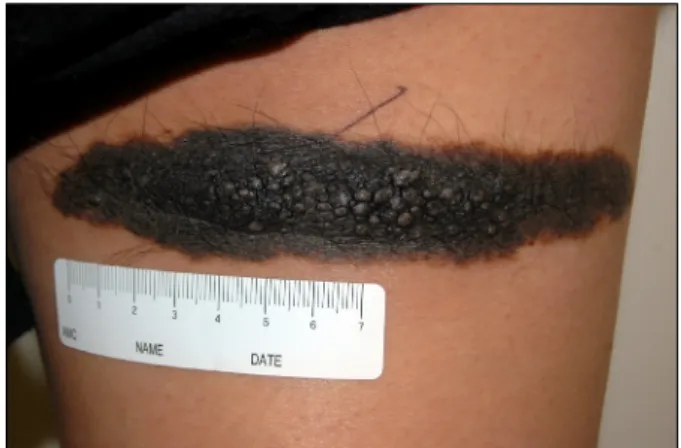
Fig. 1. Multiple papules and nodules arising in a black-colored, slightly elevated plaque on the right posterior thigh during pregnancy.
https://doi.org/10.5021/ad.2018.30.2.234
Atypical Proliferative Nodule with Melanocytic Intraepidermal Pagetoid Spreading Arising within
a Congenital Melanocytic Nevus in a Pregnant Woman
Hye-Rim Moon*, Mi Hye Lee*, Chong Hyun Won, Sung Eun Chang, Mi Woo Lee, Jee Ho Choi, Kee Chan Moon
Department of Dermatology, Asan Medical Center, University of Ulsan College of Medicine, Seoul, Korea
Dear Editor:
A 28-year-old woman at the 38th week of her first ges- tation presented with rapid enlarging papules and nodules in a 15 cm-sized black-pigmented plaque on her right pos- terior thigh. The plaque had been present since birth, and remained stable in size and pigmentation throughout her life. However, multiple clustered papules and nodules abruptly developed during the 2nd and 3rd trimester of her pregnancy (Fig. 1). To exclude the malignant melano- ma arising within a congenital melanocytic nevus (CMN), a punch biopsy was performed. The histopathologic find- ing showed epidermal proliferation of melanocytes in the background of nevoid cells. The Ki-67 labeling index of the epidermal melanocytes mildly increased as 10% to 20%, whereas dermal melanocytes showed less than 5%.
To exclude malignant melanoma, the patient underwent excision of the entire nevus. The distribution of pagetoid cells was mostly confined to the lower part of epidermis, without mitosis, necrosis and high-grade atypia (Fig. 2).
The patient finally received the definite diagnosis of be- nign proliferative nodules (PNs) with mild atypia arising in a CMN.
PNs usually represent the benign nodular proliferation of
intradermal melanocytes1. PNs arising in a CMN occasion- ally need to be distinguished from melanoma, because of clinical characteristics such as rapid proliferation, hemor- rhage, and ulceration. However, PNs have distinct histo- pathologic features: non-expanding, blending with the sur- rounding nevus, maturation, and benign prognosis2. Interestingly, our patient presented abruptly, rapidly en- larging PNs during her pregnancy. According to previous studies, the leading opinion is that pregnancy cannot in- duce significant clinical and dermoscopical changes in melanocytic nevi, except for women with dysplastic nevus syndrome3. However, Chan et al.4 reported that histo- pathologic changes in melanocytic nevi during pregnancy had tendencies of higher mitotic rates, cellular atypia, and increased proliferation. Although these studies have fo- cused on common nevi or dysplastic nevi rather than CMN, the results could partly explain the abrupt pro- liferation of atypical nevoid cells during pregnancy in our case.
Brief Report
Vol. 30, No. 2, 2018 235 Fig. 2. (A) Cellular proliferation showing no deep infiltration (H&E, ×40). (B) Proliferation of melanocytes in the background of a melanocytic nevus (H&E, ×100). (C) Intraepidermal pagetoid proliferation of melanocytes (H&E, ×200). (D) Ki-67 immunostaining showing high positivity in around 10% of the melanocytes (Ki-67, ×200). (E) Proliferation of melanocytes showing positivity for estrogen receptor (estrogen receptor β,
×40).
The development of PNs in our patient might have been associated with hormonal changes during pregnancy.
Estrogen receptor β (ER-β) expression is well known to be found in nevi and malignant melanomas, and to be in- fluenced by hormonal changes in the pregnancy and post-partum periods3. Because ER-β suppresses the pro- liferation of nevoid cells and plays a protective role by in- hibiting melanoma transformation, a decreased level of
ER-β expression in CMN during pregnancy could be as- sumed to induce the development of PNs in CMN.
However, There is not enough evidence of an association between ER-β expression level and histological atypia during pregnancy5.
In our case, although aggressive histological features in- cluding pagetoid spread of melanocytes and cellular aty- pia were mimicking malignant melanoma, the final diag-
Brief Report
236 Ann Dermatol
Received January 9, 2017, Revised March 7, 2017, Accepted for publication April 3, 2017
Corresponding author: You Chan Kim, Department of Dermatology, Ajou University School of Medicine, 164 WorldCup-ro, Yeongtong-gu, Suwon 16499, Korea. Tel: 82-31-219-5190, Fax: 82-31-219-5189, E-mail: maychan@ajou.ac.kr
This is an Open Access article distributed under the terms of the Creative Commons Attribution Non-Commercial License (http://creativecommons.org/
licenses/by-nc/4.0) which permits unrestricted non-commercial use, distribution, and reproduction in any medium, provided the original work is properly cited.
Copyright © The Korean Dermatological Association and The Korean Society for Investigative Dermatology
nosis was benign PNs based on the overall benign archi- tectural features. As shown by this case, the hormonal changes during pregnancy can be associated with various clinical and histopathological changes of CMN. However, even during pregnancy, whenever exclusion of malignant melanoma is required, proper procedures should be per- formed immediately.
CONFLICTS OF INTEREST
The authors have nothing to disclose.
REFERENCES
1. Nguyen TL, Theos A, Kelly DR, Busam K, Andea AA.
Mitotically active proliferative nodule arising in a giant congenital melanocytic nevus: a diagnostic pitfall. Am J Dermatopathol 2013;35:e16-e21.
2. Chung YL, Chang SN, Kim SC, Park WH, Chun SI.
Proliferating nodules within a congenital melanocytic nevus:
proper criteriae for surgical removal in infantile periods. Ann Dermatol 2001;13:120-122.
3. Driscoll MS, Grant-Kels JM. Nevi and melanoma in the pregnant woman. Clin Dermatol 2009;27:116-121.
4. Chan MP, Chan MM, Tahan SR. Melanocytic nevi in pregnancy: histologic features and Ki-67 proliferation index.
J Cutan Pathol 2010;37:843-851.
5. Nading MA, Nanney LB, Ellis DL. Pregnancy and estrogen receptor β expression in a large congenital nevus. Arch Dermatol 2009;145(6):691-694.
https://doi.org/10.5021/ad.2018.30.2.236
Scrotal Calcinosis in Brothers
Young Joon Park, Byung Woo Soh, You Chan Kim
Department of Dermatology, Ajou University School of Medicine, Suwon, Korea
Dear Editor:
Scrotal calcinosis (SC) is a rare pathological condition characterized by painless, hard, asymptomatic nodules on scrotal skin without any tissue injury or metabolic de- rangement. The nodules are generally skin-colored, yel- lowish or white, and consist of calcium and phosphate deposits. SC usually affects patients in childhood or early adulthood. Because of the age of onset and location of oc- currence, the condition may be a cause of embarrassment or misunderstanding. We describe the cases of two young adult patients with SC, who were brothers.
Two healthy Korean men, aged 22 and 23 years old, re- spectively, presented with scrotal nodules that gradually
increased over time. The nodules first appeared in ado- lescence and increased in size and number during the pre- vious 5 to 6 years. Both of the patients denied trauma, as- sociated symptoms, or any prior treatments. There was no history of other systemic inflammatory or metabolic disease.
Physical examination revealed multiple firm, non-tender, whitish papules on the scrotums of both patients (Fig. 1A, B). Skin biopsy was performed on both of the patients, and multiple calcium deposits and basophilic globules were found in the dermis (Fig. 1C, D). Routine laboratory ex- aminations including serum calcium and phosphorus were all within normal limits. The possibility of tumoral calcinosis was ruled out, as both the patients had rela-