접 수 일:2010. 8. 4.
채 택 일:2010. 9. 6.
교신저자:구천회
E-mail:kchob@gilhospital.com
요실금 수술 후 발생한 배뇨장애의 처치
가천의과학대학교 산부인과학교실 구천회 ․ 황병철
Management of voiding dysfunction after anti-incontinence operation
Chun Hoe Ku, M.D., Byung Chul Whang, M.D.
Department of Obstetric and Gynecology, Gachon University of Medicine and Science, Incheon, Korea
With the increasing number of surgery for incontinence, voiding dysfunction after anti-incontinence surgery will continue to be a problem.
The patient with postoperative voiding dysfunction may present with primarily storage symptoms or voiding symptoms, or a combination of both. Detailed knowledge of the preoperative voiding status may aid in the diagnosis of voiding dysfunction. Diagnosis is based on history, physical examination, urinalysis and postvoid residual volume, but additional informations from urodynamic study and cystoscopy are useful. Patients with postoperative voiding dysfunction should be initially treated conservatively with intermittent or continuous catheterization, fluid restriction, anticholinergics and pelvic floor physiotherapy. When conservative treatment fails, surgical intervention should be done. It is important to distinguish between midurethral sling and other procedures because the timing and type of intervention vary. In case of midurethral sling, loosening or cutting the tape has had excellent results. Prevention of obstruction during surgery may be the best way to avoid reoperation.
Key Words: Anti-incontinence surgery, Voiding dysfunction
서 론
복압성 요실금은 주로 수술적 치료로 만족스러운 성적을 보이며, 수술적 치료 방법은 보다 안전하고 간편하며 효과 가 우수한 방법으로 발전해 왔다. 1996년 tension-free vaginal tape (TVT)와 2001년 transobturator tape (TOT) 가 발표된 이후, 이 수술법들의 높은 성공률과 낮은 합병증 으로 요실금 수술 건수가 급속하게 증가하였으며 이에 따라 필연적으로 요실금 수술과 연관된 합병증들도 증가하고 있 다.1-7 요실금 수술 후 발생하는 배뇨장애는 환자에게 추가 적인 비용을 지출하게 하고 수술 만족도를 낮추는 요인이지
만 아직까지 진단방법 및 치료에 대해 확실하게 정립된 것 이 없다. 이에 여기에서는 요실금 수술 후 발생한 배뇨장애 의 처치에 대해 알아보고자 한다.
증상 및 정의
요실금 수술 후 나타나는 배뇨장애는 다양한 증상으로 나 타날 수 있으며, 크게 보면 저장 증상 (storage symptoms) 과 배뇨증상 (voiding symptoms)으로 나눌 수 있다. 저장 증상은 주로 방광 자극증상 (irritative symptom)으로 나타 나며 빈뇨 (frequency), 절박뇨 (urgency), 야간뇨 (noct- uria), 절박성 요실금 (urge incontinence) 등이 있으며, 배 뇨증상은 주로 폐색증상 (obstructive symptoms)으로 나 타나며 배뇨지연 (hesitancy), 배뇨 시 복부 힘주기 (void- ing with abdominal straining), 약한 소변줄기 (poor
Fig. 1. Liverpool nomogram for maximum urine flow rate in woman.
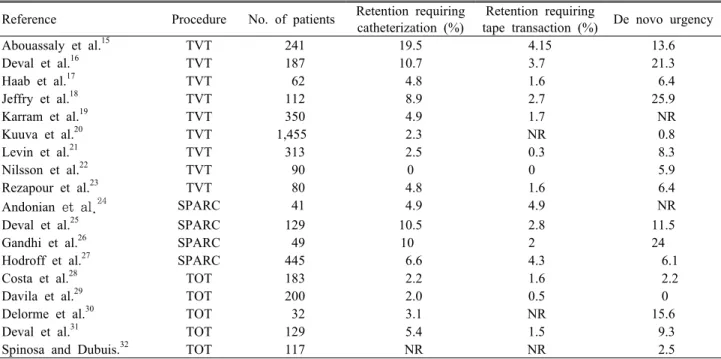
Reference Procedure No. of patients Retention requiring catheterization (%)
Retention requiring
tape transaction (%) De novo urgency
Abouassaly et al.15 TVT 241 19.5 4.15 13.6
Deval et al.16 TVT 187 10.7 3.7 21.3
Haab et al.17 TVT 62 4.8 1.6 6.4
Jeffry et al.18 TVT 112 8.9 2.7 25.9
Karram et al.19 TVT 350 4.9 1.7 NR
Kuuva et al.20 TVT 1,455 2.3 NR 0.8
Levin et al.21 TVT 313 2.5 0.3 8.3
Nilsson et al.22 TVT 90 0 0 5.9
Rezapour et al.23 TVT 80 4.8 1.6 6.4
Andonian et al.24 SPARC 41 4.9 4.9 NR
Deval et al.25 SPARC 129 10.5 2.8 11.5
Gandhi et al.26 SPARC 49 10 2 24
Hodroff et al.27 SPARC 445 6.6 4.3 6.1
Costa et al.28 TOT 183 2.2 1.6 2.2
Davila et al.29 TOT 200 2.0 0.5 0
Delorme et al.30 TOT 32 3.1 NR 15.6
Deval et al.31 TOT 129 5.4 1.5 9.3
Spinosa and Dubuis.32 TOT 117 NR NR 2.5
TVT: tension-free vaginal tape, SPARC: suprapubic arc, TOT: transobturator tape, NR: none reported.
Table 1. Voiding dysfunction after mid-urethral sling procedures stream), 잔뇨감 (incomplete emptying), 요폐 (urinary retention) 등이 있다.8 그러나 이러한 임상증상만 가지고 는 실제 요실금 수술 후 배뇨장애 환자를 구별하기는 어렵 다. Carr와 Webster9는 요실금 수술 후 배뇨장애로 요도박 리술 (urethrolysis)을 시행한 51명의 환자에서 가장 흔한 증상인 자극증상이 75%의 환자에서 나타났으며, 지속적인 요정체가 나타난 환자는 24%였다고 보고하였다. 또한 Bl- aivas와 Groutz10도 여성 방광출구폐색 (bladder outlet obstruction, BOO) 환자의 58%에서 자극증상과 폐색증상 이 같이 있으며, 자극증상만을 보이는 경우도 32%였으나
폐색 증상만 보이는 경우는 오직 10%에 불과하다고 하였다.
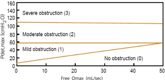
그러므로 요실금 수술 후 배뇨장애의 증상을 보이는 환자들 은 요류검사 및 잔뇨량을 측정하여 배뇨기능을 평가해야 한 다. 아직까지 배뇨장애에 대한 정의나 명확한 진단기준이 정립되지는 않았으나, 통상적으로 배뇨 전 방광용적 (배뇨 량과 잔뇨량의 합)이 최소한 150 mL 이상일 때, 최대요속이 15 mL/sec 미만 또는 잔뇨량이 50 mL를 초과하는 경우로 정의하거나 혹은 배뇨량과 최대 요속의 평균치를 나타낸 Liverpool nomogram을 이용하여 10 percentile 이하일 때 로 정의한다 (Fig. 1).11,12
유병률
요실금 수술 후 배뇨장애의 유병률은 진단 기준 및 수술 방법에 따라 다양하게 보고되고 있다. 1997년 미국 비뇨기 과학회 (American Urological Association)에서 1994년 1 월까지 시행된 요실금 수술 282편의 논문을 분석하여 발표 한 자료에 따르면, 4주 이상 지속되는 요폐가 치골뒤 걸이술 (retropubic suspension)은 3~7%, 경질 걸이술 (transv- aginal suspension)은 4~8%, 슬링 시술 (sling procedure) 은 6~11%로 보고하였으며, 영구적인 요폐는 세 시술 모두
5% 이하일 것이라고 하였다.13 Dunn 등14은 1966에서 2001 년까지 수술 후 배뇨장애 (postoperative voiding dysf- unction)에 관한 논문을 분석한 결과 Burch 질걸이술 (colposuspension)은 4~22%, Marshall-Marchetti-Kr- anz 시술은 5~20%, 침 걸이술 (needle suspension)은 5~7%, 치골질 슬링 (pubovaginal sling)은 4~10%, TVT는 24%의 배뇨장애를 보였다고 하였다. 최근 널리 이용되는 TVT, TOT 등의 중부요도 슬링 (mid-urethral sling)의 경 우 이론적으로 중부요도에 장력 없이 (tension free) 테이프 를 유치함으로써 기존의 수술보다 배뇨장애의 빈도가 현저 히 감소될 것으로 기대되었으나, 여전히 다양하게 배뇨장애 가 보고되고 있다 (Table 1).15-32
수술 후 새로 생기는 절박뇨 (de novo urgency)의 경우 치골뒤 걸이술은 8~16%, 경질 걸이술은 3~10%, 슬링 시술 은 3~11%, TVT는 1~25%, TOT는 0~15%로 보고되고 있 다. 한편, 수술 전에 절박뇨 증상을 가지고 있는 환자는 수 술 후에도 절박뇨 증상을 가질 가능성이 높으며, 치골뒤 걸 이술의 경우 36~66%, 경질 걸이술은 54%, 슬링 시술은 34~46%로 요실금 수술 후 절박뇨 증상이 지속된다고 한 다.13
기 전
모든 요실금 수술의 성공을 위해서는 어느 정도의 출구 저항 (outlet resistance)의 증가가 필요하다. 그러나 출구 저항의 증가는 복압이 증가하는 상황에서만 뚜렷하게 나타 나야 하며 배뇨 중에는 최소한으로 유지되어야 한다. 요실 금 수술 후 배뇨장애의 기전은 수술의 종류에 따라서 다를 수 있다. 치골뒤 또는 경질 걸이술의 경우 외측 요도 옆 조직 을 안정화시키므로 방광경부의 과들림 (hyperelevation)이 나 근위부 요도의 꼬임 (kinking)으로 인하여 폐색이 나타 날 수 있다. 반면, 슬링 수술의 경우 슬링 재료의 장력의 증 가로 인하여 직접적으로 요도를 압박하여 발생하며, 대부분 은 수술 시 장력 조절의 실패로 인한 과도한 견인 때문이며 슬링 재료와 주위 조직 간에 협착이 진행되어 당겨져서 발 생할 수도 있다.8 중부요도 슬링 수술의 경우 무장력으로 시 술되지만 배뇨 시 어느 정도의 요도 압박은 발생한다고 한 다. Lukacz 등33은 TVT 수술을 받은 65명의 환자의 수술 전 과 수술 1년 후 배뇨증상과 요역동학 검사를 비교하였을 때,
주관적인 배뇨증상은 변화가 없었으나 수술 전 최대 요속은 28.6 mL/sec, 수술 1년 후에는 16.3 mL/sec로 유의하게 감 소하였으며 배뇨 시간은 29.1초에서 42.6초로 유의하게 증 가하는 것으로 보고하였다. TOT 수술의 경우 유치된 테이 프의 각도가 TVT 보다 완만하여 요도 압박이 적을 것이라 생각되나, Barry 등34은 TOT 수술을 받은 83명의 환자를 대 상으로 한 전향적인 연구에서 수술 전에 비하여 수술 6~8주 후 최대요속이 유의하게 감소한다고 보고하였다.
진 단
요실금 수술 후 배뇨장애의 진단을 위해서는 먼저 수술 전 배뇨상태에 대한 자세한 정보가 도움이 될 수 있다. 수술 전 저장 및 배뇨증상의 유무, 잔뇨량 등의 정보가 유용하게 사용될 수 있으며 요역동학 검사를 시행했다면 다시 한 번 검토해 보아야 한다. 만일 수술 전 배뇨 기능이 정상인 환자 가 수술 후 잔뇨감 혹은 요폐가 발생하였다면 이는 BOO의 가능성을 강하게 시사할 수 있다.
요실금 수술 후 배뇨장애의 진단을 위해서는 철저한 병력 청취가 중요하다. 수술 후 빈뇨, 절박뇨, 야간뇨, 절박성 요 실금 등의 저장증상만 있는지 또는 배뇨지연, 약한 소변줄 기, 잔뇨감, 요폐 등의 배뇨증상만 있는지 또는 두 증상이 같이 있는지 확인하여야 하며, 수술 후 증상이 언제 나타났 는지 얼마나 지속되었는지도 확인하여야 한다.
그 후 이학적 검사를 시행하여 방광경부의 과들림 여부를 관찰하고 기침유발검사를 통해 복압성 요실금 여부를 관찰 하고 방광질 누공 여부도 확인해야 한다. 골반장기탈출이 새로 발생했는지, 기존에 골반장기탈출이 있었다면 더 심해 졌는지도 관찰해야 한다.
지속적인 저장증상을 가진 환자들은 방광경 검사를 시행 하여 슬링 재료 및 봉합사에 의한 방광 천공, 요도 미란 (urethral erosion)을 배제해야 하며 방광질 누공, 중부요 도 또는 방광경부의 과들림, 요도 협착 등을 관찰해야 한다.
폐색증상이 있는 환자들은 선별검사로서 요류검사 및 잔 뇨량을 측정한다. 배뇨장애는 앞서 기술한 기준에 따라 판 정하나, 요류 및 잔뇨량 검사로는 압력에 대한 정보를 제공 할 수 없어 비정상적인 결과를 보이는 경우 배뇨근 수축력 의 감소에 의한 것인지 아니면 방광출구 폐색에 의한 것인 지 감별할 수 없다.35 그러므로 요류 및 잔뇨량 검사에서 배
Free Qmax (mL/sec)
Fig. 2. Bladder outlet obstruction nomogram for woman.
뇨장애가 의심되는 경우 압력요류검사 (pressure-flow study)를 시행하여야 한다.
압력요류검사에서 요속의 감소와 배뇨근압의 상승이 나 타날 경우 BOO를 의심해야 한다. 현재까지 BOO에 대한 표 준화된 진단기준이 없이 여러 기준이 제시되고 있으나, 대 부분 최대요속 (Qmax)은 11~15 mL/sec 이하 그리고 최대 요속 시 배뇨근압 (PdetQmax)은 20~25 cmH2O 이상일 경 우로 진단기준을 제시하고 있다.36-38
한편, Blaivas와 Groutz10는 압력요류검사에서의 요류검 사와 최대요속 시 배뇨근압을 사용하지 않고 비침습적인 요 류검사와 최대배뇨근압을 이용한 nomogram을 여성 BOO 의 진단기준으로 제시하였다 (Fig. 2). 그들은 경요도 카테 터가 요속의 감소에 영향을 주기 때문에 압력요류검사 시 실제보다 BOO 환자가 증가할 수 있다고 하였으며, 경요도 카테터의 영향을 피할 수 있는 비침습적 최대요속 (free Qmax)과 최대배뇨근압 (Pdet.max)을 이용하여 BOO를 정 도에 따라서 경도 (mild), 중등도 (moderate), 고도 (sev- ere)로 구분하였다.
치 료
1. 보존적 방법 (Conservative management)
잔뇨량 증가 및 요폐 등 배뇨증상을 주로 호소하는 환자 는 간헐적 혹은 지속적 자가도뇨를 하며 증상의 호전을 기 다려 볼 수 있다. 소수의 환자에서는 재수술에 대한 두려움 과 요실금 재발에 대한 걱정으로 보존적인 치료를 선호하 나, 보존적인 치료로 배뇨증상이 해결되지 않는 환자들은 대부분 수술적인 방법을 선택하게 된다.
저장증상을 주로 호소하나 정상적으로 배뇨를 하는 환자
들은 우선적으로 수분제한, 항콜린성 약제, 골반근육운동 등의 치료를 한다. 이런 보존적 치료에도 불구하고 지속적 으로 저장증상이 있다면 요역동학 검사 및 방광경 검사를 시행하여 BOO를 배제하는 것이 중요하다. 만약 BOO가 존 재한다면 슬링 절단 (incision)이나 요도박리술 등을 고려 해야 한다.
일부에서는 Hegar 확장기를 이용하여 요도와 요도주위 를 이완하는 요도확장술 (urethral dilatation)을 시행하여 배뇨증상이 호전 되었다는 보고가 있으나, 연구가 제한적이 며 또한 무리하게 시행될 경우 요도에 손상을 줄 수 있으므 로 주의를 기울여야 한다.19,39
2. 수술적 방법 (Surgical intervention)
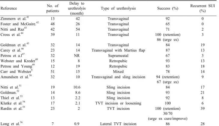
수술적 방법은 배뇨장애가 BOO에 의한 원인으로 보존적 치료에 호전되지 않을 때 시행하게 된다. 수술적 치료는 수 술 시기와 방법이 다양하므로 이전에 어떤 종류의 요실금 수술을 하였는지 구분하는 것이 중요하다.40 중부요도 슬링 의 경우 대개 수술적 치료가 보다 빨리 시행되며 덜 침습적 이나, 치골질 슬링, 경질 걸이술, 치골뒤 걸이술의 경우 대 개 수개월 후에 수술적 치료가 시행되며 수술 방법도 침습 적인 경우가 많다. 수술 방법의 경우 이전 수술이 치골뒤 걸 이술이었던 경우에는 치골뒤 요도박리술이 주로 시행되며, 치골질 슬링의 경우 경질 슬링 절단 또는 경질 요도박리술 이 시행되며, 중부요도 슬링의 경우 슬링의 이완 또는 절단 이 시행되며 각각의 수술 성적은 다음과 같다 (Table 2).41-56
1) 중부요도 슬링 이완 또는 절단 (Midurethral sling loosening or incision)
중부요도 슬링의 경우 수술 후 대부분의 환자들이 72시 간 이내에 정상적인 배뇨가 가능하므로 BOO가 의심되는 경 우 초기에 치료하는 것이 선호된다. 초기 치료는 대부분 2주 이내에 이루어지며 외래에서 국소마취하에 시행된다. 이전 절개부위를 통해 테이프를 찾은 후 right angle clamp를 이 용하여 테이프를 요도 아래쪽으로 내려주며 1~2 cm 정도 이완시킨다.54 수술 후 2주 이상 지난 경우에는 조직이 테이 프 내로 내증식 (ingrowth)하게 되어 테이프를 절단하는 경 우가 많으며 보다 광범위한 박리가 필요하므로 수술실에서
Reference No. of patients
Delay to urethrolyis (month)
Type of urethrolysis Success (%) Recurrent SUI (%)
Zimmern et al.41 13 42 Transvaginal 92 0
Foster and McGuire.42 48 26 Transvaginal 65 0
Nitti and Raz43 42 54 Transvaginal 71 2
Cross et al.44 39 11 Transvaginal 100 (retention)
86 (urge sx)
3
Goldman et al.45 32 14 Transvaginal 84 19
Carey et al.46 23 14 Transvaginal with Martius flap 87 13
Petrou et a.l47 32 NR Suprameatal 67 3
Webster and Kreder48 15 8 Retropubic 93 13
Petrou and Young49 12 19 Retropubic 83 18
Carr and Webster9 51 15 Mixed 78 14
Amundsen et al.50 32 10 Transvaginal and sling incision 94 (retention) 67 (urge sx)
9
Nitti et al.51 19 10.6 Sling incision 84 17
Goldman.52 14 8.6 Sling incision 93 21
Thiel et al.53 13 2.2 Sling incision 92 8
Klutke et al.54 17 2.1 TVT incision or loosening 100 6
Rardin et al.55 23 2 TVT incision 100 (retention)
30/70
(urge sx cure/improve)
39
Long et al.56 7 0.9 Lateral TVT incision 86 28
SUI: stress urinary incontinence, NR: none reported, TVT: tension-free vaginal tape.
Table 2. Summary of series on sling loosening/incision and urethrolysis for obstruction after incontinence surgery
시행하는 것을 고려해야 한다. 테이프의 이완 또는 절단 후 비교적 좋은 성적이 보고 되는데 Klukte 등54은 TVT 수술 후 BOO로 테이프의 이완 또는 절단한 17명의 환자 모두 폐 색증상이 완치 되었으며, 요실금이 재발된 경우는 1명이었 다고 하였다. Rardin 등55은 TVT 수술 후 지속적인 배뇨장 애를 보인 23명의 환자에게 테이프 절단을 시행했으며 6주 후에 모든 환자에서 폐색 증상은 없어졌으며, 자극 증상의 경우 30%는 완전히 없어졌으며 70%는 호전되었고, 요실금 의 재발은 13%, 부분 재발은 26%라고 보고하였다.
2) 경질 슬링 절단 (Transvaginal sling incision) 경질 슬링 절단의 경우 중부요도 슬링보다 더 오랜 기간 이 지난 후에 시행되며 보다 광범위한 박리가 필요하다. 전 신마취 혹은 부위마취 (regional anesthesia)가 필요하며 수술 전과 수술 후에 방광경을 시행하여 방광 및 요도의 미 란 및 손상을 확인해야 한다. 요도박리술보다 신경 및 연부 조직의 손상이 덜하고 박리 후 섬유화 (fibrosis)가 덜하기 때문에 우선적으로 고려해야 하며, 84~93.5%의 성공률이 보고되며 요실금의 재발은 8~21% 정도이다.50-53 경질 슬링
절단으로 슬링이 완전하게 제거가 안되거나 폐색증상이 지 속되는 경우 요도박리술을 시행하여야 한다.
3) 경질 요도박리술 (Transvaginal urethrolysis) 전질벽에 중앙 혹은 역 U자 (inverted U) 절개를 한 후 외측으로 요도주위 조직을 따라서 내골반근막 (endopelvic fascia)을 천공한 후 치골뒤 공간까지 접근한 뒤 요도가 치 골하부로부터 자유로운 움직임을 갖도록 박리한다.57 수술 전과 후에 방광경을 시행하며 대개는 수술 전에 요도와 방 광경부가 고정되어 있고 이동성 (mobility)이 없다가 요도 박리술 후에는 이동성이 복구된다. 요도 주위에 유착이 심 하거나 반복되는 요도박리술의 경우 치골하부와 요도 사이 에 Martius labial fat flap을 사용하기도 하며 이는 재발성 유착을 감소시키며 요도를 지지하는 역할을 하고 요도 손상 을 감소시키는 역할을 한다.46
4) 치골뒤 요도박리술 (Retropubic urethrolysis) 질식 접근이 불충분할 때, 이전 수술이 치골뒤 걸이술인 경우, 경질 요도박리술 시행 시 방광 천공, 누공 등의 합병
증이 발생한 경우 시행한다.48,49
결 론
최근 요실금 수술의 증가에 따라 수술 후 배뇨장애의 보 고도 늘어나고 있다. 요실금 수술 후 배뇨장애의 증상은 방 광 자극증상 및 폐색증상으로 다양하게 나타날 수 있으며 진단을 위해서는 수술 전 환자의 배뇨상태를 파악하는 것이 도움이 된다. 요실금 수술 후 배뇨장애가 발생한 경우 자세 한 병력 청취, 이학적 검사, 잔뇨량 검사를 시행하며 필요에
따라서 요역동학 검사, 방광경 검사를 시행하여야 한다. 치 료로는 우선 자가됴뇨, 수분제한, 항콜린성약제, 골반근육 운동 등의 보존적 치료를 시행하며 효과가 없을 경우 수술 적 치료를 한다. 이전 요실금 수술에 따라서 수술 시기 및 방 법이 달라지나 최근 중부요도 슬링의 경우 빠른 시기에 테 이프를 이완하거나 절단하는 방법으로 좋은 성적이 보고되 고 있다. 요실금 수술 후 배뇨장애를 예방하는 가장 좋은 방 법은 수술 시 과도한 장력을 피하는 것이므로, 항상 이를 염 두에 두고 수술을 시행하여야 하겠다.
참고문헌
1. Ulmsten U, Henriksson L, Johnson P, Varhos G. An ambulatory surgical procedure under local anesthesia for treatment of female urinary incontinence. Int Urogynecol J Pelvic Floor Dysfunct 1996; 7: 81-5.
2. de Leval J. Novel surgical technique for the treatment of female stress urinary incontinence: transobturator vaginal tape inside-out. Eur Urol 2003; 44: 724-30.
3. Nilsson CG, Palva K, Rezapour M, Falconer C.
Eleven years prospective follow-up of the tension-free vaginal tape procedure for treatment of stress urinary incontinence. Int Urogynecol J Pelvic Floor Dysfunct 2008; 19: 1043-7.
4. Agostini A, Bretelle F, Franchi F, Roger V, Cravello L, Blanc B. Immediate complications of tension-free vaginal tape (TVT): results of a French survey. Eur J Obstet Gynecol Reprod Biol 2006; 124: 237-9.
5. Tamussino KF, Hanzal E, Kolle D, Ralph G, Riss PA.
Tension-free vaginal tape operation: results of the Austrian registry. Obstet Gynecol 2001; 98: 732-6.
6. deTayrac R, Deffieux X, Droupy S, Chauveaud- Lambling A, Calvanese-Benamour L, Fernandez H. A prospective randomized trial comparing tension-free vaginal tape and transobturator suburethral tape for surgical treatment of stress urinary incontinence. Am J Obstet Gynecol 2004; 190: 602-8.
7. Porena M, Costantini E, Frea B, Giannantoni A, Ranzoni S, Mearini L, et al. Tension-free vaginal tape versus transobturator tape as surgery for stress urinary incontinence: results of a multicentre randomised trial. Eur Urol 2007; 52: 1481-90.
8. Gomelsky A, Nitti VW, Dmochowski RR. Manag- ement of obstructive voiding dysfunction after incon- tinence surgery: lessons learned. Urology 2003; 62:
391-9.
9. Carr LK, Webster GD. Voiding dysfunction follow- ing incontinence surgery: diagnosis and treatment with retropubic or vaginal urethrolysis. J Urol 1997;
157: 821-3.
10. Blaivas JG, Groutz A. Bladder outlet obstruction nomogram for women with lower urinary tract symptomatology. Neurourol Urodyn 2000; 19: 553- 64.
11. Haylen BT, Law MG, Frazer M, Schulz S. Urine flow rates and residual urine volumes in urogynecology patients. Int Urogynecol J Pelvic Floor Dysfunct 1999; 10: 378-83.
12. Costantini E, Mearini E, Pajoncini C, Biscotto S, Bini V, Porena M. Uroflowmetry in female voiding disturbances. Neurourol Urodyn 2003; 22: 569-73.
13. Leach GE, Dmochowski RR, Appell RA, Blaivas JG, Hadley HR, Luber KM, et al. Female Stress Urinary Incontinence Clinical Guidelines Panel summary re- port on surgical management of female stress urinary incontinence. The American Urological Association. J Urol 1997; 158: 875-80.
14. Dunn JS Jr, Bent AE, Ellerkman RM, Nihira MA, Melick CF. Voiding dysfunction after surgery for stress incontinence: literature review and survey results. Int Urogynecol J Pelvic Floor Dysfunct 2004;
15: 25-31.
15. Abouassaly R, Steinberg JR, Lemieux M, Marois C, Gilchrist LI, Bourque JL, et al. Complications of ten- sion-free vaginal tape surgery: a multi-institutional review. BJU Int 2004; 94: 110-3.
16. Deval B, Jeffry L, Al Najjar F, Soriano D, Darai E.
Determinants of patient dissatisfaction after a ten- sion-free vaginal tape procedure for urinary incon- tinence. J Urol 2002; 167: 2093-7.
17. Haab F, Sananes S, Amarenco G, Ciofu C, Uzan S, Gattegno B, et al. Results of the tension-free vaginal tape procedure for the treatment of type II stress uri- nary incontinence at a minimum followup of 1 year. J Urol 2001; 165: 159-62.
18. Jeffry L, Deval B, Birsan A, Soriano D, Darai E.
Objective and subjective cure rates after tension-free vaginal tape for treatment of urinary incontinence.
Urology 2001; 58: 702-6.
19. Karram MM, Segal JL, Vassallo BJ, Kleeman SD.
Complications and untoward effects of the ten- sion-free vaginal tape procedure. Obstet Gynecol 2003; 101: 929-32.
20. Kuuva N, Nilsson CG. A nationwide analysis of com- plications associated with the tension-free vaginal tape (TVT) procedure. Acta Obstet Gynecol Scand 2002; 81: 72-7.
21. Levin I, Groutz A, Gold R, Pauzner D, Lessing JB, Gordon D. Surgical complications and medium-term outcome results of tension-free vaginal tape: a pro- spective study of 313 consecutive patients. Neurourol Urodyn 2004; 23: 7-9.
22. Nilsson CG, Kuuva N, Falconer C, Rezapour M, Ulmsten U. Long-term results of the tension-free vaginal tape (TVT) procedure for surgical treatment of female stress urinary incontinence. Int Urogynecol J Pelvic Floor Dysfunct 2001; 12(Suppl 2): S5-8.
23. Rezapour M, Ulmsten U. Tension-Free vaginal tape (TVT) in women with mixed urinary incontinence--a long-term follow-up. Int Urogynecol J Pelvic Floor Dysfunct 2001; 12(Suppl 2): S15-8.
24. Andonian S, Chen T, St-Denis B, Corcos J.
Randomized clinical trial comparing suprapubic arch sling (SPARC) and tension-free vaginal tape (TVT):
one-year results. Eur Urol 2005; 47: 537-41.
25. Deval B, Levardon M, Samain E, Rafii A, Cortesse A, Amarenco G, et al. A French multicenter clinical trial of SPARC for stress urinary incontinence. Eur Urol 2003; 44: 254-8.
26. Gandhi S, Abramov Y, Kwon C, Beaumont JL, Botros S, Sand PK, et al. TVT versus SPARC: com- parison of outcomes for two midurethral tape procedures. Int Urogynecol J Pelvic Floor Dysfunct 2006; 17: 125-30.
27. Hodroff MA, Sutherland SE, Kesha JB, Siegel SW.
Treatment of stress incontinence with the SPARC sling: intraoperative and early complications of 445 patients. Urology 2005; 66: 760-2.
28. Costa P, Grise P, Droupy S, Monneins F, Assenmach- er C, Ballanger P, et al. Surgical treatment of female stress urinary incontinence with a trans-obturator- tape (T.O.T.) Uratape: short term results of a pro- spective multicentric study. Eur Urol 2004; 46:
102-6.
29. Davila GW, Johnson JD, Serels S. Multicenter experi- ence with the Monarc transobturator sling system to treat stress urinary incontinence. Int Urogynecol J Pelvic Floor Dysfunct 2006; 17: 460-5.
30. Delorme E, Droupy S, de Tayrac R, Delmas V.
Transobturator tape (Uratape): a new minimally-in- vasive procedure to treat female urinary incontine- nce. Eur Urol 2004; 45: 203-7.
31. Deval B, Ferchaux J, Berry R, Gambino S, Ciofu C, Rafii A, et al. Objective and subjective cure rates after trans-obturator tape (OBTAPE) treatment of female urinary incontinence. Eur Urol 2006; 49: 373-7.
32. Spinosa JP, Dubuis PY. Suburethral sling inserted by the transobturator route in the treatment of female stress urinary incontinence: preliminary results in 117 cases. Eur J Obstet Gynecol Reprod Biol 2005; 123:
212-7.
33. Lukacz ES, Luber KM, Nager CW. The effects of the tension-free vaginal tape on voiding function: a pro- spective evaluation. Int Urogynecol J Pelvic Floor Dysfunct 2004; 15: 32-8.
34. Barry C, Naidu A, Lim Y, Corsitaans A, Muller R, Rane A. Does the MONARC transobturator suburet- hral sling cause post-operative voiding dysfunction? A prospective study. Int Urogynecol J Pelvic Floor Dysfunct 2006; 17: 30-4.
35. Schafer W, Abrams P, Liao L, Mattiasson A, Pesce F, Spangberg A, et al. Good urodynamic practices: uro- flowmetry, filling cystometry, and pressure-flow studies. Neurourol Urodyn 2002; 21: 261-74.
36. Chassagne S, Bernier PA, Haab F, Roehrborn CG, Reisch JS, Zimmern PE. Proposed cutoff values to de- fine bladder outlet obstruction in women. Urology 1998; 51: 408-11.
37. Lemack GE, Zimmern PE. Pressure flow analysis may aid in identifying women with outflow obstruction. J Urol 2000; 163: 1823-8.
38. Defreitas GA, Zimmern PE, Lemack GE, Shariat SF.
Refining diagnosis of anatomic female bladder outlet obstruction: comparison of pressure-flow study pa- rameters in clinically obstructed women with those of normal controls. Urology 2004; 64: 675-9.
39. Mishra VC, Mishra N, Karim OM, Motiwala HG.
Voiding dysfunction after tension-free vaginal tape: a conservative approach is often successful. Int Urog- ynecol J Pelvic Floor Dysfunct 2005; 16: 210-4.
40. Ghoniem G, Abdelwahab H, Elmissiry M, Khater U.
Surgical choices for the treatment of bladder outlet obstruction after sling procedures. J Pelvic Med Surg 2008; 14: 369-74.
41. Zimmern PE, Hadley HR, Leach GE, Raz S. Female urethral obstruction after Marshall-Marchetti-Krantz operation. J Urol 1987; 138: 517-20.
42. Foster HE, McGuire EJ. Management of urethral ob- struction with transvaginal urethrolysis. J Urol 1993;
150: 1448-51.
43. Nitti VW, Raz S. Obstruction following anti-incon- tinence procedures: diagnosis and treatment with transvaginal urethrolysis. J Urol 1994; 152: 93-8.
44. Cross CA, Cespedes RD, English SF, McGuire EJ.
Transvaginal urethrolysis for urethral obstruction af- ter anti-incontinence surgery. J Urol 1998; 159:
1199-201.
45. Goldman HB, Rackley RR, Appell RA. The efficacy of urethrolysis without re-suspension for iatrogenic urethral obstruction. J Urol 1999; 161: 196-8.
46. Carey JM, Chon JK, Leach GE. Urethrolysis with Martius labial fat pad graft for iatrogenic bladder out- let obstruction. Urology 2003; 61: 21-5.
47. Petrou SP, Brown JA, Blaivas JG. Suprameatal trans- vaginal urethrolysis. J Urol 1999; 161: 1268-71.
48. Webster GD, Kreder KJ. Voiding dysfunction follo- wing cystourethropexy: its evaluation and manage- ment. J Urol 1990; 144: 670-3.
49. Petrou SP, Young PR. Rate of recurrent stress urinary incontinence after retropubic urethrolysis. J Urol 2002; 167: 613-5.
50. Amundsen CL, Guralnick ML, Webster GD.
Variations in strategy for the treatment of urethral obstruction after a pubovaginal sling procedure. J Urol 2000; 164: 434-7.
51. Nitti VW, Carlson KV, Blaivas JG, Dmochowski RR.
Early results of pubovaginal sling lysis by midline sling incision. Urology 2002; 59: 47-51.
52. Goldman HB. Simple sling incision for the treatment of iatrogenic urethral obstruction. Urology 2003; 62:
714-8.
53. Thiel DD, Pettit PD, McClellan WT, Petrou SP.
Long-term urinary continence rates after simple sling incision for relief of urinary retention following fascia lata pubovaginal slings. J Urol 2005; 174: 1878-81.
54. Klutke C, Siegel S, Carlin B, Paszkiewicz E, Kirkemo A, Klutke J. Urinary retention after tension-free vagi- nal tape procedure: incidence and treatment. Urology 2001; 58: 697-701.
55. Rardin CR, Rosenblatt PL, Kohli N, Miklos JR, Heit M, Lucente VR. Release of tension-free vaginal tape for the treatment of refractory postoperative voiding dysfunction. Obstet Gynecol 2002; 100: 898-902.
56. Long CY, Lo TS, Liu CM, Hsu SC, Chang Y, Tsai EM.
Lateral excision of tension-free vaginal tape for the treatment of iatrogenic urethral obstruction. Obstet Gynecol 2004; 104: 1270-4.
57. Leach GE, Raz S. Modified Pereyra bladder neck sus- pension after previously failed anti-incontinence surgery. Surgical technique and results with long- term follow-up. Urology 1984; 23: 359-62.
= 국문초록 =
최근 요실금 수술이 증가함에 따라 수술 후 배뇨장애가 문제가 되고 있다. 요실금 수술 후 배뇨장애를 가진 환자는 주로 저장증 상 또는 배뇨증상이 나타나며 진단을 위해서는 수술 전 환자의 배뇨상태를 파악하는 것이 도움이 된다. 요실금 수술 후 배뇨 장애의 진단은 병력 청취, 이학적 검사, 요검사, 잔뇨량 검사를 기초로 하며 요역동학 검사, 방광경 검사를 이용한 정보도 유용하다. 치료로는 우선 자가됴뇨, 수분제한, 항콜린성약제, 골반근육운동 등의 보존적 치료를 시행하며 효과가 없을 경우 수술적 치료를 한다. 이전 요실금 수술이 중부요도 슬링인지 다른 시술인지에 따라서 수술 시기 및 방법이 달라지므로 이를 구별하는 것이 중요하다. 최근 중부요도 슬링의 경우 빠른 시기에 테이프를 이완하거나 절단하는 방법으로 좋은 성적이 보 고되고 있다. 요실금 수술 후 배뇨장애를 예방하는 가장 좋은 방법은 수술 시 과도한 장력을 피하는 것이므로, 항상 이를 염두에 두고 수술을 시행하여야 하겠다.
중심단어: 요실금 수술, 배뇨장애