https://doi.org/10.5468/ogs.20071 pISSN 2287-8572 · eISSN 2287-8580
Introduction
Vitamin D is known to have a major influence on bone health through regulation of calcium phosphorus homeo- stasis. Recently, the female reproductive system has been defined as one of the non-classical target organs of vitamin D.
Vitamin D receptor expression was identified in ovarian gran- ulosa cells as well as other female reproductive organs, in- cluding endometrium and the uterus. These findings suggest that vitamin D may have a potential role in female reproduc- tion [1-3]. Several studies have demonstrated a direct effect of vitamin D on ovarian folliculogenesis and steroidogenesis in animal and human cell-line studies; vitamin D receptor null mutant mice have impaired folliculogenesis and vitamin D
Serum vitamin D levels and ovarian reserve markers in secondary amenorrhea patients: Is there a link?
Gyung-Mee Kim, MD, PhD 1 , Gyun-Ho Jeon MD, PhD 2
Departments of
1Psychiatry,
2Obstetrics and Gynecology, Inje University Haeundae Paik Hospital, Inje University College of Medicine, Busan, Korea
Objective
To investigate whether serum 25-hydroxyvitamin D [25(OH)D] level is associated with ovarian reserve markers in secondary amenorrhea (SA) patients.
Methods
Sixty-three women diagnosed with SA were recruited during 12 months from the initiation of this prospective observational study. Serum 25(OH)D levels, serum anti-Müllerian hormone (AMH) levels and antral follicle count (AFC) were estimated in study participants and ovarian reserve markers were compared between participants with vitamin D deficiency and those with normal vitamin D levels.
Results
Of the 63 participants, 27 (42.9%) were vitamin D deficient (<20 ng/mL) and 36 (57.1%) had normal vitamin D levels.
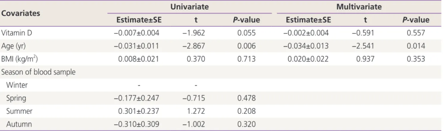
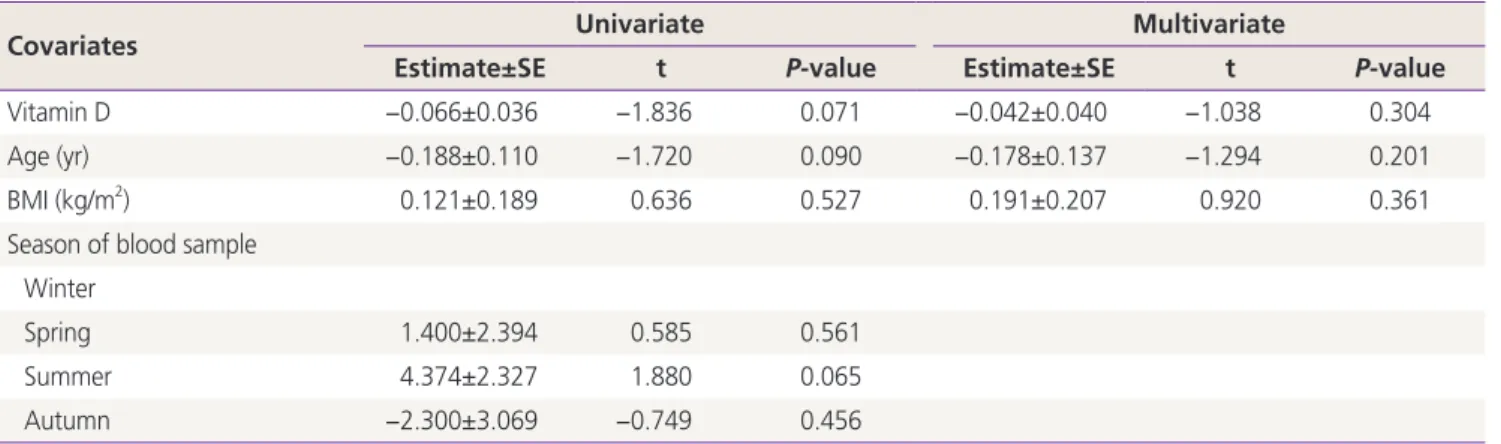
The mean AMH levels and AFC were 10.86±8.94 µ/L and 15.23±7.65 in the vitamin D deficient group, and 7.24±5.62 µ/L and 12.30±6.95 in the normal vitamin D group. Univariate and multivariate linear regression analysis of log
10transformed AMH and AFC with serum 25(OH)D adjusted for age and body mass index confirmed no association between vitamin D levels and AMH levels or AFC. There was also no correlation between serum 25(OH)D and AMH levels or AFC in all participants. However, participants with vitamin D deficiency had an increased chance of having polycystic ovarian syndrome (PCOS) as cause of SA than those with normal vitamin D levels (adjusted odds ratio, 7.559;
95% confidence interval, 1.28–44.65; P=0.026) after adjustment for clinical factors by logistic regression model.
Conclusion
There was no correlation between serum 25(OH)D levels and ovarian reserve markers in SA patients, but vitamin D deficiency may be linked to PCOS patients.
Keywords: Anti-Mullerian hormone; Amenorrhea; Ovarian reserve; 25-hydroxyvitamin D; Polycystic ovarian syndrome
Received: 2020.03.19. Revised: 2020.05.13. Accepted: 2020.06.01.
Corresponding author: Gyun-Ho Jeon, MD, PhD
Department of Obstetrics and Gynecology, Inje University Haeundae Paik Hospital, Inje University College of Medicine, 875 Haeun-daero, Haeundae-gu,Busan 48108, Korea
E-mail: [email protected] https://orcid.org/0000-0003-1128-1656
Articles published in Obstet Gynecol Sci are open-access, distributed under the terms of the Creative Commons Attribution Non-Commercial License (http://creativecommons.
org/licenses/by-nc/3.0/) which permits unrestricted non-commercial use, distribution, and reproduction in any medium, provided the original work is properly cited.
Copyright © 2020 Korean Society of Obstetrics and Gynecology
stimulated steroidogenesis in human ovarian cells [4,5].
Anti-Müllerian hormone (AMH) is produced by granulosa cells of small antral and pre-antral follicles, and along with antral follicle counts (AFCs), is a well-known representa- tive marker for ovarian reserve. AMH plays a crucial role in folliculogenesis and is not affected by the menstrual cycle, making it the most widely used ovarian marker. Although it is still unclear how vitamin D affects female reproductive function, interest in a potential relationship between vitamin D and ovarian reserve markers, particularly AMH, has in- creased since reports on the effect of vitamin D on follicular development have accumulated. Moreover, the presence of a functional vitamin D response element in the AMH gene pro- motor region was demonstrated [6] and experimental results in animal and human granulosa cells have also reported that vitamin D affects AMH signaling [7,8]. These basic research studies have indicated that vitamin D deficiency may alter go- nadal function through abnormal AMH signaling. However, discrepancy exists in clinical study findings under various set- tings, where some suggest a significant association between vitamin D levels and ovarian reserve, while others have not found any correlation between vitamin D and ovarian reserve markers [9,10]. Considering these contradictory results, we performed this prospective observational study to examine serum 25-hydroxyvitamin D [25(OH)D] levels in patients with secondary amenorrhea (SA) and investigate the relationship between serum 25(OH)D levels and ovarian reserve markers (i.e., serum AMH and AFC) in these women.
Materials and methods
1. Study design and participants
This study was conducted as a prospective cohort study for 12 months from March 2018 to February 2019. The study population was comprised of patients evaluated by a single reproductive endocrinologist for symptoms of SA. A total of 78 participants were initially recruited to the study. Three participants refused to participate in the study and 12 partici- pants were excluded due to the study’s exclusion criteria. The exclusion criteria of this study were as follows: 1) women who had taken hormonal medication including oral contraceptives or vitamin D supplements within the previous 6 months; 2) women who had ovarian surgery, chemotherapy or radio- therapy; 3) women who took medication and/or were diag-
nosed with a systemic disease that can affect menstruation (e.g., psychoaffective medicine, thyroid hormone, diabetes, hyperprolactinemia); 4) women who refused to participate this study. Patients’ information including age, parity, height, body weight, cause of SA, season of blood sampling (i.e., to account for seasonal changes in vitamin D), current medica- tions and other past medical history were recorded. Gyneco- logic ultrasonography was also performed on their first study visit. To determine cause of SA, polycystic ovarian syndrome (PCOS) was diagnosed on the basis of Rotterdam criteria [11], and primary ovarian insufficiency (POI) was regarded as 2 recordings of serum follicular stimulating hormone (FSH) levels of more than 40 IU/L at least one month apart in a woman aged under 40 years (i.e., >2 standard deviation [SD]
less than the mean menopausal age) [12]. All patients whose etiology of SA was not determined, excluding women with POI and PCOS, were deemed unexplained chronic anovula- tion [13]. PCOS in adolescents was only diagnosed in women with at least 2 years after menarche and who had SA, hyper- androgenemia, and increased ovarian volume on ultrasound [14].
Venous blood samples of all participants were taken for the measurement of serum 25(OH)D and AMH on the same day, prior to treatment for SA. Vitamin D deficiency was defined as serum 25(OH)D levels <20 ng/mL based on the Endocrine Society clinical practice guidelines [15].
2. Serum vitamin D, hormones and ovarian reserve markers measurements
1) Serum 25-hydroxyvitamin D
Each participant’s venous sample was drawn into a serum sep- aration tube (SST) and serum 25(OH)D was measured using a DIA source 25 OH Vitamin D3 radioimmunoassay kit (DIA- source ImmunoAssays S.A., Louvain-La-Neuve, Belgium) with immunoassay device (gamma 5) and presented in ng/mL.
The total imprecision coefficient of variance was 5.38% at a concentration level of 6.35 ng/mL and 4.96% at 37.71 ng/mL.
2) Serum follicular stimulating hormone
Each participant’s venous sample was drawn into an SST and
serum FSH was measured using an Elecsys FSH electroche-
miluminescence immunoassay kit (Roche Diagnostics GmbH,
Mannheim, Germany) with immunoassay device (Cobas e
801) and presented in mIU/mL. The total imprecision coef-
ficient of variance was 3.1% at a concentration level of 48.8 mIU/mL and 2.3% at 17.4 mIU/mL.
3) Anti-Müllerian hormone and antral follicle counts Blood drawn into the SST was centrifuged within 1 hour (3,000 rpm for 10 minutes). The separated serum was mea- sured for AMH level by ECLIA method with Elecsys AMH kit (Roche Diagnostics GmbH) on a Cobas e 601 immunoas- say analyzer and presented in ng/mL. The total imprecision coefficient of variance was 3.5% at a concentration level of 0.042 ng/mL and 3.4% at 0.20 ng/mL. An AFC was mea- sured by one gynecologist on the participant’s first visit in all individuals with gynecologic ultrasonography. AFC repre- sented the total number of all antral follicles, from 2 mm to 10 mm in size, in both ovaries.
3. Statistical analysis
Categorical variable data are presented as a frequency with percentage and continuous variables are presented as a group mean±SD. Differences in study participants’ charac- teristics were compared across subgroups with a χ
2test or
Fisher’s exact test for categorical variables and independent t-test or Mann-Whitney’s U test for continuous variables, as appropriate. An analysis of variance with Duncan’s post hoc test or Kruskal-Wallis test with Dunn’s post hoc test were also employed, as appropriate. To check if data were normally distributed, we used a Shapiro-Wilk test. Partial cor- relation coefficients controlling for age and body mass index (BMI) were estimated to investigate the linear relationship between 2 continuous variables. Univariate linear regression analysis between each variable (age, BMI, vitamin D, season), and multivariate linear regression analysis with all covari- ates, were performed to identify the regression coefficients for factors related to the log
10AMH, AFC, PCOS and POI.
For data visualization, box plot and scatter plot were also displayed. Finally, logistic regression analysis was performed to identify whether vitamin D deficiency is associated with PCOS as a cause of SA. All statistical analyses were carried out using SPSS 24.0 (SPSS Statistics for Windows 24.0; IBM Corp., Armonk, NY, USA) statistical software and P-values less than 0.05 was considered statistically significant.
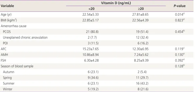
Table 1. Patients’ demographic variables
Variable Vitamin D (ng/mL)
P-value
<20 ≥20
Age (yr) 22.54±5.33 27.81±8.65 0.014
a)BMI (kg/m
2) 22.85±5.17 22.56±4.39 0.823
a)Amenorrhea cause
PCOS 21 (80.8) 19 (51.4) 0.454
b)Unexplained chronic anovulation 2 (7.7) 12 (32.4)
POI 3 (11.5) 6 (16.2)
AFC 15.23±7.65 12.30±6.95 0.119
c)AMH 10.86±8.94 7.24±5.62 0.130
a)FSH 6.30±4.28 8.25±9.39 0.392
a)Season of blood sample 0.128
b)Autumn 6 (23.1) 2 (5.4)
Spring 9 (34.6) 11 (29.7)
Summer 6 (23.1) 16 (43.2)
Winter 5 (19.2) 8 (21.6)
Values are presented as mean±standard deviation or number (%).
BMI, body mass index; PCOS, polycystic ovarian syndrome; POI, primary ovarian insufficiency; AFC, antral follicle count; AMH, anti-Müllerian hormone; FSH, follicular stimulating hormone.
a)