CASE REPORT
Copyright © 2019 The Korean Retina Society
This is an Open Access article distributed under the terms of the Creative Commons Attribution Non-Commercial License (http://creativecommons.org/licenses/by-nc/3.0/) which permits unrestricted non-commercial use, distribution, and reproduction in any medium, provided the original work is properly cited.
pISSN 2508-1926 eISSN 2508-3589
엑스염색체관련 연소망막층간분리 환자에서 외상 후 발생한 망막박리
X-linked Juvenile Retinoschisis with Rapid Progression to Retinal Detachment after Trauma
곽지용1, 임형택2, 김 민1, 이승규1, 변석호1, 김성수1, 이성철1
Jiyong Kwak1, Tyler Hyungtaek Rim2, Min Kim1, Christopher Seungkyu Lee1, Suk Ho Byeon1, Sung Soo Kim1, Sung Chul Lee1
1연세대학교 의과대학 안과학교실 시기능연구소, 2듀크-싱가포르 국립대학교 의과대학 안과학교실 시과학 프로그램, 싱가포르 안연구소,
싱가포르 국립 안센터
1Department of Ophthalmology, Severance Hospital, Institute of Vision Research, Yonsei University College of Medicine, Seoul, Korea
2Duke-NUS Medical School, Ophthalmology & Visual Sciences Academic Clinical Program (EYE ACP), Singapore Eye Research Institute, Singapore National Eye Centre, Singapore
Purpose: To report the case of a 4-year-old male with X-linked juvenile retinoschisis (XLRS) who presented with total retinal detachment of the right eye and inner retinal detachment of the left eye after trauma.
Case summary: A 4-year-old male patient presented to the emergency department with acute headache and was initially diagnosed with bilateral retinal detachment at a different hospital. Initial intraocular pressure (IOP) was high in the right eye. B-scan ultrasound and optical coherence tomography revealed total retinal detachment of the right eye and inner retinal detachment of the inferior half of the left eye. He was diagnosed with XLRS via genetic analysis, and treatment with acetazolamide stabilized the IOP and prevented further progression of retinoschisis.
Conclusions: We describe a rare case of XLRS with severe bilateral retinal detachment after eye trauma. Retinal detachment in one eye eventually stabilized with the use of acetazolamide, whereas his fellow eye, which was more severely affected, progressed to phthisis. We hope to collect and analyze more evidence to understand the apparent susceptibility to eye trauma in patients with XLRS to protect their vision in the future.
Keywords: Acetazolamide; Retinal detachment; Trauma; X-linked juvenile retinoschisis
Address reprint requests to Tyler Hyungtaek Rim, MD, MBA
Duke-NUS Medical School, Ophthalmology & Visual Sciences Academic Clinical Program (EYE ACP), Singapore Eye Research Institute, Singapore National Eye Centre, #11 Third Hospital Avenue, Singapore 168751, Singapore Tel: 65-9044-5980, Fax: 82-2-2227-7802
E-mail: [email protected]
Received: 2019. 7. 25 Revised: 2019. 9. 11 Accepted: 2019. 9. 19 Journal of Retina 2019;4(2):103-106
https://doi.org/10.21561/jor.2019.4.2.103
104
JOURNAL OF RETINA
https://doi.org/10.21561/jor.2019.4.2.103
Introduction
X-linked juvenile retinoschisis (XLRS) is a bilateral, inher- ited, progressive retinal degenerative disease that is likely present at birth. It is caused by mutation of the XLRS1 gene located on the short arm of the X chromosome, Xp22, which encodes a protein known as retinoschisin (RS1). It is the most common form of juvenile-onset retinal degeneration in males and is characterized by cystoid changes in the fovea, with the cysts arranged in a stellate pattern [1]. Patients can suffer vision loss ranging from mild to severe depending on the degree of schisis, which can be determined via clinical examination and optical coherence tomography (OCT). Due to the variable phenotypic nature of the condition, diagnosis is challenging, and the condition often does not present until school age [2]. There is currently no approved treatment for XLRS, and management involves treating complications such as vitreous hemorrhage and retinal detachment [3].
Herein, we present a case of XLRS with bilateral severe reti- nal detachment after trauma in a 4-year-old boy.
Case Report
A 4-year-old boy presented at the emergency department after being diagnosed with retinal detachments in both eyes at another facility. Three days prior to presentation, the pa- tient had mild fever and sudden onset of headache and had
undergone conjunctival injection of the right eye the day before presenting at our facility. His parents stated that he may have accidentally hit his eyes with a hose earlier on the day of arrival to the emergency department. There was no other traumatic event to his eyes. His past medical history was unremarkable except ureteropelvic junction obstruction.
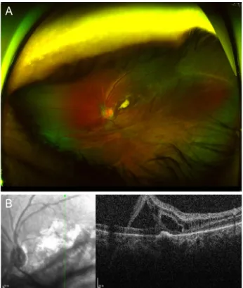
The family history of juvenile XLRS was unknown. Initial visual acuity (VA) testing showed the ability to fix-and-fol- low in both eyes. Intraocular pressure (IOP) was 69.4 mmHg in the right eye and 13.6 mmHg in the left, as determined via an ICare tonometer (ICare®, Tiolat, Helsinki, Finland). Fun- dus examination revealed hazy fundus in the right eye and choroidal effusion with inferior retinal detachment in the left (Fig. 1A, B). In the left eye, OCT confirmed retinoschisis of the inner retina involving the inferior macula (Fig. 1C, G).
B-scan ultrasonography revealed total retinal detachment in the right eye and inferior retinal detachment in the left. Brain magnetic resonance imaging from the previous hospital also depicted total retinal detachment in the right eye (Fig. 1H).
To rule out traumatic, infectious, genetic, congenital, and neoplastic causes of retinal detachment, the patient was admitted and underwent a uveitis workup and further brain and orbital magnetic resonance imaging, and the pediatric department was consulted. Ofloxacin q.i.d., prednisolone 1%
every 4 hours, latanoprost b.i.d., and dorzolamide/timolol b.i.d. eye drops in both eyes were initially prescribed. The day after admission, the patient’s IOP in the right eye dropped to 38 mmHg. Two days after admission, his VA was no light
Figure 1. Wide-angle fundus photography of right (A) and left (B) eyes. (C) B-scan ultrasound image of the right eye showed total retinal detach- ment with crystalline lens dislocation in the inferior region. (D) B-scan ultrasound image of the left eye revealed retinal detachment. (E, F) Optical coherence tomography of the left eye showed retinoschisis. (G) Slit-lamp photography demonstrated aphakia in the right eye. (H) Axial view of brain magnetic resonance imaging showed total inner retinal detachment in the right eye.
A
B
C
D
E
F
G
H
105
Kwak JY. Severe XLRS after trauma
https://doi.org/10.21561/jor.2019.4.2.103
perception in the right eye and counting fingers at 10 cm in the left eye. B-scan ultrasonography and slit-lamp examina- tion revealed a dislocated crystalline lens in the right eye (Fig. 1D-F). Detailed eye examination was performed under general anesthesia and revealed total retinal detachment in the right eye and inferior retinoschisis in the left. IOP was 12 mmHg in the right eye and 33 mmHg in the left. The condition of the right eye stabilized, and the patient was dis- charged with instructions for short-term follow up.
At the 1 week follow-up appointment, the patient’s VA was no light perception in the right eye and 0.1 in the left eye with IOP 16-18 mmHg in the right eye and 31.4 mmHg in the left. The patient was re-admitted for further evaluation.
He complained of right eye pain, and contrast-enhanced orbital computed tomography was performed and did not reveal any definite signs of cellulitis or any other periorbital inflammation. The IOP remained in the high 20 seconds in the left eye, and acetazolamide 50 mg q.i.d. mixed with food was prescribed. Four days later, the IOP in the left eye had stabilized in the mid-teens. The patient was discharged with
acetazolamide 75 mg q.i.d. Approximately 1 month later, the patient’s VA was 0.2 in the left eye. The IOP was 26 mmHg in the right eye and 20.6 mmHg in the left. Next-generation sequencing (NGS), which was performed to detect poten- tial genetic eye disorders, revealed RS1 gene mutation. No significant change in OCT was apparent, and acetazolamide 75 mg q.i.d. was maintained. At the 3 month follow-up ap- pointment, the patient’s VA was unchanged, and the IOP remained in the low to mid-teens in both eyes. OCT did not depict significant progression or improvement of retinoschi- sis.
Five months after the initial presentation, his VA was no light perception in the right eye and counting fingers at 30 cm in the left. OCT revealed slight improvement of ret- inoschisis in the left eye. The IOP was 17.4 mmHg in the right eye and 14.5 mmHg in the left. Acetazolamide was dis- continued after the patient complained of indigestion. Two weeks after discontinuation of acetazolamide, the IOP was 21 mmHg in the right eye and 16.7 mmHg in the left. Fundus examination and OCT results were stable (Fig. 2), and acet- azolamide administration was not reinitiated.
Discussion
In the present case of severe XLRS, the patient’s VA was im- paired by retinal detachments in both eyes. Based on initial OCT of the left eye, it is assumed that both eyes had severe retinoschisis. Trauma via a water hose to the eyes combined with underlying XLRS may have triggered total retinal de- tachment with crystalline lens dislocation and hemorrhage.
Posterior lens dislocation and vitreous hemorrhage, which occurred along with retinal detachment, could lead to high IOP, as was observed in this case [4,5]. In addition, uveitis precipitated by ocular trauma could induce high IOP. The right eye progressed to phthisis, and the left eye remained stable without surgical treatment. Although the patient’s condition had not deteriorated at the 6 month follow-up ap- pointment, the therapeutic options were limited.
One therapeutic approach in XLRS is acetazolamide. A number of reports describe cystoid macular edema that re- sponded to acetazolamide in patients with XLRS [6-8]. In one reported case, acetazolamide administration resulted in improved VA and improvement in cystoid macular edema [8]. In the report, acetazolamide still had some effect on the A
Figure 2. (A) Wide-angle fundus photography and (B) optical co- herence tomography of the left eye approximately 8 months after the patient’s initial visit.
B
106
JOURNAL OF RETINA
https://doi.org/10.21561/jor.2019.4.2.103 foveal schisis when it was reinitiated after recurrence of reti-
noschisis.
Acetazolamide is a carbonic anhydrase inhibitor that may alter fluid transport across the retinal pigment epithelium by increasing fluid outflow, and it may reduce the fluid con- tained in the macular schisis [6,8]. In a randomized prospec- tive study of 11 patients, Gurbaxani et al. [6] demonstrated that acetazolamide improved vision and reduced subretinal fluid in XLRS patients. In another report, Zhang et al. [7]
showed that acetazolamide responses and schisis size were strongly associated. They hypothesized that the disease may respond differently to the drug at each stage, depending on condition of the retinal pigment epithelium [7].
In the present case, there were two reasons for the use of acetazolamide. One reason was to reduce the retinoschisis, as explained above. However, because the patient initially presented with total retinal detachment in the right eye and severe inferior inner retinal detachment in the left eye, we hypothesize that the pumping function of the retinal pigment epithelium was highly compromised at the time of acetazol- amide administration. Hence, we speculate that the effect of acetazolamide was not sufficient to completely resolve the retinoschisis. The second reason for use of acetazolamide was to control the increased IOP in the right eye. After the IOP stabilized, it remained stable at subsequent follow-up visits.
We believe this is the first report of severe and rapid progression of XLRS to total retinal detachment after eye trauma. Because of the patient’s age and the severity of his eye condition at initial presentation, we performed NGS to screen for genetic disorders that may be associated with his condition. Designed by ophthalmologists and laboratory medicine specialists, the NGS panel specifically evaluated targeted sequencing of 429 genes related to retinopathy, and the only clinically significant mutation was in the RS1 gene (c.276G > C, p.Trp92Cys). The mechanisms behind the rap- id progression of the patient’s condition are unknown. One group recently studied the molecular mechanisms associated
with progression of XLRS based on RS1 secretion pattern in vitro [9]. The authors categorized patients into two groups based on RS1 secretion profile, and the results revealed that disease severity was not dependent on secretion profile.
Thus, more intricate molecular mechanisms may be involved in determination of disease severity. Therefore, further re- search is required to elucidate the mechanisms involved in the disease and to facilitate development of a highly effective treatment for XLRS patients with severe complications.
References
1. Leng T. Two cases of X-linked retinoschisis with different spectral domain optical coherence tomography findings. Clini Ophthal- mol 2012;6:1563-5.
2. Murro V, Caputo R, Bacci GM, et al. Case report of an atypical early onset X-linked retinoschisis in monozygotic twins. BMC Ophthalmol 2017;17:19.
3. Ansari WH, Browne AW, Singh RP. Juvenile X-linked retinoschisis responsive to intravitreal corticosteroids. Am J Ophthalmol Case Rep 2017;5:48-51.
4. González-Castaño C, Castro J, Alvarez-Sánchez M. Subluxation of the lens: etiology and results of treatment. Arch Soc Esp Oftalmol 2006;81:471-8.
5. Rojas L, Ortiz G, Gutiérrez M, Corredor S. Ghost cell glaucoma related to snake poisoning. Arch Ophthalmol 2001;119:1212-3.
6. Gurbaxani A, Wei M, Succar T, et al. Acetazolamide in retinoschi- sis: a prospective study. Ophthalmology 2014;121:802-3.
7. Zhang L, Reyes R, Lee W, et al. Rapid resolution of retinoschisis with acetazolamide. Doc Ophthalmol 2015;131:63-70.
8. Ghajarnia M, Gorin MB. Acetazolamide in the treatment of X-linked retinoschisis maculopathy. Arch Ophthalmol 2007;125:571-3.
9. Sudha D, Neriyanuri S, Sachidanandam R, et al. Understanding variable disease severity in X-linked retinoschisis: does RS1 secretory mechanism determine disease severity? PLoS One 2018;13:e0198086.