449
서 론
유방암은 조기에 원격전이가 잘 일어나는 고형종양으로 환자의 생존율은 원격전이의 유무에 의해서 많이 결정된 다. 현재까지 이 원격전이나 재발을 줄이고 생존율을 증가 시키기 위하여 항암 화학요법과 호르몬 조절요법이 사용되 고 있으며 현재의 예후 인자로는 종양의 크기와 임파선 전 이가 주된 지표로 사용되고 조직학적 분화도, 호르몬 수용 체의 발현 유무나 c-erb-B2 등이 치료의 결정에 도움이 되고 있다.(1-5) 그러나 재발의 대부분은 유방암의 진단 당시에 다른 장기에 미세 전이가 일어나고 이 종양세포가 추후에 계속 자라거나 보조치료로부터 살아 남아서 임상적으로 의 미를 가지는 크기로 자라면 전이나 재발로 판정하게 되는 것으로 알려져 있으며, 따라서 암의 생물학적 특징을 조기 에 알기 위하여 타 장기에 미세 전이가 존재하는가를 판정 하려는 노력이 꾸준히 있어 왔다. 유방암 환자에서 골수 미
유방암 환자에서 Cytokeratin 19와 Mammaglobin을 이용한 골수미세전이의 측정과 예후적 가치
아주대학교 의과대학 외과학교실, 1박희붕 외과, 2아주대학교 의과대학 병리학교실
정용식․이상림․정인호․윤태일․안상익․박희붕1․임현이2․김혜진․소의영․김명욱
책임저자:김명욱, 경기도 수원시 영통구 원천동 산 5번지 ꂕ 442-721, 아주대학교 의과대학 외과학교실 Tel: 031-219-5200, Fax: 031-19-5755
E-mail: [email protected]
접수일:2004년 11월 30일, 게재승인일:2005년 4월 14일 2004년도 대한외과학회 춘계학술대회에서 발표되었음.
Prognostic Value of Bone Marrow Micro- metastasis Detected by Nested RT-PCR for Cytokeratin 19 and Mammaglobin in Breast Cancer
Yong-Sik Jung, M.D., Sang-Lim Lee, M.D., In-Ho Jeong, M.D., Tae-Il Yoon, M.D., Sang-Ick Ahn, M.D., Hee Boong Park, M.D.1, Hyun-Ee Yim, M.D.2, Hye-Jin Kim, Euy-Young Soh, M.D. and Myung-Wook Kim, M.D.
Purpose: Breast cancers frequently undergo distant metas- tasis during the early phase, on which the survival of patients is greatly dependent. It has been suggested that the occurrence of micrometastasis relates with other prognostic features of breast cancer, such as lymph node metastasis and the presence of vascular invasion. The aim of this study was to examine the presence of keratin-19 and mammaglo- bin mRNA in bone marrow aspirates obtained from breast cancer patients, and their possible correlation with tumor staging and disease free survival.
Methods: Bone marrow samples were obtained from 254 breast cancer patients at the time of surgery. We separated the mononuclear fraction from the samples and carried out nested reverse transcriptase polymerase chain reaction for the detection of keratin-19 and mammaglobin mRNA using two different pairs of primers. We also studied the possible correlations between the tumor size, nodal involvement, stage, and distant metastasis.
Results: Seventy-five of the 254 samples were studied for cytokeratin 19 and the others for cytokeratin and mammaglo- bin. The median follow-up time was 21.1 months. Sixty-five (26%) of the 254 samples were cytokeratin 19 positive and 25 (14.3%) of the 175 were mammaglobin positive. Eight
cases (12.3%) in the cytokeratin positive group showed a recurrent disease in distant organs. Whereas, six (3.2%) out of 185 cytokeratin negative patients had distant recurrences.
Mammaglobin positivity was not correlated with distant metastasis. The stage, nodal status, and estrogen receptor were independent of bone marrow micrometastasis.
Conclusion: Bone marrow micrometastasis, detected by nested RT-PCR for cytokeratin 19, could be a useful predic- tive marker for the distant metastasis of breast cancer. (J Korean Surg Soc 2005;68:449-456)
Key Words: Breast carcinoma, Cytokeratin-19, Mammaglo- bin, Micrometastasis, Nested RT-PCR, Recur- rence
중심 단어: 유방암, 골수미세전이, 역전사 중합 효소 반응, 싸이토 케라틴 19, 맘마글로빈 ꠏꠏꠏꠏꠏꠏꠏꠏꠏꠏꠏꠏꠏꠏꠏꠏꠏꠏꠏꠏꠏꠏꠏꠏꠏꠏꠏꠏꠏꠏꠏꠏꠏꠏꠏꠏꠏꠏꠏꠏꠏꠏꠏꠏꠏꠏꠏꠏꠏꠏ Department of Surgery, Ajou University, School of Medicine,
1Park Hee Boong Breast Clinic, 2Department of Pathology, Ajou University School of Medicine, Suwon, Korea
ꠏꠏꠏꠏꠏꠏꠏꠏꠏꠏꠏꠏꠏꠏꠏꠏꠏꠏꠏꠏꠏꠏꠏꠏꠏꠏꠏꠏꠏꠏꠏꠏꠏꠏꠏꠏꠏꠏꠏꠏꠏꠏꠏꠏꠏꠏꠏꠏꠏꠏꠏꠏꠏꠏꠏꠏꠏꠏꠏꠏꠏꠏꠏꠏꠏꠏꠏꠏꠏꠏꠏꠏꠏꠏꠏꠏꠏꠏꠏꠏꠏꠏꠏꠏꠏꠏꠏꠏꠏꠏꠏꠏꠏꠏꠏꠏꠏꠏꠏꠏꠏꠏꠏꠏꠏꠏꠏꠏꠏꠏꠏꠏꠏꠏꠏ 세전이에 대해서는 Redding 등에 의해 처음 시도되었으며
여러 연구에서 예후와 관계가 있음이 제시되었다.(6,7) 그러 나 초기의 연구에서는 미세전이의 발견율이 낮아서 그 후 로 민감도를 높이기 위해 많은 노력이 지속되었다. 현재 두 가지 방법이 널리 쓰이는데 골수 세포에서 유핵 세포 분획 을 분리하여 상피세포에 존재하고 골수세포에는 존재하지 않는 단백질에 대한 면역화학염색법을 이용하여 전이세포 를 발견하는 방법이 있고 다른 하나는 특정 단백질의 생성 에 필요한 m-RNA를 역전사 중합효소 연쇄반응법(RT-PCR) 에 의해서 발견하는 방법이다.(8-10) 또한 골수에서 미세전 이를 발견하는데 이용되는 표지자로는 cytokeratin과 mam- maglobin, CEA 등이 널리 이용되고 있으며 연구자에 따라 각기 다른 결과들이 보고되고 있어 이에 대한 연구는 아직 논란의 여지가 있는 것이 사실이다.
저자들은 RT-PCR법을 개선하여 감수성을 높인 nested RT-PCR법을 이용하여 cytokeratin 19와 mammaglobin에 대 해 골수 내에서의 미세전이를 검사하였으며 골수 미세 전 이 여부와 임상적 특징과의 관련성에 대해 연구하였으며 원격전이와의 관련성에 대해 연구하였다.
방 법
환자 대상은 2001년 6월부터 2003년 12월까지 아주대학 교병원에 내원하여 유방암으로 수술 받은 환자 중 골수 keratin-19 혹은 mammaglobin에 대한 RT-PCR법을 시행한 환자 254명을 대상으로 하여 검사결과와 임상결과를 비교 하였다.
실험방법은 수술 전 골수 천자를 시행하여 골수를 채취 한 후 유핵 세포를 분리하고 이들 세포에서 acid guanidine phenol chloroform 방법을 이용하여 total RNA를 추출한 후 cytokeratin-19 혹은 mammaglobin에 대해 각기 다른 두 쌍의 시발체(primer)를 이용하여 nested RT-PCR을 시행하였다 (Table 1). Nested RT-PCR 후 증폭된 DNA를 2% agarose gell 에 전기영동하여 확인하였다. Total RNA에 대한 control은 베 타 액틴(β-actin)을 이용하였다. 자세한 과정은 다음과 같다.
1) RNA 추출
유방암 환자로부터 채취한 골수 10 ml을 lymphoprep (Nycomed) 동량에 띄운 후 2,700 rpm에서 20분간 원심분리 하여 세포분획을 취하여 -70oC에 보관하였다. Trizol (Gibco BRL)을 첨가하여 total RNA 추출 후 분광광도계를 이용하 여 260과 280 nm에서 측정하여 순도와 농도를 측정하였다.
2) Oligonucleotide primer 합성
네 가지의 oligonucleotide primer는 cytokeratin 19 DNA에 상보적으로 결합하는 DNA 단편으로서 external primer set인 K19ES (external sense) 5'-AGGTGGATTCCGCTCCGGGCA-
3', K19EA (external antisense) 5'-GCTCGAGGGACAGGAA GAT-3'는 RT-PCR에 사용되어 461개 염기 쌍의 산물을 합 성하게 되며 inner primer set인 K19IS (internal sense) 5'- GACATGCGAAGCCAATATGAGG-3', K19IA (internal anti- sensense) 5'-GCTGCCTTGGAAGACACACT-3'는 nested PCR 에 사용되어 최종적으로 221개 염기 쌍의 PCR 산물을 합성 하는데 사용하였다. Mammaglobin의 DNA에 상보적으로 결 합하는 primer set는 external sense 5'-GAAGTTGCTGATG GTCCTCATGCTGGC-3', external antisense는 5'-GTCACCA TACCCTGCAGTTCTGTGAGC-3'가 사용되어 325개의 염기 쌍을 합성하게 되며 internal sense로 5'-GTCCCAGCACTG CTACGCAGGCTC-3', internal antisense로 5'-CACCTCAAC ATTGCTCAGAGTTTCATCCG-3'로 사용되어 201개 염기 쌍의 산물을 합성하는데 사용되었다.
3) RT-PCR과 nested PCR
PCR 반응액(50 mM KCl, 10 mM Tris-HCl (pH 9.0), 0.1%
Triton X-100, 200 uM dNTP, 2.5 mM MgCl2, 0.1 mM DTT, 60 U M-MLV reverse transcriptase, 0.5 U taq polymerase, 12 Table 1. Relationship between expression of bone marrow cyto-
keratin 19 and age and TNM staging
ꠚꠚꠚꠚꠚꠚꠚꠚꠚꠚꠚꠚꠚꠚꠚꠚꠚꠚꠚꠚꠚꠚꠚꠚꠚꠚꠚꠚꠚꠚꠚꠚꠚꠚꠚꠚꠚꠚꠚꠚꠚꠚꠚꠚꠚꠚꠚꠚꠚꠚꠚꠚꠚꠚꠚ All patients CK 19 (+)* CK 19 (-)
ꠏꠏꠏꠏꠏꠏꠏꠏꠏꠏꠏꠏꠏꠏꠏꠏꠏꠏꠏꠏꠏꠏꠏꠏꠏꠏꠏꠏꠏꠏꠏꠏꠏꠏꠏꠏꠏ P value
N=66 N=188
N=254
(26%) (74%)
ꠏꠏꠏꠏꠏꠏꠏꠏꠏꠏꠏꠏꠏꠏꠏꠏꠏꠏꠏꠏꠏꠏꠏꠏꠏꠏꠏꠏꠏꠏꠏꠏꠏꠏꠏꠏꠏꠏꠏꠏꠏꠏꠏꠏꠏꠏꠏꠏꠏꠏꠏꠏꠏꠏꠏ
Age, mean 46 44.2 46.5
(25∼80) (25∼74) (28∼80) NS Stage
DCIS 16 (6.3%) 1 (6.2%) 15 (93.8%)
NS
I 58 (22.8%) 15 (25.9%) 43 (74.1%) II 126 (49.6%) 35 (27.8%) 91 (72.2%) III 43 (16.6%) 12 (27.9%) 31 (72.1%) IV 4 (1.6%) 1 (25.0%) 3 (75.0%) T stage
Tis 16 (6.3%) 1 (6.2%) 15 (93.8%) T1 80 (31.5%) 19 (23.8%) 61 (76.2%) T2 139 (54.7%) 41 (29.5%) 98 (70.5%) NS T3 7 (2.7%) 2 (28.6%) 5 (71.4%) T4 7 (2.7%) 1 (14.3%) 6 (85.7%) N stage
N0 150 (59.1%) 39 (26.0%) 111 (74.0%) N1 57 (22.4%) 14 (24.6%) 43 (75.4%) N2 18 (7.1%) 4 (22.2%) 14 (77.8%) NS N3 24 (9.4%) 7 (29.2%) 17 (70.8%) ꠏꠏꠏꠏꠏꠏꠏꠏꠏꠏꠏꠏꠏꠏꠏꠏꠏꠏꠏꠏꠏꠏꠏꠏꠏꠏꠏꠏꠏꠏꠏꠏꠏꠏꠏꠏꠏꠏꠏꠏꠏꠏꠏꠏꠏꠏꠏꠏꠏꠏꠏꠏꠏꠏꠏ
*CK-19 = cytokeratin 19.
ꠏꠏꠏꠏꠏꠏꠏꠏꠏꠏꠏꠏꠏꠏꠏꠏꠏꠏꠏꠏꠏꠏꠏꠏꠏꠏꠏꠏꠏꠏꠏꠏꠏꠏꠏꠏꠏꠏꠏꠏꠏꠏꠏꠏꠏꠏꠏꠏꠏꠏꠏꠏꠏꠏꠏꠏꠏꠏꠏꠏꠏꠏꠏꠏꠏꠏꠏꠏꠏꠏꠏꠏꠏꠏꠏꠏꠏꠏꠏꠏꠏꠏꠏꠏꠏꠏꠏꠏꠏꠏꠏꠏꠏꠏꠏꠏꠏꠏꠏꠏꠏꠏꠏꠏꠏꠏꠏꠏꠏꠏꠏꠏꠏꠏꠏ U RNase inhibitor, 10 mM sense primer, 10 mM antisense
primer)에 total RNA 1 ug과 DEPC처리 DW를 넣어 반응액의 최종량을 30 ul로 하였다. DNA thermal cycler (Perkin Elmer Cetus)를 이용하여 42oC에서 1시간 반응하여 cDNA를 합성 하고 denaturation (94oC, 1분), annealing (57oC, 1.5분), exten- sion (72oC, 2분)의 PCR cycle을 35회 반복한 후 마지막 extension은 10분간 유지시켜 RT-PCR을 수행하였다. 이 시 료를 2 ul를 취하여 위와 동일한 조건으로 nested RT-PCR을 수행하였다. 이 시료를 2% agarose gel에서 전기영동하여 ethidium bromide 염색 후 UV illuminator 하에서 밴드를 확 인하였다.
4) 민감도와 특이도
검사의 특이도 확인을 위해서 14명의 골수 이식 공여자 에게서 채취된 골수를 이용하였으며 mRNA의 검출의 대조 군으로 모든 세포에서 발현되는 β-actin의 RNA를 사용하 였다. Nested RT-PCR의 감수성을 확인하기 위하여 MCF 7 과 SKBR3 세포주를 이용하였다. Cytokeratin 19 mRNA의 민감도 확인을 위해 MCF 7 세포주를 이용하였고 Mam- maglobin의 민감도 확인을 위해 SKBR3 세포주를 이용하였 다. 1,000개의 세포주를 건강한 자원자의 10 ml의 말초혈액 에 투여한 뒤 5 ml의 샘플에 5 ml의 말초혈액을 계속 투여 하여 세포주의 수가 말초혈액 5 ml당 3개가 될 때까지 희석 시켰으며 각각의 희석된 5 ml의 말초혈액에서 RNA를 추출 하여 nested RT-PCR을 시행하였다.
5) 환자의 추적관찰
환자는 3개월 간격으로 진찰을 시행하고 말초 혈액검사와 흉부 촬영, 복부초음파 검사와 골 주사를 6개월 간격으로 시행하여 재발여부를 검사하였다. 통계 처리는 SPSS (version 11.0)를 이용하였으며 기존의 예후인자와 재발과의 관계에 대해 Pearson chi square test로, 골수미세전이와 재발
과의 관계를 Kaplan-Meier법을 이용하여 log-rank test로 통 계적 의의를 검사하였으며 P value가 0.05 이하일 때 의미가 있는 것으로 판정하였다.
결 과
1) 민감도와 특이도
14명의 골수 이식 공여자에게서 채취된 골수를 이용한 특이도 검사에서 cytokeratin 19 및 mammaglobin이 한 예에 서도 검출되지 않아 nested RT-PCR의 검사 특이도는 100%
였다(Fig. 1).
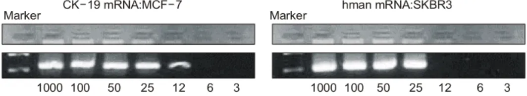
MCF 7과 SKBR 3 세포주를 이용한 민감도 검사에서 희 석된 5 ml의 말초혈액에서 MCF 7 세포 12개와 SKBR 3 세 포 25개를 검출할 수 있어 cytokeratin 19의 민감도가 좀 더 높음을 알 수 있었다(Fig. 2).
2) 임상양상 및 발현율
총 254명의 유방암 환자가 본 연구에서 분석되었으며 여 자가 252명, 남자가 2명이었다. 환자의 나이는 25세부터 80 세까지였으며 평균 나이는 46세였다. 환자의 병기는 관상 피내암이 16명, 1기암 58명, 2기 126명, 3기 43명, 4기 4명이 었으며 나머지 7명은 정확한 병기가 파악되지 않았다.
연구 초기의 76명은 cytokeratin 19만을 측정하였고 나머 지 178명에 대해서는 cytokeratin 19와 mammaglobin 두 가지 를 함께 측정하였다.
골수에서 cytokeratin 19가 양성인 군은 254명 중 66명으 로 26%였고 mammaglobin이 양성인 군은 178명 중 27명으 로 15.2%였다. cytokeratin 19와 mammaglobin이 함께 발현된 군은 11명으로 6.2%였다.
3) 종양의 병기와 골수 cytokeratin 19 결과의 비교 관상피내암 16예 중 1예(6.3%)에서 양성을 보였으며 1기
Fig. 1. Nested RT-PCR for β-actin, cytokeratin-19, mammaglobin in control group (A) and patients bone marrow sam- ples (B). β-actin shows pos- itive result in all samples of normal bone marrow trans- plant donors but cytokeratin- 19 and mammaglobin are not.
CK 19- Mamm B actin-
221 bp 202 bp 154 bp Marker 1 2 3 4 5 6 7 8 9 10 11 12 13
1 2 3 4 5 6 7 8 9 10 11 12 14
M 13 15 P Np
CK 19-
Mamm
221 bp
202 bp
A
B
ꠏꠏꠏꠏꠏꠏꠏꠏꠏꠏꠏꠏꠏꠏꠏꠏꠏꠏꠏꠏꠏꠏꠏꠏꠏꠏꠏꠏꠏꠏꠏꠏꠏꠏꠏꠏꠏꠏꠏꠏꠏꠏꠏꠏꠏꠏꠏꠏꠏꠏꠏꠏꠏꠏꠏꠏꠏꠏꠏꠏꠏꠏꠏꠏꠏꠏꠏꠏꠏꠏꠏꠏꠏꠏꠏꠏꠏꠏꠏꠏꠏꠏꠏꠏꠏꠏꠏꠏꠏꠏꠏꠏꠏꠏꠏꠏꠏꠏꠏꠏꠏꠏꠏꠏꠏꠏꠏꠏꠏꠏꠏꠏꠏꠏꠏ
58예 중 15예(25.9%), 2기 126예 중 35예(27.8%), 3기 43예 중 12예(27.9%), 4기 4예 중 1예(25%)에서 골수에서 cytoker- atin 19가 발현되었으며 병기에 따른 차이는 없었다(Table 1).
세부적으로도 T1 병변에서 19예(23.8%), T2 병변에서 41 예(29.5%), T3 병변에서 2예(28.6%), T4 병변에서 1예(14.3%) 로 종양의 크기와 골수미세전이 사이의 통계적 유의성은 관찰되지 않았으며 임파선 전이와도 관계가 없는 것으로 나타났다. 다른 예후 인자인 호르몬 수용체나 c-erb B2의 발 현 여부와 골수 cytokeratin 19의 발현 여부는 관계가 없었으 며 조직학적 분화도와 골수 cytokeratin 19의 발현간에도 차
이가 없었다.
4) 종양의 병기와 골수 mammaglobin 결과의 비교 관상피내암 9예 모두 골수 mammaglobin 발현이 관찰되 지 않았으며 1기 42명 중 9명(21.4%), 2기 82명 중 11명 (13.4%), 3기 36명 중 5명(13.9%), 4기 3명 중 2명(66.7%)에 서 골수 미세전이가 관찰되어 병기와 mammaglobin의 발현 간에는 통계적인 유의성이 관찰되지 않았다(Table 2).
종양의 크기와 골수 mammaglobin의 발현은 T1에서 11명 (20%), T2에서 14명(14.3%), T3에서 1명(14.3%), T4에서 1명 (20%)으로 관계가 없었으며 임파선 전이와도 통계적 유의 성이 관찰되지 않았다. 기타 호르몬 수용체, c-erb B2의 발 현 및 조직학적 분화도도 골수 mammaglobin의 발현에 영향 을 주지 않는 것으로 나타났다.
5) 골수 미세 전이와 원격전이와의 관계
총 254예 중 진단 당시 종양의 병기가 4기였던 4명을 제 외한 250명에 대해 재발여부를 조사하였다. 추적관찰 기간 은 5개월에서 33개월이었으며 평균 관찰 기간은 15개월이 었다. 이 기간 중 18예의 재발이 관찰되었으며 이중 원격전 이는 14예에서 관찰되었다. 원격전이 부위는 골 전이가 9예 로 가장 많았으며 폐 전이 3예, 뇌 전이 1예, 간 전이 1예였 으며 수술 후 재발까지의 평균 기간은 12.9개월(4∼32개월) 이였다. 병기에 따라서는 관상피내암과 1기암에서는 원격 Fig. 2. Sensitivity test of nested RT-PCR for cytokeratin 19 and mammaglobin. One thousand cells of the breast cancer cell line MCF7 and SKBR3 were transferred into 10 ml of peripheral blood of healthy volunteers and diluted with 5ml of peripheral blood until reaching a calculated concentration of 3 cells in 5 ml of peripheral blood. Cytokeratin-19 could be detected in 12 cells dilution and mammaglobin could be detected in 25 cells dilution.
Marker Marker
1000 100 50 25 12 6 3 1000 100 50 25 12 6 3 CK 19 mRNA:MCF 7- - hman mRNA:SKBR3
Table 2. Relationship between expression of bone marrow mam- maglobin and age and TNM staging
ꠚꠚꠚꠚꠚꠚꠚꠚꠚꠚꠚꠚꠚꠚꠚꠚꠚꠚꠚꠚꠚꠚꠚꠚꠚꠚꠚꠚꠚꠚꠚꠚꠚꠚꠚꠚꠚꠚꠚꠚꠚꠚꠚꠚꠚꠚꠚꠚꠚꠚꠚꠚꠚꠚꠚ All patients Hmam (+)* Hmam (-)
ꠏꠏꠏꠏꠏꠏꠏꠏꠏꠏꠏꠏꠏꠏꠏꠏꠏꠏꠏꠏꠏꠏꠏꠏꠏꠏꠏꠏꠏꠏꠏꠏꠏꠏꠏꠏꠏ P value
N=27 N=151
N=178
(15.2%) (84.8%)
ꠏꠏꠏꠏꠏꠏꠏꠏꠏꠏꠏꠏꠏꠏꠏꠏꠏꠏꠏꠏꠏꠏꠏꠏꠏꠏꠏꠏꠏꠏꠏꠏꠏꠏꠏꠏꠏꠏꠏꠏꠏꠏꠏꠏꠏꠏꠏꠏꠏꠏꠏꠏꠏꠏꠏ
Age, mean 46.5 46.6 46.5
(25∼80) (25∼80) (28∼64) NS Stage
DCIS 9 (5.1%) 0 (0.0%) 9 (100%) I 42 (23.6%) 9 (21.4%) 33 (78.6%) II 82 (46.1%) 11 (13.4%) 71 (86.6%) NS III 36 (20.2%) 5 (13.9%) 31 (86.1%) IV 3 (2.0%) 2 (66.7%) 1 (33.3%) T stage
Tis 9 (5.1%) 0 (0.0%) 9 (100%) T1 55 (30.9%) 11 (20.0%) 44 (80.0%) T2 98 (55.1%) 14 (14.3%) 84 (85.7%) NS T3 7 (3.9%) 1 (14.3%) 6 (85.7%) T4 5 (2.8%) 1 (20.0%) 4 (80.0%) N stage
N0 104 (58.4%) 13 (12.5%) 91 (87.5%) N1 37 (20.8%) 6 (16.2%) 31 (83.8%) N2 15 (8.4%) 3 (20.0%) 12 (80.0%) NS N3 19 (10.7%) 4 (21.1%) 15 (78.9%)
ꠏꠏꠏꠏꠏꠏꠏꠏꠏꠏꠏꠏꠏꠏꠏꠏꠏꠏꠏꠏꠏꠏꠏꠏꠏꠏꠏꠏꠏꠏꠏꠏꠏꠏꠏꠏꠏꠏꠏꠏꠏꠏꠏꠏꠏꠏꠏꠏꠏꠏꠏꠏꠏꠏꠏ
*hmam = mammaglobin.
Table 3. Incidence of distant metastasis according to stage ꠚꠚꠚꠚꠚꠚꠚꠚꠚꠚꠚꠚꠚꠚꠚꠚꠚꠚꠚꠚꠚꠚꠚꠚꠚꠚꠚꠚꠚꠚꠚꠚꠚꠚꠚꠚꠚꠚꠚꠚꠚꠚꠚꠚꠚꠚꠚꠚꠚꠚꠚꠚꠚꠚꠚ
Number Number of
Stage of distant P value
cases
metastasis
ꠏꠏꠏꠏꠏꠏꠏꠏꠏꠏꠏꠏꠏꠏꠏꠏꠏꠏꠏꠏꠏꠏꠏꠏꠏꠏꠏꠏꠏꠏꠏꠏꠏꠏꠏꠏꠏꠏꠏꠏꠏꠏꠏꠏꠏꠏꠏꠏꠏꠏꠏꠏꠏꠏꠏ
DCIS 16 0 0.003
Stage I 58 0
Stage II 126 10
Stage III 43 4
ꠏꠏꠏꠏꠏꠏꠏꠏꠏꠏꠏꠏꠏꠏꠏꠏꠏꠏꠏꠏꠏꠏꠏꠏꠏꠏꠏꠏꠏꠏꠏꠏꠏꠏꠏꠏꠏꠏꠏꠏꠏꠏꠏꠏꠏꠏꠏꠏꠏꠏꠏꠏꠏꠏꠏ
Total 243 14
ꠏꠏꠏꠏꠏꠏꠏꠏꠏꠏꠏꠏꠏꠏꠏꠏꠏꠏꠏꠏꠏꠏꠏꠏꠏꠏꠏꠏꠏꠏꠏꠏꠏꠏꠏꠏꠏꠏꠏꠏꠏꠏꠏꠏꠏꠏꠏꠏꠏꠏꠏꠏꠏꠏꠏ
ꠏꠏꠏꠏꠏꠏꠏꠏꠏꠏꠏꠏꠏꠏꠏꠏꠏꠏꠏꠏꠏꠏꠏꠏꠏꠏꠏꠏꠏꠏꠏꠏꠏꠏꠏꠏꠏꠏꠏꠏꠏꠏꠏꠏꠏꠏꠏꠏꠏꠏꠏꠏꠏꠏꠏꠏꠏꠏꠏꠏꠏꠏꠏꠏꠏꠏꠏꠏꠏꠏꠏꠏꠏꠏꠏꠏꠏꠏꠏꠏꠏꠏꠏꠏꠏꠏꠏꠏꠏꠏꠏꠏꠏꠏꠏꠏꠏꠏꠏꠏꠏꠏꠏꠏꠏꠏꠏꠏꠏꠏꠏꠏꠏꠏꠏ 전이가 관찰되지 않았으며 2기와 3기에서만 관찰되어 통계
적 의의가 있는 것으로 나타났다(Table 3). Cytokeratin 19를 측정한 250명 중 골수에서 음성인 185명 중 6예에서 원격 전이가 관찰되었으며 양성인 65명 중 8예에서 원격 전이가 관찰되어 골수 미세 전이 여부와 원격 전이 간에 통계적으 로 유의한 차이를 나타내었다(Table 4A). Mammaglobin을 측정한 175명 중 골수에서 음성인 150명 중 10명, 양성인 25명 중 1명에서 원격전이가 관찰되어 통계적인 유의성은 없었다(Table 4B).
골수 미세 전이와 원격 전이와의 상관 관계를 log rank
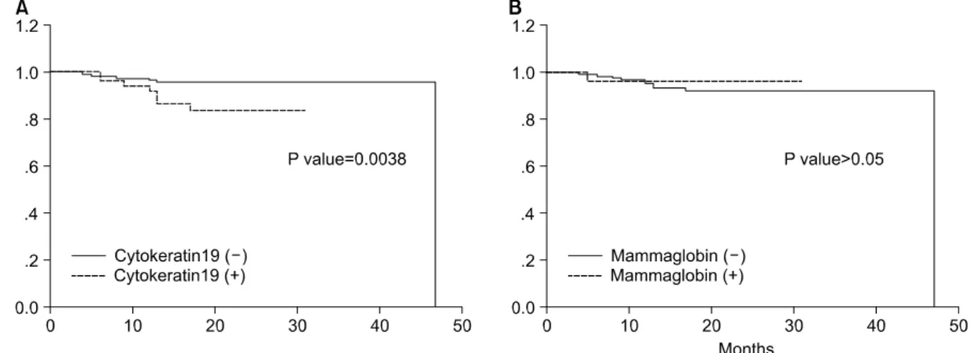
test를 이용하여 무전이 생존율을 구하였다. 15개월 간의 평 균 추적관찰 기간 동안 골수cytokeratin 19 음성 군에서는 96.8%, 골수 cytokeratin 19 양성 군에서는 87.7%로 통계적으 로 유의한 차이를 나타내었으며 골수mammaglobin의 발현 여부에 따라서는 무전이 생존율에 차이가 없었다(Fig. 3).
병기에 따른 무전이 생존율도 관상피내암이 100%, 1기암이 100%, 2기암 92%, 3기암 90%로 병기에 따라 낮아지는 양상 을 보였으나 통계적인 의의는 없는 것으로 관찰되었다.
고 찰
현재까지 알려진 유방암의 예후 인자로는 액와부 임파선 전이를 들 수 있다. 그러나 임파선에 전이가 없는 유방암 환자의 25∼30%에서 원격 전이가 관찰되어 기존의 예후 인 자 외에 생존율에 영향을 미치는 다른 인자를 찾기 위한 노력이 계속되어 왔으며 최근 들어 면역화학염색법이나 역 전사 효소 중합 반응 등을 이용하여 골수나 말초혈액에서 암세포를 검출하고자 하는 시도가 꾸준히 진행되어 오고 있다.(11,12)
면역화학염색법을 통한 연구는 오랜 역사를 가지고 있으 며 많은 연구가 이를 기반으로 해서 진행되어 왔으나 초기 의 연구에 사용되었던 항체들은 정상 골수 세포의 세포막 에도 반응을 보여 10% 가까운 위양성률을 보이므로 특이도 가 낮다는 단점이 있으며 이후 사용된 cytokeratin family (CK-8, CK-18, CK-19, CK-20) 등을 통해 향상되기는 했으나 항체의 다양성과 정상 골수 세포와의 교차 반응, 판독상의 문제 등으로 인해 만족할 만한 민감도와 특이도를 보여주 지 못하는 것이 사실이다.(13-15)
역전사 중합 효소 반응법을 이용한 방법은 미세전이를 발견하는 가장 예민한 방법이며 106∼107개의 골수세포 중 에서 한 개의 암세포를 발견할 수 있는 것으로 알려져 있다.
Fig. 3. Distant metastasis free survival according to expression of bone marrow cytokeratin 19 (A) and mammaglobin (B). Distant metastasis free survival curve by cytokeratin 19 (A) shown statistically significant difference between groups but mammaglobin was not (B).
Table 4. Incidence of distant metastasis according to expression of cytokeratin 19 (Table A) and mammaglobin (Table B) A.
ꠚꠚꠚꠚꠚꠚꠚꠚꠚꠚꠚꠚꠚꠚꠚꠚꠚꠚꠚꠚꠚꠚꠚꠚꠚꠚꠚꠚꠚꠚꠚꠚꠚꠚꠚꠚꠚꠚꠚꠚꠚꠚꠚꠚꠚꠚꠚꠚꠚꠚꠚꠚꠚꠚꠚ Number of
Number
distant P value of cases
metastasis
ꠏꠏꠏꠏꠏꠏꠏꠏꠏꠏꠏꠏꠏꠏꠏꠏꠏꠏꠏꠏꠏꠏꠏꠏꠏꠏꠏꠏꠏꠏꠏꠏꠏꠏꠏꠏꠏꠏꠏꠏꠏꠏꠏꠏꠏꠏꠏꠏꠏꠏꠏꠏꠏꠏꠏ
CK-19 negative 185 6 0.019
CK-19 positive 65 8
ꠏꠏꠏꠏꠏꠏꠏꠏꠏꠏꠏꠏꠏꠏꠏꠏꠏꠏꠏꠏꠏꠏꠏꠏꠏꠏꠏꠏꠏꠏꠏꠏꠏꠏꠏꠏꠏꠏꠏꠏꠏꠏꠏꠏꠏꠏꠏꠏꠏꠏꠏꠏꠏꠏꠏ
Total 250 14
ꠏꠏꠏꠏꠏꠏꠏꠏꠏꠏꠏꠏꠏꠏꠏꠏꠏꠏꠏꠏꠏꠏꠏꠏꠏꠏꠏꠏꠏꠏꠏꠏꠏꠏꠏꠏꠏꠏꠏꠏꠏꠏꠏꠏꠏꠏꠏꠏꠏꠏꠏꠏꠏꠏꠏ
B.
ꠚꠚꠚꠚꠚꠚꠚꠚꠚꠚꠚꠚꠚꠚꠚꠚꠚꠚꠚꠚꠚꠚꠚꠚꠚꠚꠚꠚꠚꠚꠚꠚꠚꠚꠚꠚꠚꠚꠚꠚꠚꠚꠚꠚꠚꠚꠚꠚꠚꠚꠚꠚꠚꠚꠚ Number of
Number
distant P value of cases
metastasis
ꠏꠏꠏꠏꠏꠏꠏꠏꠏꠏꠏꠏꠏꠏꠏꠏꠏꠏꠏꠏꠏꠏꠏꠏꠏꠏꠏꠏꠏꠏꠏꠏꠏꠏꠏꠏꠏꠏꠏꠏꠏꠏꠏꠏꠏꠏꠏꠏꠏꠏꠏꠏꠏꠏꠏ
Mammaglobin negative 150 10 0.61
Mammaglobin positive 25 1
ꠏꠏꠏꠏꠏꠏꠏꠏꠏꠏꠏꠏꠏꠏꠏꠏꠏꠏꠏꠏꠏꠏꠏꠏꠏꠏꠏꠏꠏꠏꠏꠏꠏꠏꠏꠏꠏꠏꠏꠏꠏꠏꠏꠏꠏꠏꠏꠏꠏꠏꠏꠏꠏꠏꠏ
Total 175 11
ꠏꠏꠏꠏꠏꠏꠏꠏꠏꠏꠏꠏꠏꠏꠏꠏꠏꠏꠏꠏꠏꠏꠏꠏꠏꠏꠏꠏꠏꠏꠏꠏꠏꠏꠏꠏꠏꠏꠏꠏꠏꠏꠏꠏꠏꠏꠏꠏꠏꠏꠏꠏꠏꠏꠏ
ꠏꠏꠏꠏꠏꠏꠏꠏꠏꠏꠏꠏꠏꠏꠏꠏꠏꠏꠏꠏꠏꠏꠏꠏꠏꠏꠏꠏꠏꠏꠏꠏꠏꠏꠏꠏꠏꠏꠏꠏꠏꠏꠏꠏꠏꠏꠏꠏꠏꠏꠏꠏꠏꠏꠏꠏꠏꠏꠏꠏꠏꠏꠏꠏꠏꠏꠏꠏꠏꠏꠏꠏꠏꠏꠏꠏꠏꠏꠏꠏꠏꠏꠏꠏꠏꠏꠏꠏꠏꠏꠏꠏꠏꠏꠏꠏꠏꠏꠏꠏꠏꠏꠏꠏꠏꠏꠏꠏꠏꠏꠏꠏꠏꠏꠏ 그러나 유방암 자체의 특이적인 표지자가 적고,(16,17) 골수
세포에서도 낮은 양의 cytokeratin 메신저 RNA가 검출이 된 다는 점,(18) 그리고 골수 채취 과정에서 오염으로 인한 위 양성이 있을 수 있다는 점 등이 결점으로 지적된다.(19) 역 전사 중합 효소 반응법을 이용한 골수 미세전이 측정에는 cytokeratin과 mammaglobin, CEA 등이 널리 이용되고 있으 며 이 중 가장 널리 이용되는 표지자는 cytokeratin이다. 본 연구에서 골수전이의 표지자로 사용한 것은 cytokeratin 19 로서 상피세포에서만 발현되는 것으로 골수세포에서는 정 상적으로 발현되지 않는다. 이러한 cytokeratin에는 8,18,19 등이 있으며 이중 keratin(8,18)은 위양성이 많은 것으로 알 려져 있고 keratin 19는 유방암에서 높은 특이를 나타내고 혈액 및 골수에서 민감도가 높아 미세전이의 표지자로 적 합하며 실제로 Schoenfeld 등(20)의 연구에서 면역화학염색 법과 비교하여 10배 가까운 민감도를 나타내는 것으로 보 고되었다. 여러 연구에서 보고된 골수에서의 cytokeratin의 발현율은 30∼49% 정도이며 평균적으로 30% 전후로 알려 져 있다.(21,22) 본 연구에서도 26%의 발현율을 보여 비슷 한 결과를 보였다. Silva 등(23)은 본 연구와 같은 nested RT-PCR법을 이용하여 49%의 발현율을 보고하였으나 민감 도 검사에서 일반적인 역전사 중합효소 연쇄반응법에서와 유사한 수준을 나타냈고 검사 대상의 수가 적어 실제로 nested RT-PCR법이 전통적인 역전사 중합효소 연쇄반응법 에 비해 뛰어난 민감도를 나타낸다고 보기는 어렵다.
Mammaglobin을 이용한 말초 혈액에서의 미세전이는 Zach 등(24)에 의해서 최초로 보고되었으며 cytokeratin에 비 해 유방 조직에 대한 특이도가 높아 미세 전이 측정의 유용 한 대안으로 많이 연구되고 있다.(25) 역전사 중합 효소 반 응법을 이용한 방법에서 골수 혹은 말초혈액에서 mam- maglobin의 양성률은 9∼54% 정도로 연구에 따라 다양하게 나타나며 이는 연구에 원격전이가 있는 유방암의 포함 여 부에 의해서 차이가 나타나는 것으로 생각된다.(26-28) 골수 미세전이와 기존의 예후 인자들간의 연관성에 대해 서는 여러 연구 간에 다른 결과를 보여 아직 논란의 여지가 있다. Mansi 등(29)은 골수 미세전이의 발현이 종양의 크기, 임파선 전이, 호르몬 수용체의 발현과 연관이 있다고 보고 하였고, Gebauer 등(30)은 임파선 전이와 관련이 있다고 보 고하였다. 그러나 다른 연구에서는 기존의 예후 인자와 연 관성이 관찰되지 않는다고 보고하여 추후 더 많은 연구가 더 필요할 것이다. 본 연구에서도 종양의 크기나 임파선 전 이 등에 상관 없이 비슷한 발현을 보였으며 호르몬 수용체, 조직학적 등급, 혈관침윤과도 연관이 없는 것으로 나타났 다. 관상피내암 16예 중 1예에서 cytokeratin 19가 발현되었 으며 이는 채취 과정에서 피부로부터 오염이 되었거나 혹 은 종양의 일부에서 침윤성 유방암이 존재하고 있을 가능 성을 시사하는 소견이라고 할 수 있다.
종양의 크기, 임파선 전이, 호르몬 수용체의 발현, 조직학
적 등급 등은 중요한 예후 인자들이나 본 연구에서는 생존 율 곡선간에 유의한 차이를 나타내지 못하였다. 이는 추적 관찰 기간이 짧고 재발의 예가 적기 때문인 것으로 생각된 다. 그럼에도 불구하고 cytokeratin이 골수에서 발현된 경우 3배 이상의 원격전이를 보이고 무전이 생존율에 있어서도 통계적으로 유의한 차이를 나타내는 것을 볼 때 이러한 골 수미세전이가 원격전이, 특히 조기 재발의 경우에 있어서 중요한 예후인자임을 의미한다고 생각한다. Mammaglobin 과 예후에 대한 연구는 아직 없으며 본 연구에서는 골수에 서의 mammaglobin의 발현이 무병생존율과 관련이 없는 것 으로 나타났다. 다른 연구에서도 골수에서 cytokeratin의 발 현과 무전이 생존율과의 관계는 대체적으로 일치하는 결과 를 보여 주고 있으며 미세전이가 동반된 경우 나쁜 예후를 보이는 것으로 알려져 있다.(30) 그러나 대부분의 연구가 면역조직화학염색법을 이용하였으며 연구에 따라 다른 항 체를 이용하였고 적은 환자 수와 다른 치료 방법 등으로 인해 직접적인 비교는 힘든 것이 사실이다. Cytokeratin 19 와 mammaglobin이 동반 발현된 경우가 본 연구에서 11예에 서 관찰이 되었으나 예후와는 상관성이 관찰되지 않았으며 mammaglobin의 예후적 가치에 대해서는 더 많은 연구가 진 행되어야 할 것으로 생각된다.
향후에 관심이 가는 부분은 위양성을 최소화하는 것과 보조적인 전신 치료의 결정에 적용 여부이다. 위양성을 줄 이기 위한 기술적인 방법으로는 피부절개를 통해서 오염될 수 있는 가능성을 최소화하고 여러 가지 표지자를 함께 검 사하는 방법 등이 있으며 치료 방법의 결정에 대한 연구는 앞으로 진행해야 할 과제라고 할 수 있다.
결 론
Cytokeratin 19를 이용한 골수에서의 미세전이 측정은 유 방암의 원격전이와 밀접한 관계가 있었으며 짧은 추적관찰 기간 내에서도 통계적으로 유의한 차이를 보여 조기에 발 생하는 원격 전이에 대한 예후인자로서의 가치를 갖는 것 으로 생각된다. 또한 기존의 예후 인자와 cytokeratin 19의 발현은 관계가 없는 것으로 나타나 독립적인 예후 인자일 가능성이 있다고 생각되며 장기적인 추적관찰을 통해 재발 의 예가 늘어난다면 더 명확해질 것으로 보인다. Mam- maglobin은 원격전이 및 생존율 곡선에서 통계적인 의미가 없는 것으로 나타났으며 이에 대한 추가적인 연구 또한 필 요할 것으로 생각된다.
REFERENCES
1) Rochefordiere ADL, Asselain B, Campana F, Scholl SM, Fenton J, Vilcoq JR, et al. Age as prognosis factor in pre- menopausal breast carcinoma. Lancet 1993;341:1039-43.
ꠏꠏꠏꠏꠏꠏꠏꠏꠏꠏꠏꠏꠏꠏꠏꠏꠏꠏꠏꠏꠏꠏꠏꠏꠏꠏꠏꠏꠏꠏꠏꠏꠏꠏꠏꠏꠏꠏꠏꠏꠏꠏꠏꠏꠏꠏꠏꠏꠏꠏꠏꠏꠏꠏꠏꠏꠏꠏꠏꠏꠏꠏꠏꠏꠏꠏꠏꠏꠏꠏꠏꠏꠏꠏꠏꠏꠏꠏꠏꠏꠏꠏꠏꠏꠏꠏꠏꠏꠏꠏꠏꠏꠏꠏꠏꠏꠏꠏꠏꠏꠏꠏꠏꠏꠏꠏꠏꠏꠏꠏꠏꠏꠏꠏꠏ 2) Fisher B, Bauer M, Wickerham DL, Redmond CK, Fisher ER.
Relation of number of positive axillary lymph nodes to the prognosis of patients with primary breast cancer. An NSABP update. Cancer 1983;52:1551-7.
3) Fisher ER, Redmond C, Fisher B, Bass G. Pathologic findings from national surgical adjuvant breast and bowel project (NSABP). Prognostic discriminants for 8-year survival for node-negative invasive breast cancer patients. Cancer 1990;65:
2121-8.
4) Rudolph P, Olsson H, Bonatz G, Ratjen V, Bolte H, Baldetorp B, et al. Correlation between p53, c-erbB-2, and topoisomerase II alpha expression, DNA ploidy, hormonal receptor status and proliferation in 356 node negative breast carcinomas: prog- nostic implications. J Patho 1999;187:207-16.
5) Harada Y, Katagiri T, Ito I, Akiyama F, Sakamoto G, Kasumi F, et al. Genetic studies of 457 breast cancers. Clinico- pathologic parameters compared with genetic alterations. Can- cer 1994;74:2281-6.
6) Redding WH, Coombes RC, Monaghan P, Clink HM, Imrie SF, Dearnaley DP, et al. Detection of micrometastases in pa- tients with primary breast cancer. Lancet 1983;2:1271-4.
7) Cote RJ, Rosen PP, Lesser ML, Old LJ, Osborne MP. Pre- diction of early relapse in patients with operable breast cancer by detection of occult bone marrow micrometastases. J Clin Oncol 1991;9:1749-56.
8) Raj GV, Moreno JG, Gomella LG. Utilization of polymerase chain reaction technology in the detection of solid tumors.
Cancer 1998;82:1419-42.
9) Luppi M, Morselli M, Bandieri E, Bandieri E, Fedrico M, Marasca R, et al. Sensitive detection of circulating breast can- cer cells by reverse-transcriptase polymerase chain reaction of maspin gene. Ann Oncol 1996;7:619-24.
10) Datta YH, Adams PT, Drobyski WR, Ethier SP, Terry VH, Roth MS. Sensitive detection of occult breast cancer by the reverse-transcriptase polymerase chain reaction. J Clin Oncol 1994;12:475-82.
11) Adair F, Berg J, Joubert L, Robbins GF. Long-term follow up of breast cancer patients: the 30-year report. Cancer 1974;
33:1145-50.
12) Carter CL, Allen C, Henson DE. Relation of tumor size, lymph node status, and survival in 24740 breast cancer cases. Cancer 1989;63:181-7.
13) Brugger W, Buhring HJ, Grunebach F, Vogel W, Kaul S, Muller R, et al. Expression of MUC-1 epitopes on normal bone marrow: implications for the detection of micrometastatic tumor cells. J Clin Oncol 1999;17:1535-44.
14) Braun S, Muller M, Hepp F, Schlimok G, Riethmuller G, Pantel K. Re: Micrometastatic breast cancer cells in bone mar- row at primary surgery: prognostic value in comparison with nodal status. J Natl Cancer Inst 1998;90:1099-101.
15) Thor A, Viglione MJ, Ohuchi N, Simpson J, Steis R, Cousar J, et al. Comparison of monoclonal antibodies for the detection
of occult breast carcinoma metastases in bone marrow. Breast Cancer Res Treat 1988;11:133-45.
16) Zippelius A, Kufer P, Honold G, Kollermann MW, Oberneder R, Schlimok G, et al. Limitations of reverse-transcriptase polymerase chain reaction analyses for detection of micro- metastatic epithelial cancer cells in bone marrow. J Clin Oncol 1997;15:2701-8.
17) Johnson PW, Burchill SA, Selby PJ. The molecular detection of circulating tumour cells. Br J Cancer 1995;72:268-76.
18) Berger U, Bettelheim R, Mansi JL, Easton D, Coombes RC, Neville AM. The relationship between micrometastases in the bone marrow, histopathologic features of the primary tumor in breast cancer and prognosis. Am J Clin Pathol 1988;90:1-6.
19) Yamamoto N, Kato Y, Yanagisawa A, Ohta H, Takahashi T, Kitagawa T. Predictive value of genetic diagnosis for cancer micrometastasis: histologic and experimental appraisal. Cancer 1997;80:1393-8.
20) Schoenfeld A, Kruger KH, Gomm J, Sinnett HD, Gazet JC, Sacks N, et al. The detection of micrometastases in the periph- eral blood and bone marrow of patients with breast cancer using immunohistochemistry and reverse transcriptase poly- merase chain reaction for keratin 19. Eur J Cancer 1997;33:
854-61.
21) Naume B, Borgen E, Kvalheim G, Karesen R, Qvist H, Sauer T, et al. Detection of isolated tumor cells in bone marrow in early-stage breast carcinoma patients: comparison with preop- erative clinical parameters and primary tumor characteristics.
Clin Cancer Res 2001;7:4122-9.
22) Braun S, Pantel K, Muller P, Janni W, Hepp F, Kentenich CR, et al. Cytokeratin-positive cells in the bone marrow and survival of patients with stage I, II, or III breast cancer. N Engl J Med 2000;342:525-33.
23) Silva JM, Dominguez G, Silva J, Garcia JM, Sanchez A, Rodriguez O, et al. Detection of epithelial messenger RNA in the plasma of breast cancer patients is associated with poor prognosis tumor characteristics. Clinical Cancer Research 2001;7:2821-5.
24) Zach O, Kasparu H, Krieger O, Hehenwarter W, Girschikofsky M, Lutz D. Detection of circulating mammary carcinoma cells in the peripheral blood of breast cancer patients via a nested reverse transcriptase polymerase chain reaction for mamma- globin mRNA. J Clin Oncol 1999;17:2015-9.
25) Watson M, Dintzis S, Darrow C, Voss LE, DiPersio J, Jensen R, et al. Mammaglobin expression in primary, metastatic, and occult breast cancer. Caner Res 1999;59:3028-31.
26) Gruenewald K, Haun M, Urbanek M, Fliegl M, Mueller- Holzner E, Gunsilius E, et al. Mammaglobin gene expression:
a superior marker of breast cancer cells in peripheral blood in comparison to epidermal growth-factor receptor and cyto- keratin-19. Laboratory Investigation 2000;80:1071-7.
27) Silva AL, Tome MJ, Correia AE, Passos-Coelho JL. Human mammaglobin RT-PCR assay for detection of occult breast
ꠏꠏꠏꠏꠏꠏꠏꠏꠏꠏꠏꠏꠏꠏꠏꠏꠏꠏꠏꠏꠏꠏꠏꠏꠏꠏꠏꠏꠏꠏꠏꠏꠏꠏꠏꠏꠏꠏꠏꠏꠏꠏꠏꠏꠏꠏꠏꠏꠏꠏꠏꠏꠏꠏꠏꠏꠏꠏꠏꠏꠏꠏꠏꠏꠏꠏꠏꠏꠏꠏꠏꠏꠏꠏꠏꠏꠏꠏꠏꠏꠏꠏꠏꠏꠏꠏꠏꠏꠏꠏꠏꠏꠏꠏꠏꠏꠏꠏꠏꠏꠏꠏꠏꠏꠏꠏꠏꠏꠏꠏꠏꠏꠏꠏꠏ cancer cells in hematopoietic products. Ann Oncol 2002;13:
422-9.
28) Bossolasco P, Ricci C, Farina G, Soligo D, Pedretti D, Scanni A, et al. Detection of micrometastatic cells in breast cancer by RT-PCR for the mammaglobin gene. Cancer Detect Prev 2002;26:60-3.
29) Mansi JL, Easton D, Berger U, Gazet JC, Ford HT, Dearnaley
D, et al. Bone marrow micrometastases in primary breast can- cer: prognostic significance after 6 years' follow-up. Eur J Cancer 1991;27:1552-5.
30) Gebauer G, Fehm T, Merkle E, Beck EP, Lang N, Jager W.
Epithelial cells in bone marrow of breast cancer patients at time of primary surgery: clinical outcome during long-term follow-up. J Clin Oncol 2001;19:3669-74.