Proposal of a Selective Prophylaxis Strategy Based on Risk Factors to Prevent Early and Late Pneumocystis jirovecii
Pneumonia after Renal Transplantation
Ho Lee, M.D.1, Ahram Han, M.D.1, Chanjoong Choi, M.D.1, Sanghyun Ahn, M.D.1, Sang-il Min, M.D.1, Seung-Kee Min, M.D.1, Hajeong Lee, M.D.2, Yon Su Kim, M.D.2,
Jaeseok Yang, M.D.1,3 and Jongwon Ha, M.D.1,3
Departments of Surgery1, Internal Medicine2, Transplantation Research Institute3, Seoul National University College of Medicine, Seoul, Korea
Background: Currently, trimethoprim-sulfamethoxazole is used for Pneumocystis jirovecii pneumonia (PJP) prophylaxis, but it is associated with frequent adverse effects. This study evaluated the efficacy and safety of the current protocol and proposes an individualized risk-based prophylaxis protocol.
Methods: The PJP incidence and risk factors during the first 6 months (early PJP) and afterwards (late PJP) was assessed in renal transplant recipients with (prophylaxis group) and without (no-prophylaxis group) 6-month PJP prophylaxis.
Results: In 578 patients, there were 39 cases of PJP during a median follow-up of 51 months. Renal adverse events were encountered frequently during trimethoprim-sulfamethoxazole prophylaxis, leading to premature discontinuation. Patients without the pro- phylaxis had a significantly higher incidence of early PJP (n=27, 6.6%) compared to patients with the prophylaxis (n=0). The in- cidence of late PJP was 2.2%, without between-group differences. The factors associated with early PJP were preoperative desensi- tization and acute rejection within 1 month, whereas late PJP was associated with age, deceased donor transplant, and acute rejection requiring antithymocyte globulin treatment.
Conclusions: Based on the simulation results of several risk-based scenarios, the authors recommend universal prophylaxis up to 6 months post-transplant and extended selective prophylaxis in patients aged ≥57 years and those with a transplant from deceased donors.
Key Words: Selective prophylaxis, Kidney transplantation, Pneumocystis jirovecii pneumonia 중심 단어: 선택적 예방, 신장이식, Pneumocystis jirovecii 폐렴
Received October 18, 2018 Revised October 29, 2018 Accepted October 29, 2018 Corresponding author: Jongwon Ha
Department of Surgery, Seoul National University College of Medicine, 101 Daehak-ro, Jongno-gu, Seoul 03080, Korea Tel: 82-2-2072-2991, Fax: 82-2-766-3975
E-mail: [email protected]
INTRODUCTION
Pneumocystis jirovecii causes opportunistic respiratory in- fections in immunocompromised hosts(1). The importance
of P. jirovecii pneumonia (PJP) in solid organ transplant pa- tients has been increasingly recognized over the past three decades owing to its increasing incidence. Without prophy- laxis, 4% to 16% of patients experience PJP after renal transplantation. Although the mortality rate has improved greatly from 50% in 1990s, about 14% of renal transplant patients die owing to PJP(2). Currently, universal prophy- laxis is recommended after all solid organ transplantations, including renal transplantation(3).
The first-line agent for PJP prevention is trimetho- prim-sulfamethoxazole (TMP-SMX). After solid organ
transplantation, guidelines recommend administering 80/400 mg daily TMP-SMX or 160/800 mg thrice a week(3-5).
Although TMP-SMX is highly effective in preventing PJP(6), it is far from ideal. Given the high rate of adverse reactions, TMP-SMX is frequently discontinued during therapy. Mitsides et al.(7) reported that 38% of renal trans- plant recipients experienced adverse reaction after TMP- SMX administration, leading to its discontinuation. Another controversial issue regarding PJP prophylaxis in renal allog- raft recipients is the therapy duration. While the European Renal Association recommends at least 4 months of prophy- laxis after transplantation, 3 to 6 months are recommended in the Kidney Disease Improving Global Outcomes guide- line(4,5). The latest guideline from the American Society of Transplantation recommends 6 to 12 months of prophy- laxis for all solid organ transplant recipients(3). Recent rec- ommendations of prolonged prophylaxis stem from data showing increased risk of infection after completing pro- phylaxis, especially within 2 years post-transplantation(8).
PJP outbreaks even years after transplantation have also been increasingly reported worldwide(9). Hence, consider- ing the high PJP-associated mortality and morbidity, some even advocate lifelong prophylaxis(10). However, consider- ing the aforementioned adverse reactions of TMP-SMX and emergence of TMP-SMX-resistant PJP, an individualized risk-based prophylaxis may be a more rational approach.
Here, to establish a patient-centred PJP prophylaxis pro- tocol based on individual risk after renal transplantation, we evaluated the risk factors of PJP development at different post-transplantation periods. Furthermore, to correctly eval- uate the limitation of current PJP prophylaxis protocol, we investigated the incidence and risk factors of adverse re- actions causing premature TMP-SMX discontinuation and proposed a selective protocol based on the risk factors of PJP at different time points.
MATERIALS AND METHODS
1. Study population and design
This single-centre study retrospectively evaluated the risk factors of PJP development during the first 6 months (early PJP) and afterwards (late PJP) in renal transplant recipients with (prophylaxis group) and without (no-prophylaxis group)
6 months of PJP prophylaxis. A total of 578 patients aged
≥18 years who underwent kidney transplantation at the Seoul National University Hospital from January 2011 to December 2015 were included. Patients who underwent si- multaneous transplantation of the liver, heart, or pancreas and those with documented poor compliance with im- munosuppressive therapy were excluded. The data collection and analysis of this study were approved by the Institutional Review Board (IRB) of the Seoul National University Hospital (IRB no.: H-1708-044-876). Informed consent was waived due to the retrospective non-interventional study design.
2. PJP prophylaxis and diagnosis
From 2011 to 2015, PJP prophylaxis for renal transplant patients was not a routine practice in our centre. Although routine prophylaxis after renal transplantation was recom- mended worldwide, agreement on its implementation was not reached in our centre due to the drug’s frequent adverse reactions. During the study period, PJP prophylaxis was ad- ministered according to the preference of the primary physician. For those with prophylaxis, patients were started on daily low-dose oral TMP-SMX (80 mg TMP/400 mg SMX) within 1 month after kidney transplantation. The prophylaxis was continued for 6 months. Second line agents were not an option in our institution during the study peri- od, because of its high cost (pentamidine) or unavailability (dapsone and meprone).
PJP diagnosis was based on clinical symptoms (fever, non-productive cough, shortness of breath, or night sweats), characteristic features on chest computed tomography (patchy ground glass opacity), and microbiologic confirmation.
Microbiologic diagnosis was made by identifying the organ- ism, P. jirovecii, in respiratory samples (sputum, trans-tra- cheal aspirate, or bronchioalveolar lavage fluid) through im- munofluorescent staining or detection of P. jirovecii DNA through polymerase chain reaction.
3. Immunosuppressive regimen
During the study period, most of the recipients received basiliximab as the induction therapy. In selected immuno- logically high-risk patients with panel reactive antibody (PRA) over 80% or high mean fluorescence intensity (MFI)
of donor specific antibody (DSA), antithymocyte globulin (ATG) was administered instead.
After renal transplantation, patients were maintained on triple immunosuppressive regimen consisting of tacrolimus, mycophenolate mofetil or mycophenolic acid, and steroids.
Tacrolimus was administered twice a day, with an initial dose of 0.075 mg/kg and adjusted according to the daily se- rum concentration thereafter. The target trough level of ta- crolimus was 10 to 12 ng/mL during the first 3 months post-operation, 8 to 10 ng/mL until 6 months, 6 to 8 ng/mL until 12 months, and 4 to 6 ng/mL thereafter. Mycophenolate mofetil or mycophenolic acid was administered at fixed dos- ages (500 or 360 mg, respectively; two times a day). After intraoperatively administering 500-mg methylprednisolone, steroid was rapidly tapered from 1 mg/kg/day methyl- prednisolone to 5 mg/day oral prednisone within 4 weeks.
Highly sensitized patients in the deceased donor waiting list (PRA >30%) or patient scheduled to receive graft from ABO incompatible (ABOi) or human leukocyte antigen (HLA) incompatible living donor underwent desensitization therapy prior to transplant. In living donor transplantation candidates, rituximab was administered in a single dose for 2 to 3 weeks before renal transplantation. The dose of ritux- imab was 200 mg in ABOi patients, and 375 mg/m2 if otherwise. Plasmapheresis was performed for three to six sessions until MFI of DSA dropped below 3,000 or until the isoagglutinin titre decreased to <1:16 in ABOi patients.
Intravenous immunoglobulin (IVIG) was administered (0.1 g/kg) after each session of plasmapheresis.
Acute rejection episodes were treated with 500 mg/day of methylprednisolone for 3 to 6 days and tapered afterwards.
In patients with acute T-cell mediated rejection where there was no clinical improvement with steroid therapy, 1.5 mg/kg/day of ATG was administered for up to 3 weeks. PJP prophylaxis was not administered during the steroid therapy, but was administered during ATG therapy.
4. Data collection
Clinical data, including demographics, past medical his- tory, and transplantation characteristics (donor type, history of previous renal transplantation, degree of HLA mismatch, presence of DSA, ABOi, immunosuppressive regimen), were collected. Each patient’s post-transplantation infection his-
tory, including PJP prophylaxis administration, PJP devel- opment and severity, and cytomegalovirus (CMV) infection, and graft outcomes including rejection and graft loss, were also documented. PJP was defined as severe if patient’s blood gas examination revealed hypoxemia (PaO2 <60 mmHg while breathing room air) or a widened alveo- lar-to-arterial oxygen difference ([A-a] DO2 ≥45 mmHg).
For those who received TMP-SMX for PJP prophylaxis, presence and type of adverse reaction were searched for and recorded. Estimated glomerular filtration rate (eGFR) was calculated by using Modification of Diet in Renal Disease (MDRD) equation: eGFR=186×(serum Cr)−1.154×(age)−0.203× 1.212 (if patient is black)×0.742 (if patient is female).
5. Statistical analysis
Baseline demographic and transplant characteristics of the study population were analysed using descriptive statistics.
Chi-square or Fisher’s exact test was performed to compare dichotomous variables, and independent sample t-test or a Mann-Whitney U test was used for continuous variables. To evaluate the risk factors of PJP development and factors as- sociated with occurrence of adverse reaction after TMP- SMX prophylaxis, multivariate binary logistic regression was performed with factors with P<0.10 in the univariate analysis. To examine whether various clinicopathologic fac- tors were associated with late PJP development, univariate and multivariate analyses with Cox regression were used.
Subsequently, we used acute rejection or CMV infection as a time-varying covariate to include only those episodes that precede PJP. To estimate the effect of several potential risk-based protocols, the following parameters were calcu- lated assuming 100% efficacy of TMP-SMX prophylaxis as described by de Boer et al.(11): proportion of prevented PJP cases, residual incidence, proportion of patients treated unnecessarily, and number required to treat to prevent one case. Receiver operator curve (ROC) analysis was per- formed to determine the optimal cut-off value of the con- tinuous variables for a simplified risk-based protocol. All statistical analyses were performed using SPSS ver. 22 (IBM Co., Armonk, NY, USA) and R 3.4.1 (http://www.r-project.
org). P<0.05 was considered statistically significant.
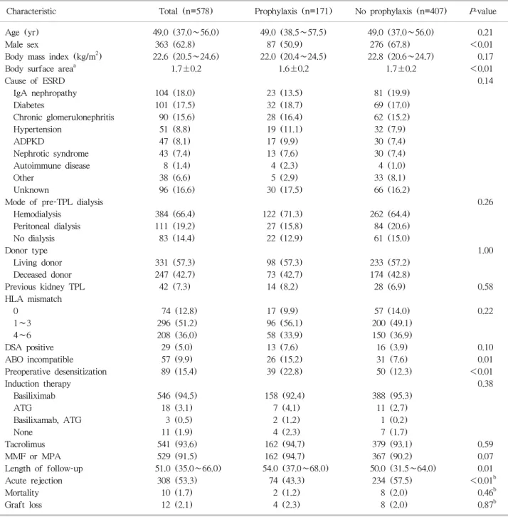
Table 1. Demographics and baseline characteristics
Characteristic Total (n=578) Prophylaxis (n=171) No prophylaxis (n=407) P-value
Age (yr) 49.0 (37.0∼56.0) 49.0 (38.5∼57.5) 49.0 (37.0∼56.0) 0.21
Male sex 363 (62.8) 87 (50.9) 276 (67.8) <0.01
Body mass index (kg/m2) 22.6 (20.5∼24.6) 22.0 (20.4∼24.5) 22.8 (20.6∼24.7) 0.17
Body surface areaa 1.7±0.2 1.6±0.2 1.7±0.2 <0.01
Cause of ESRD 0.14
IgA nephropathy 104 (18.0) 23 (13.5) 81 (19.9)
Diabetes 101 (17.5) 32 (18.7) 69 (17.0)
Chronic glomerulonephritis 90 (15.6) 28 (16.4) 62 (15.2)
Hypertension 51 (8.8) 19 (11.1) 32 (7.9)
ADPKD 47 (8.1) 17 (9.9) 30 (7.4)
Nephrotic syndrome 43 (7.4) 13 (7.6) 30 (7.4)
Autoimmune disease 8 (1.4) 4 (2.3) 4 (1.0)
Other 38 (6.6) 5 (2.9) 33 (8.1)
Unknown 96 (16.6) 30 (17.5) 66 (16.2)
Mode of pre-TPL dialysis 0.26
Hemodialysis 384 (66.4) 122 (71.3) 262 (64.4)
Peritoneal dialysis 111 (19.2) 27 (15.8) 84 (20.6)
No dialysis 83 (14.4) 22 (12.9) 61 (15.0)
Donor type 1.00
Living donor 331 (57.3) 98 (57.3) 233 (57.2)
Deceased donor 247 (42.7) 73 (42.7) 174 (42.8)
Previous kidney TPL 42 (7.3) 14 (8.2) 28 (6.9) 0.58
HLA mismatch
0 74 (12.8) 17 (9.9) 57 (14.0) 0.22
1∼3 296 (51.2) 96 (56.1) 200 (49.1)
4∼6 208 (36.0) 58 (33.9) 150 (36.9)
DSA positive 29 (5.0) 13 (7.6) 16 (3.9) 0.10
ABO incompatible 57 (9.9) 26 (15.2) 31 (7.6) 0.01
Preoperative desensitization 89 (15.4) 39 (22.8) 50 (12.3) <0.01
Induction therapy 0.38
Basiliximab 546 (94.5) 158 (92.4) 388 (95.3)
ATG 18 (3.1) 7 (4.1) 11 (2.7)
Basilixamab, ATG 3 (0.5) 2 (1.2) 1 (0.2)
None 11 (1.9) 4 (2.3) 7 (1.7)
Tacrolimus 541 (93.6) 162 (94.7) 379 (93.1) 0.59
MMF or MPA 529 (91.5) 162 (94.7) 367 (90.2) 0.07
Length of follow-up 51.0 (35.0∼66.0) 54.0 (37.0∼68.0) 50.0 (31.5∼64.0) 0.01
Acute rejection 308 (53.3) 74 (43.3) 234 (57.5) <0.01b
Mortality 10 (1.7) 2 (1.2) 8 (2.0) 0.46b
Graft loss 12 (2.1) 4 (2.3) 8 (2.0) 0.87b
Data are presented as median (interquartile range), number (%), or mean±standard deviation.
Abbreviations: ESRD, end stage renal disease; IgA, immunoglobulin A; ADPKD, autosomal dominant polycystic kidney disease; TPL, transplantation; HLA, human leukocyte antigen; DSA, donor specific antibody; ATG, antithymocyte globulin; MMF, mycophenolate acid;
MPA, mycophenolate mofetil.
aCalculated using the Du Bois formula (body surface area=0.007184×W0.425×H0.725); bP-values from log rank test.
RESULTS
1. Patient characteristics
In total, 578 renal transplant recipients were included, of
which 241 (41.7%) were started on TMP-SMX within 1 month post-transplant to prevent PJP and 171 (29.6%; pro- phylaxis group) completed the 6-month prophylaxis course.
Patients who prematurely discontinued the drug (n=70,
Table 2. Clinical presentation of the PJP infection in the two groups with and without 6 months TMP-SMX prophylaxis Total case of PJP Total (n=39) Prophylaxis (n=6) No prophylaxis (n=33) P-value
Severe PJPa 18 (46.2) 3 (50.0) 15 (45.5) 0.84
PJP needing ventilator care 9 (23.1) 1 (16.7) 8 (24.2) 1.00
PJP related death 4 (10.3) 1 (16.7) 3 (9.1) 0.50
Data are presented as number (%).
Abbreviations: PJP, Pneumocystis jirovecii pneumonia; TMP-SMX, trimethoprim-sulfamethoxazole.
aSevere PJP defined as PJP showing hypoxemia (PaO2 <60 mmHg while breathing room air) or a widened alveolar-to-arterial oxygen difference ([A-a] DO2 ≥45 mmHg) in blood gas examination.
Fig. 1. Cumulative incidence of Pneumocystis jirovecii pneumonia (PJP) infection after renal transplantation.
12.1%) were included in the no-prophylaxis group with pa- tients who were not administered prophylactic TMP-SMX (n=337, 58.3%). Baseline demographic characteristics of each group are shown in Table 1. The prophylaxis and no-prophylaxis group significantly differed in the pro- portion of male sex (50.9% vs. 67.8%), rate of ABOi trans- plantation (15.2% vs. 7.6%), preoperative desensitization sta- tus (22.8% vs. 12.3%), and rate of acute rejection (43.3% vs.
57.5%).
2. Incidence and outcomes of PJP
Overall, there were 39 cases of PJP (six and 33 cases in the prophylaxis and no-prophylaxis groups, respectively) during a median follow-up of 51 months (interquartile range, 35.0 to 66.0) (Fig. 1). Calculated incidence rate of
PJP in patients with and without prophylaxis was 8.3 and 21.7 per 1,000 patient year, respectively. Among 39 patients with confirmed PJP, 18 (46.2%) was categorized as severe PJP based on its presentation, and nine (23.1%) required ventilator care (Table 2). In four patients, PJP was lethal;
thus, PJP mortality rate was 10.3%. Of note, all mortality cases were from late PJP.
3. Incidence and risk factors of early PJP
The large difference in the incidence rate between the groups was due to the effective PJP prevention during the prophylaxis period in the prophylaxis group. Twenty-seven patients (6.6%) developed PJP within 6 months post-trans- plant in the no-prophylaxis group, whereas the prophylaxis group had none. The first case of early PJP documented oc-
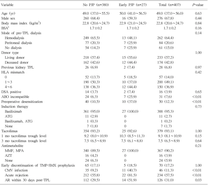
Table 3. Risk factors of early PJP infection among no prophylaxis group
Variable No PJP (n=380) Early PJP (n=27) Total (n=407) P-value
Age (yr) 49.0 (37.0∼55.5) 50.0 (41.0∼56.5) 49.0 (37.0∼56.0) 0.63
Male sex 260 (68.4) 16 (59.3) 276 (67.8) 0.44
Body mass index (kg/m2) 22.8 (20.6∼24.7) 22.9 (21.0∼24.5) 22.8 (20.6∼24.7) 0.84
BSAa 1.7±0.2 1.7±0.2 1.7±0.2 0.16
Mode of pre-TPL dialysis 0.14
Hemodialysis 249 (65.5) 13 (48.1) 262 (64.4)
Peritoneal dialysis 77 (20.3) 7 (25.9) 84 (20.6)
No dialysis 54 (14.2) 7 (25.9) 61 (15.0)
Donor type 1.00
Living donor 218 (57.4) 15 (55.6) 233 (57.2)
Deceased donor 162 (42.6) 12 (44.4) 174 (42.8)
Previous kidney TPL 26 (6.9) 2 (7.4) 28 (6.8) 0.97
HLA mismatch 0.42
0 52 (13.7) 5 (18.5) 57 (14.0)
1∼3 190 (50.3) 10 (37.0) 200 (49.1)
4∼6 138 (36.3) 12 (44.4) 150 (36.9)
DSA positive 14 (3.7) 2 (7.4) 16 (3.9) 0.65
ABO incompatible 24 (6.3) 7 (25.9) 31 (7.6) <0.01
Preoperative desensitization 40 (10.5) 10 (37.0) 50 (12.3) <0.01
Induction therapy 0.73
Basiliximab 361 (95.0) 27 (100.0) 388 (95.3)
ATG 11 (2.9) 0 11 (2.7)
Basilixamab, ATG 1 (0.3) 0 1 (0.2)
None 7 (1.8) 0 7 (1.7)
Tacrolimus 354 (93.2) 25 (92.6) 379 (93.1) 1.00
1 mo tacrolimus trough level 9.2 (8.0∼10.9) 10.3 (8.5∼11.3) 9.3 (8.1∼10.9) 0.15
3 mo tacrolimus trough level 7.5 (6.5∼8.9) 7.5 (6.1∼8.8) 7.5 (6.5∼8.9) 0.64
Antimetabolite 0.21
MMF, MPA 340 (89.5) 27 (100.0) 367 (90.2)
AZT 16 (4.2) 0 16 (3.9)
None 24 (6.3) 0 24 (5.9)
Early discontinuation of TMP-SMX prophylaxis 65 (17.1) 5 (18.5) 70 (17.2) 1.00
CMV infection 35 (9.2) 11 (40.7) 46 (11.3) <0.01
Acute rejection 212 (55.8) 22 (81.5) 234 (57.5) <0.01
AR within 30 days post-TPL 112 (29.5) 14 (51.9) 126 (31.0) 0.03
Data are presented as median (interquartile range), number (%), or mean±standard deviation.
Abbreviations: PJP, Pneumocystis jirovecii pneumonia; BSA, body surface area; TPL, transplantation; HLA, human leukocyte antigen;
DSA, donor specific antibody; ATG, antithymocyte globulin; MMF, mycophenolate acid; MPA, mycophenolate mofetil; AZT, azathioprine;
TMP-SMX, trimethoprim-sulfamethoxazole; CMV, cytomegalovirus; AR, acute rejection.
bCalculated using the Du Bois formula (BSA=0.007184×W0.425×H0.725).
curred 50 days after transplant.
Factors associated with early PJP in the no-prophylaxis group were ABOi transplantation, preoperative desensitiza- tion, CMV infection within 6 months, and acute rejection (Table 3). To identify risk factors that may stratify pa- tients’ risk, thereby stratifying the individual need of PJP prophylaxis during the first 6 months, we performed a mul- tivariate analysis including significant factors from uni-
variate analysis that were identifiable at 1 month post-trans- plant (ABOi transplantation, preoperative desensitization, acute rejection within 1 month). Analysis revealed pre- operative desensitization (odds ratio [OR], 4.59; 95% con- fidence interval [CI], 1.95 to 10.82; P=0.001) and acute re- jection within 1 month (OR, 2.31; 95% CI, 1.03 to 5.17;
P=0.04) as significant predictive factors of subsequent early PJP.
Table 4. Risk factors of late PJP infection
Variable
Univariate Cox Multivariate
OR 95% CI P-value OR 95% CI P-value
Age (yr) 1.11 1.04∼1.18 0.02 1.09 1.03∼1.19 <0.01
Male sex 1.06 0.19∼5.80 0.95
Body mass index (kg/m2) 0.99 0.83∼1.17 0.89
BSAa 0.80 0.04∼16.77 0.89
Deceased donor TPL 6.80 1.49∼31.05 0.01 4.89 1.05∼22.89 0.04
Previous kidney TPL 2.59 0.57∼11.80 0.22
DSA positive 0.05 0.01∼4,090.65 0.60
ABO incompatible 0.84 0.11∼6.48 0.87
Preoperative desensitization 1.10 0.24∼5.029 0.90
Basiliximab induction 21.75 0.01∼19,300 0.60
Tacrolimus
6 mo tacrolimus trough level 0.66 1.32 0.70
MMF or MPA 1.03 0.13∼7.95 0.98
6 mo TMP-SMX prophylaxis 2.37 0.77∼7.36 0.14
Early PJP 0.05 0.01∼11,899 0.63
AR within post-TPL 6 mo 0.57 0.16∼2.11 0.40
CMV within post-TPL 6 mo 1.50 0.19∼11.62 0.70
CMV infection 4.13 1.24∼13.72 0.02
Previous CMV infectionb 2.67 0.59∼12.33 0.22
Acute rejectionb 0.65 0.17∼2.46 0.53
AR needing ATGb 7.56 0.96∼59.89 0.06 8.88 1.11∼71.07 0.04
Antibody mediated rejectionb 0.05 0.00∼8.74×10⁹ 0.80
No. of AR 0.94 0.51∼1.75 0.85
No. of biopsy proven AR 1.79 0.96∼3.31 0.07
Abbreviations: PJP, Pneumocystis jirovecii pneumonia; OR, odds ratio; CI, confidence interval; BSA, body surface area; TPL, transplantation; DSA, donor specific antibody; MMF, mycophenolate acid; MPA, mycophenolate mofetil; TMP-SMX, trimethoprim- sulfamethoxazole; AR, acute rejection; CMV, cytomegalovirus; ATG, antithymocyte globulin.
aCalculated using the Du Bois formula (BSA=0.007184×W0.425×H0.725); bFactors as time-varying covariate.
4. Estimated efficacy of the risk-based prophylaxis strategy to prevent early PJP
We simulated the efficacy of the risk-based strategy, which excludes early routine prophylaxis in patients with low risk, i.e., patients without preoperative desensitization and acute rejection within 1 month post-transplant (Supplementary Table 1). When simulated with the data from the no-prophylaxis group, the proposed strategy will reduce the number of patients with unnecessary prophylaxis (93% to 34%) compared to the universal prophylaxis strat- egy, but would result in 2.2% residual incidence of early PJP.
5. Incidence and risk factors of late PJP
Six patients each from the prophylaxis and no-prophy-
laxis groups developed PJP at 6 months post-transplant. The rate of late PJP was higher in the prophylaxis group (3.5% vs.
1.5%), but without statistical difference (Table 4). The ear- liest case of PJP in the prophylaxis group developed 51 days after completing prophylaxis. All late PJP cases developed within 2 years post-transplant, and none had recurrent PJP in our study population.
The two groups were combined in the subsequent analysis for risk factors of late PJP. Factors associated with late PJP were age, deceased donor transplant, steroid-resistant acute rejection requiring ATG, and number of biopsy proven acute rejection (Table 4). In multivariate Cox regression analysis, age (OR, 1.09; 95% CI, 1.03 to 1.19; P<0.01), de- ceased donor transplantation (OR, 4.89; 95% CI, 1.05 to 22.89; P=0.04), and steroid resistant acute rejection requir-
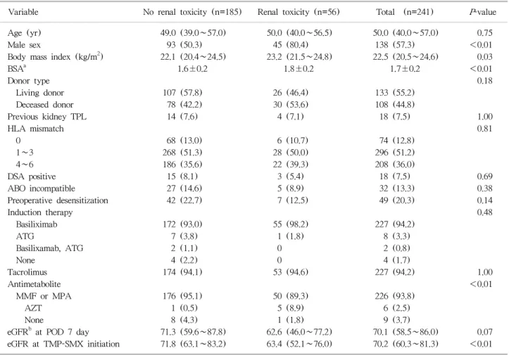
Table 5. Risk factors of renal toxicity after prophylactic use of TMP-SMX
Variable No renal toxicity (n=185) Renal toxicity (n=56) Total (n=241) P-value
Age (yr) 49.0 (39.0∼57.0) 50.0 (40.0∼56.5) 50.0 (40.0∼57.0) 0.75
Male sex 93 (50.3) 45 (80.4) 138 (57.3) <0.01
Body mass index (kg/m2) 22.1 (20.4∼24.5) 23.2 (21.5∼24.8) 22.5 (20.5∼24.6) 0.03
BSAa 1.6±0.2 1.8±0.2 1.7±0.2 <0.01
Donor type 0.18
Living donor 107 (57.8) 26 (46.4) 133 (55.2)
Deceased donor 78 (42.2) 30 (53.6) 108 (44.8)
Previous kidney TPL 14 (7.6) 4 (7.1) 18 (7.5) 1.00
HLA mismatch 0.81
0 68 (13.0) 6 (10.7) 74 (12.8)
1∼3 268 (51.3) 28 (50.0) 296 (51.2)
4∼6 186 (35.6) 22 (39.3) 208 (36.0)
DSA positive 15 (8.1) 3 (5.4) 18 (7.5) 0.69
ABO incompatible 27 (14.6) 5 (8.9) 32 (13.3) 0.38
Preoperative desensitization 42 (22.7) 7 (12.5) 49 (20.3) 0.14
Induction therapy 0.48
Basiliximab 172 (93.0) 55 (98.2) 227 (94.2)
ATG 7 (3.8) 1 (1.8) 8 (3.3)
Basilixamab, ATG 2 (1.1) 0 2 (0.8)
None 4 (2.2) 0 4 (1.7)
Tacrolimus 174 (94.1) 53 (94.6) 227 (94.2) 1.00
Antimetabolite <0.01
MMF or MPA 176 (95.1) 50 (89.3) 226 (93.8)
AZT 1 (0.5) 5 (8.9) 6 (2.5)
None 8 (4.3) 1 (1.8) 9 (3.7)
eGFRb at POD 7 day 71.3 (59.6∼87.8) 62.6 (46.0∼77.2) 70.1 (58.5∼86.0) 0.07
eGFR at TMP-SMX initiation 71.8 (63.1∼83.2) 63.4 (52.1∼76.0) 70.2 (60.3∼81.3) <0.01 Data are presented as median (interquartile range), number (%), or mean±standard deviation.
Abbreviations: TMP-SMX, trimethoprim-sulfamethoxazole; BSA, body surface area; TPL, transplantation; HLA, human leukocyte antigen;
DSA, donor specific antibody; ATG, antithymocyte globulin; MMF, mycophenolate acid; MPA, mycophenolate mofetil; AZT, azathioprine;
eGFR, estimated glomerular filtration rate; POD, post-operative day.
aCalculated using the Du Bois formula (BSA=0.007184×W0.425×H0.725); bCalculated by using Modification of Diet in Renal Disease (MDRD) equation: eGFR=186×(serum Cr)−1.154×(age)−0.203×1.212 (if patient is black)×0.742 (if patient is female).
ing ATG treatment (OR, 8.88; 95% CI, 1.11 to 71.07;
P=0.04) were significantly associated with late PJP.
6. Estimated efficacy of the risk-based prophylaxis strategy to prevent late PJP
Several prophylaxis strategies to prevent late PJP are pro- posed based on risk factors (Supplementary Table 2).
Theoretically, extended prophylaxis in patients aged ≥57 years and transplant from deceased donor would prevent 67% of the late PJP cases, resulting in the residual incidence of 0.7%. PJP risk in the proposed patient group for whom prophylaxis is indicated would be 10.3%, and the number required to treat to prevent one case would be 9.5. The
cut-off age of 57 years was derived from the ROC curve analysis (Supplementary Fig. 1).
7. Adverse reaction related to TMP-SMX prophylaxis TMP-SMX prophylaxis were prematurely discontinued in 29% (n=70) of 241 patients who had PJP prophylaxis of- fered initially. The main reasons for discontinuation were TMP-SMX-related adverse reaction including increased cre- atinine levels (n=59), hyperkalaemia (n=1), cytopenia (n=5), and nausea (n=1). Among the remaining seven pa- tients, five had TMP-SMX discontinued due to increased creatinine levels, later revealed to be linked to other causes (rejection, n=1; bacterial infection, n=2; CMV infection,
n=1; other viral infection, n=1), but the drug was not resumed. In two patients, the reason for discontinuation was not specified. Risk factors of increased creatinine levels causing premature drug discontinuation was male sex, high body surface area, concomitant use of azathioprine, and lower eGFR at TMP-SMX initiation in univariate analysis (Table 5). In multivariate analysis, the significant risk factors were male sex (OR, 4.24; 95% CI, 1.94 to 9.26; P<0.01), concomitant use of azathioprine (OR, 58.7; 95% CI, 2.78 to 1,240; P<0.01), and lower eGFR at TMP-SMX initiation (OR, 0.96; 95% CI, 0.94 to 0.98; P<0.01) (Supplementary Table 3).
DISCUSSION
To establish an effective and safe prophylaxis strategy, one should carefully balance the risks and benefits of a pro- phylaxis regimen. This study evaluated the effect of current PJP prophylaxis protocol on the rate of early and late PJP after renal transplantation and the adverse reactions asso- ciated with prophylactic TMP-SMX. Moreover, to propose a selective risk-based protocol, we investigated PJP risk fac- tors at different time points post-transplantation.
Generally, universal prophylaxis is recommended in pa- tient groups with PJP incidence of more than 3% to 5%(12).
Cumulative rate of early PJP in our patients undergoing contemporary immunosuppressive therapy was 6.1% without prophylaxis. PJP prophylaxis with TMP-SMX was highly effective; thus, none had early infection in the prophylaxis group. However, we frequently encountered TMP-SMX-as- sociated adverse reactions. Although adverse events asso- ciated with PJP prophylaxis in non- Human immuno- deficiency virus (HIV) patients were rare in the meta-anal- ysis by Stern et al.(6), 29% of our patients experienced ad- verse events, leading to early prophylaxis discontinuation.
These frequent adverse reactions were the main reason for the delay in implementing universal PJP prophylaxis at our centre. Our adverse event rate is similar to those of Mitsides et al.(7) (38%) and Urbancic et al.(13) (35%).
The most frequent adverse reaction leading to premature prophylaxis termination was increased serum creatinine lev- el, and factors associated with this were male sex, con- comitant azathioprine use, and lower eGFR at TMP-SMX
initiation. Increased serum creatinine levels are frequently attributed to the inhibition of tubular creatinine secretion by the TMP component of the drug and thus are suggested to occur without real change in glomerular filtration rate(14). However, not all increases in creatinine levels caused by TMP-SMX are benign, and tubulointerstitial nephritis, acute tubular necrosis, and acute kidney injury cases have been reported, especially when therapeutic doses are used(15). In the post-renal transplant setting, the in- crease in creatinine during TMP-SMX prophylaxis leads to difficulty in differentiating whether it is caused by in- hibition of creatinine excretion by TMP, tubulointerstitial nephritis caused by SMX, or other problems unrelated to TMP-SMX, including graft rejection or infection. Hence, regardless of the reversibility and benign nature of majority of the cases with increased serum creatinine, it frequently necessitates drug discontinuation and subsequent evaluation for its cause. In contrast, mere observation without evalua- tion may mask rejection or infection episodes requiring ur- gent treatments.
Given the high rates of adverse reaction and afore- mentioned clinical complexity caused by TMP-SMX, a more selective risk-based approach in PJP prophylaxis may be more beneficial. If a patient group for whom routine pro- phylaxis can be safely avoided can be identified, it would lower the risk of adverse reaction, reduce cost, and reduce the development of microbial resistance in the long term.
Here, we demonstrated that different patient groups were at risk of developing PJP at different time points after renal transplantation. The risk factors of PJP development during the first 6 months, which is the period of highest risks of PJP after renal transplant, were acute rejection within 30 days post-transplant and pre-transplant desensitization. Only the factors available at 30 days post-transplant were chosen because previous reports recommend prophylaxis to be start- ed within 1 month post-transplant based on the finding that PJP are rare during the first month. Delaying the PJP pro- phylaxis 1 month post-transplant results in stable renal function, and thereby reducing the number of cases with renal toxicity resulting in premature discontinuation. Acute rejection has been frequently identified as a risk factor of early PJP in previous reports and is related to increased bur- den of immunosuppression during treatment of rejection.
Additionally, our data show that preoperative desensitiza- tion confers additional risk for PJP. Preoperative desensiti- zation regimen consists of rituximab in combination with plasmapheresis and/or IVIG. The association between T-cell immunosuppression and PJP is well known, whereas that be- tween B-cell depletion therapy and PJP has only been de- scribed in a few recent reports(16). Although the mecha- nism is unclear, B-cells may be important in clearing pneu- mocystis infection by participating in the early priming of CD4+ T-cells(17).
There were 12 cases of PJP after 6 months post-trans- plant in our mixed study population of renal recipients with or without prophylaxis. All late PJP cases developed within 2 years post-transplant, and the rates were not affected by the previous prophylaxis. Iriart et al.(8) recently showed that 6-month prophylaxis prevented PJP in the first year and that the second year post-transplant was the period of highest risk. Our data also show that most of the late PJP cases occur within 2 years, but the rate of PJP was higher in 6 to 12 months post-transplant than in the second year (Fig. 1). The discrepancy may have been caused by the dif- ference in transplant type (mixed solid organ transplant re- cipients vs. kidney transplant recipients), immunosuppression regimen, and possible regional difference in the risk of de novo infection. While more studies are needed to define the temporal change in the risk of PJP after prophylaxis, it is noteworthy that PJP can occur early (51 days in our study) after discontinuing PJP prophylaxis. Although documented severity of PJP was not different between early and late PJPs, all four mortality cases were from late PJPs, possibly indicating the clinical importance of preventing late PJPs.
Factors associated with late PJP were old age, transplant from deceased donor, and ATG treatment for steroid re- sistant acute rejection. Age and ATG were also suggested as risk factors for late pneumocystis infection in other re- cent studies that evaluated the risk factors of late PJP (Supplementary Table 4)(8,18,19). By depleting T-cells, ATG increases host susceptibility to opportunistic infection, including PJP. Although the mechanism of how old age in- creases PJP risk is less clear, age-related immune dysfunc- tions, including thymic impairment, are possible underlying mechanisms(20). Other changes in T-cell function may also contribute to increased risks of infection in the elderly(21).
In contrast, donor source is rarely linked to PJP. One ex- planation for the increased risk of late PJP in deceased do- nor transplant compared to living donor transplant would be the higher burden of cumulative immunosuppression due to higher number of acute rejection and delayed graft func- tion; however, this needs to be evaluated in further studies.
Additionally, the CMV infection is a factor that deserves mention. The association between CMV infection and PJP has been frequently demonstrated elsewhere(8,18,19), sug- gesting that immune-modulating functions of CMV may have a role in PJP. We also observed a high rate of con- current CMV and PJP infection that was frequently ob- served in our study population (Supplementary Fig. 2). The CMV infection was also associated with late PJP when a COX regression analysis was performed, with the CMV in- fection as a fixed covariate. However, the association was not significant when the CMV infection was treated as a time-dependent covariate. Our data suggest that CMV is not a risk factor for subsequent PJP, and thus does not necessa- rily predict subsequent PJP. It is more likely that both CMV and PJP reflect a high degree of immunosuppression.
We simulated the effectiveness of several prophylaxis strategies designed based on the risk factors of PJP identi- fied in the present study. Estimates after simulation showed that while the strategy avoiding routine TMP-SMX prophy- laxis within 6 months post-transplant in low risk patients would prevent two-thirds of the early PJP cases in our pop- ulation, residual incidence of early PJP would not be negli- gible (2.2%). Thus we recommend universal PJP prophy- laxis during the first 6 months after transplantation.
Meanwhile, combination of age criteria and donor source seemed promising in discriminating patients with high risks of late PJP in which extended prophylaxis may be applied.
Selective prophylaxis in patients aged ≥57 years and trans- plant from deceased donor until 2 years post-transplant would result in 0.7% of late PJP incidence while avoiding TMP-SMX use in 87% of the population. The selective pro- phylaxis we propose here are similar with the one of the scenarios proposed by de Boer et al.(11). By analysing the risk factor of overall PJP development in patients without PJP prophylaxis, de Boer et al.(11) proposed that 2 to 6 months universal prophylaxis and extended selective pro- phylaxis in >55 years of age or those with rejection as an
optimal prophylaxis strategy. Our study differ in that we have separately evaluated the risk factors of PJP at differ- ent time points after transplantation, and in that we have incorporated those with PJP prophylaxis in the analysis for the late PJP. Regardless of the difference, de Boer et al.(11) and our study group have reached a similar prophylactic strategy.
While we started this study to propose a selective prophy- laxis strategy during the first 6 months because of the high rate of adverse event, we were unable to define a group of patients whose risk of PJP was low enough that PJP pro- phylaxis during the first 6 months could be safely avoided.
As PJP prophylaxis in the early transplant period seems un- avoidable, future studies on less toxic prophylactic agent, as well as the accurate diagnosis and strategical management of TMP-SMX related adverse reaction are warranted.
The current study has several limitations, including the inherent limitation owing to its non-randomized retro- spective single-centre design. The prophylaxis and no-pro- phylaxis groups were not randomly selected, and the two groups differed significantly in several baseline clin- icopathologic properties. As prophylaxis virtually eliminated early PJP in the prophylaxis group, the risk factors of early PJP were evaluated only in the no-prophylaxis group.
Although the incidence of early PJP was not associated with the factors that differed between the two groups, such dif- ference should be taken into account when interpreting and applying our results. The overall incidence of PJP was high (6.6%) and generally, may not be applicable in centers with a lower incidence rate. While this was mainly due to the high rate of early PJP caused by the lack of prophylaxis, considering that the rate of late PJP in the prophylaxis group (2.2%) was higher compared with Western countries, who showed an incidence rate of 0.3% to 1.8% after pro- phylaxis(8,19,22), there is also a possibility that the inherent rate of PJP in our center is high. Because the net effect of a prophylaxis strategy depends greatly on the incidence of infection, antimicrobial resistance profile of the organ- ism, and drug-tolerability of the target population, local characteristics should be considered before generalizing the results of our study. Last, as we did not have a common protocol on the indication of prophylaxis and management of adverse events during the study period, the decision to
start or stop prophylaxis was left to the treating physician, and thus, may have been biased by individual preferences.
Without definite criteria of a renal adverse event (i.e., de- gree of rise in creatinine increase), and when to stop pro- phylaxis, we may have overestimated the rate of renal ad- verse events.
CONCLUSION
Different patient groups are at risk of developing PJP during early and late periods after transplantation. Adverse renal events were frequently encountered during TMP-SMX prophylaxis, leading to premature discontinuation. After modelling different prophylaxis scenarios, we proposed a universal prophylaxis of up to 6 months post-transplant combined with extended selective prophylaxis in patients aged ≥57 years and transplants from deceased donors as a possible strategy to effectively prevent PJP after renal transplantation.
CONFLICTS OF INTEREST
No potential conflict of interest relevant to this article was reported.
ACKNOWLEDGEMENTS
This study was supported by Seoul National University Hospital Clinical Research Institute (0420173070 [2017-1392]).
REFERENCES
1) Thomas CF Jr, Limper AH. Pneumocystis pneumonia. N Engl J Med 2004;350:2487-98.
2) Iriart X, Bouar ML, Kamar N, Berry A. Pneumocystis pneu- monia in solid-organ transplant recipients. J Fungi (Basel) 2015;1:293-331.
3) Martin SI, Fishman JA; AST Infectious Diseases Community of Practice. Pneumocystis pneumonia in solid organ transplantation. Am J Transplant 2013;13 Suppl 4:272-9.
4) EBPG Expert Group on Renal Transplantation. European best practice guidelines for renal transplantation. Section IV: long-term management of the transplant recipient.
IV.7.1 Late infections. Pneumocystis carinii pneumonia.
Nephrol Dial Transplant 2002;17 Suppl 4:36-9.
5) Kidney Disease: Improving Global Outcomes (KDIGO) Transplant Work Group. KDIGO clinical practice guideline for the care of kidney transplant recipients. Am J Transplant 2009;9 Suppl 3:S1-155.
6) Stern A, Green H, Paul M, Vidal L, Leibovici L. Prophylaxis for Pneumocystis pneumonia (PCP) in non-HIV im- munocompromised patients. Cochrane Database Syst Rev 2014;10:CD005590.
7) Mitsides N, Greenan K, Green D, Middleton R, Lamerton E, Allen J, et al. Complications and outcomes of trimetho- prim-sulphamethoxazole as chemoprophylaxis for pneu- mocystis pneumonia in renal transplant recipients.
Nephrology (Carlton) 2014;19:157-63.
8) Iriart X, Challan Belval T, Fillaux J, Esposito L, Lavergne RA, Cardeau-Desangles I, et al. Risk factors of Pneumocystis pneumonia in solid organ recipients in the era of the com- mon use of posttransplantation prophylaxis. Am J Transplant 2015;15:190-9.
9) Yiannakis EP, Boswell TC. Systematic review of outbreaks of Pneumocystis jirovecii pneumonia: evidence that P. jir- ovecii is a transmissible organism and the implications for healthcare infection control. J Hosp Infect 2016;93:1-8.
10) Goto N, Takahashi-Nakazato A, Futamura K, Okada M, Yamamoto T, Tsujita M, et al. Lifelong prophylaxis with trimethoprim-sulfamethoxazole for prevention of outbreak of Pneumocystis jirovecii pneumonia in kidney transplant recipients. Transplant Direct 2017;3:e151.
11) de Boer MG, Kroon FP, le Cessie S, de Fijter JW, van Dissel JT. Risk factors for Pneumocystis jirovecii pneumonia in kidney transplant recipients and appraisal of strategies for selective use of chemoprophylaxis. Transpl Infect Dis 2011;13:559-69.
12) Fishman JA. Prevention of infection caused by Pneumocystis carinii in transplant recipients. Clin Infect Dis 2001;33:
1397-405.
13) Urbancic KF, Ierino F, Phillips E, Mount PF, Mahony A,
Trubiano JA. Taking the challenge: a protocolized approach to optimize Pneumocystis pneumonia prophylaxis in renal transplant recipients. Am J Transplant 2018;18:462-6.
14) Delanaye P, Mariat C, Cavalier E, Maillard N, Krzesinski JM, White CA. Trimethoprim, creatinine and creatinine- based equations. Nephron Clin Pract 2011;119:c187-93.
15) Fraser TN, Avellaneda AA, Graviss EA, Musher DM. Acute kidney injury associated with trimethoprim/sulfamethoxazole.
J Antimicrob Chemother 2012;67:1271-7.
16) Alexandre K, Ingen-Housz-Oro S, Versini M, Sailler L, Benhamou Y. Pneumocystis jirovecii pneumonia in patients treated with rituximab for systemic diseases: report of 11 cases and review of the literature. Eur J Intern Med 2018;
50:e23-4.
17) Opata MM, Hollifield ML, Lund FE, Randall TD, Dunn R, Garvy BA, et al. B lymphocytes are required during the early priming of CD4+ T cells for clearance of pneumocystis infection in mice. J Immunol 2015;195:611-20.
18) Lee SH, Huh KH, Joo DJ, Kim MS, Kim SI, Lee J, et al.
Risk factors for Pneumocystis jirovecii pneumonia (PJP) in kidney transplantation recipients. Sci Rep 2017;7:1571.
19) Faure E, Lionet A, Kipnis E, Noel C, Hazzan M. Risk factors for Pneumocystis pneumonia after the first 6 months follow- ing renal transplantation. Transpl Infect Dis 2017;19:
e12735.
20) Schurmann M, Schurmann D, Schindler R, Meisel C, Liman P, Kruse J, et al. Impaired thymic function and CD4+ T lymphopenia, but not mannose-binding lectin deficiency, are risk factors for Pneumocystis jirovecii pneumonia in kidney transplant recipients. Transpl Immunol 2013;28:
159-63.
21) Haynes L, Maue AC. Effects of aging on T cell function.
Curr Opin Immunol 2009;21:414-7.
22) Wang EH, Partovi N, Levy RD, Shapiro RJ, Yoshida EM, Greanya ED. Pneumocystis pneumonia in solid organ trans- plant recipients: not yet an infection of the past. Transpl Infect Dis 2012;14:519-25.
Prophylaxis strategy (0∼6 mo)
Total no.
of patient (Ntot)
Incidence (I)
No. of cases that would have occurred without
prophylaxis (Nnorm)
No. of cases receiving prophylaxis (Npro)
Cases prevented
with prophylaxis
(Nprev)
proportion of patients with PCP prevented
(Pprev)
proportion of patients treated unnecessarily
(Ppt)
Incidence after pophylaxis
(Ires)
No. of needed to treat to
prevent 1 case (NNTP)
No prophylaxis 407 6.6% 27 0 0 0 0 6.6% -
All patients 407 6.6% 27 407 27 1 0.93 0% 14.2
Patients with
≥1 risk factors
407 6.6% 27 155 18 0.67 0.34 2.2% 8.1
Abbreviation: PCP, Pneumocystis pneumonia.
Supplementary Table 2. Estimated effects of different selective extended prophylactic strategies on late PJP
Prophylaxis strategy (12∼24 mo)
Total no.
of patient (Ntot)
Incidence (I)
No. of cases that would have occurred without
prophylaxis (Nnorm)
No. of cases receiving prophylaxis (Npro)
Cases prevented
with prophylaxis
(Nprev)
Estimated proportion
|of patients with PJP prevented
(Pprev)
Estimated proportion of patients treated unnecessarily
(Ppt)
Incidence after prophylaxis
(Ires)
No. of needed to treat to
prevent 1 case (NNTP)
No prophylaxis 578 2.1% 12 0 0 0 0 2.1% -
All patients 578 2.1% 12 578 12 1 0.98 0 46.7
Age ≥57 yr 578 2.1% 12 137 9 0.75 0.22 0.5% 14.8
DDKT 578 2.1% 12 247 10 0.83 0.41 0.4% 23.9
ATG treatment for AR
578 2.1% 12 14 1 0.08 0.02 1.9% 13.6
Age ≥57 yr or DDKT
578 2.1% 12 306 11 0.92 0.49 0.2% 26.1
Age ≥57 yr and DDKT
578 2.1% 12 78 8 0.67 0.12 0.7% 9.5
All patients with
≥1 risk factors
578 2.1% 12 315 11 0.92 0.54 0.2% 28.3
All patients with
≥2 risk factors
578 2.1% 12 81 8 0.67 0.13 0.7% 9.8
Abbreviations: PJP, Pneumocystis jirovecii pneumonia; DDKT, deceased donor kidney transplantation; ATG, antithymocyte globulin; AR, acute rejection.