서 론
비만은 에너지 섭취와 소비 사이의 불균형으로 인 한 비정상적이거나 과도한 지방의 축적이 발병의 원
인이다[1,2]. 비만은 대사 증후군, 제2형 당뇨병, 고 콜레스테롤 혈증, 다양한 혈관성 질환 등 여러 장기 에 합병증을 유발할 수 있는 위험 인자이며, 전 세계 적으로 비만의 유병률이 높아지고 있는 추세이다
* Corresponding author
Phone: +82-51-890-3319 (Y.H.Choi), +82-51-890-1594 (H.J.Hwang) E-mail: [email protected] (Y.H.Choi), [email protected] (H.J.Hwang)
This is an open-access journal distributed under the terms of the Creat ive Commons Attribution Non-Commercial License
(http://creativecommons.org/licenses/by-nc/4.0/)
https://doi.org/10.15433/ksmb.2021.13.1.001 ISSN 2383-5400 (Online)
3T3-L1 및 B16F10 세포에서 청각 메탄올 추출물에 의한 지방 세포 분화 및 멜라닌 생성의 억제 효과
Inhibition of adipogenesis and melanogenesis by methanol extract of Codium fragile (Suringar) Hariot in 3T3-L1
adipocytes and B16F10 melanocytes
최은옥1, 최영현2,*, 황혜진3.*
Eun-Ok Choi1, Yung Hyun Choi2,* and Hye-Jin Hwang3,*
1박사후연구원, 동의대학교 항노화연구소, 부산 47340, 대한민국
2교수, 동의대학교 한의과대학 생화학교실, 부산 47227, 대한민국
3교수, 동의대학교 의료·보건·생활대학 식품영양학과, 부산 47340, 대한민국
1Anti-Aging Research Center, Dongeui University, Busan 47340, Korea
2Department of Biochemistry, College of Korean Medicine, Dong-eui University, Busan 47227, Korea
3Department of Food and Nutrition, Dong-eui University, Busan 47340, Korea
(Received 10 May 2021, Revised 17 May 2021, Accepted 17 May 2021)
Abstract Codium fragile (Suringar) Hariot, a green alga of the Codiales family, has been reported to have several bioactive properties, including antioxidant and anti-inflammatory properties.
However, its antiobesity and whitening effects and their underlying mechanisms are unclear. This study aimed to evaluate the antiobesity and melanogenesis inhibitory effects of C. fragile using methanol extracts of C. fragile (MECF). The results of this study revealed that MECF inhibited the accumulation of lipid droplets and triacylglycerol in differentiated 3T3-L1 adipocytes, which was associated with the inhibition of the expression of adipogenesis-related transcription factors, such as peroxisome proliferator-activated receptor γ, CCAAT/enhancer-binding protein-α (C/EBPα), and C/EBPβ, which function as the key regulators of adipogenesis. Also, MECF reduced tyrosinase activity and melanin content in B16F10 cells as well as the expression of tyrosinase, tyrosinase-related protein-1 (TRP-1), TRP-2, and microphthalmia-related transcription factor in the presence of α-melanocyte-stimulating hormone. Taken together, our findings suggest that the extract of C. fragile could be considered a promising functional ingredient for the pre- vention and treatment of obesity and skin pigmentation in the food and cosmetic industry.
Keywords : Codium fragile, 3T3-L1 adipocytes, anti-obesity, B16F10 melanocytes, anti-melanogenesis
[3,4]. 현재 비만의 치료에 널리 처방되고 있는 대표 적인 항비만제가 orlistat와 sibutramine이다[5,6].
Orlistat는 췌장이나 다른 소화계에서 생성되는 lipase 의 억제제로서 장에서의 지질 흡수를 차단하여 지질 대사를 억제하고 지질 축적을 감소시킨다[7]. 반면 sibutramine은 serotonin과 norepinephrine 흡수를 억제 하여 식욕을 저하시켜 지질 저장 및 합성을 억제함 으로서 체중의 감소를 유도한다[8,9]. 그러나 이들 항비만제는 지방변을 포함한 불쾌감과 간 기능의 저 해를 포함한 다양한 부작용을 동반한다[6,9]. 특히 sibutramine은 장기간 사용 시 심근 경색 및 뇌졸중을 포함한 심각한 심혈관 질환을 유발할 수 있다[10].
따라서 부작용을 유발하지 않고 효율적인 비만의 예 방과 치료를 위한 새로운 식의약 자원의 발굴이 요 구된다.
한편, 인간의 피부색은 멜라닌(melanin)의 양과 이 들이 피부에 분산되는 정도에 따라 다르며 햇빛 노 출 정도와 환경의 영향도 받는다[11,12]. 멜라닌은 동물과 식물에서 발견되는 고분자량 화합물이며 인 간의 눈, 피부 및 머리 색깔을 결정하는 중요한 색소 이다. 멜라닌은 유해한 자외선으로부터 피부를 보호 할 수도 있지만, 과도한 멜라닌 생성은 색소 침착을 포함한 미용 및 건강상의 문제를 유발할 수 있다 [13,14]. 최근, 과도한 멜라닌 생성으로 인한 문제를 해결하기 위해 많은 의료 및 화장품 산업에서 ty- rosinase 활성 억제제에 초점을 맞추고 있다[15,16].
그러나 현재 사용되고 있는 arbutin, kojic acid, hydro- quinone, ascorbic acid를 포함한 다양한 피부 미백제 는 피부에 잘 침투하지 못하며 장기간 사용시 피부 자극, 염증, 가려움, 색소 침착과 같은 부작용이 발생 할 수 있다[17,18]. 따라서 인체 피부의 과도한 색소 침착을 부작용없이 안전하게 예방하고 치료할 수 있 는 효과적인 미백제 발굴이 요구되고 있다.
최근 다양한 생리 활성 성분이 포함된 해조류는 잠재적인 식의약 소재의 주요 공급원으로 많은 주목 을 받고 있다. 그동안 축적된 연구들에 의하면 해조 류는 항산화, 항염증, 항암, 항비만 및 미백 효능과 같은 유익한 생물학적 활성을 가지고 있음을 알 수 있다[19-22]. 해조류 중, 청각(Codium fragile (Suringar) Hariot)은 식용 녹조류의 일종(Codiales family)으로 아시아, 유럽 및 오세아니아의 해안 지
역에 널리 분포하며, 우리나라에서는 전통적으로 장 염, 수종, 배뇨 장애 등의 치료 등을 위해서도 사용되 어 왔다[23]. 최근 연구에 의하면 청각 추출물 또는 대사산물은 항산화, 항염증, 면역 조절 등을 포함한 다양한 생리활성을 가지고 있는 것으로 밝혀지고 있 다[24-28]. 비록 Kolsi et al. [29]에 의하여 청각 유래 황화 다당류가 항산화 활성과 연계된 지질 생성 저 하 효과를 가진다는 보고는 있었지만, 지방 세포의 분화를 억제할 수 있는지에 대한 연구와 미백 효능 에 관한 구체적인 기전 연구는 이루어진 바 없다.
따라서 본 연구에서는 대표적인 in vitro 항비만 및 미백 효능 평가 모델을 이용하여 청각 추출물의 비 만세포 분화 억제 및 멜라닌 생성 억제 효과를 평가 하였다.
재료 및 방법 1. 시료 준비
본 연구에 사용된 청각 메탄올 추출물(methanol extracts of C. fragile, MECF)은 제주테크노파크(Jeju Technopark) 내 제주생물자원지원센터의 유용생물 자원추출물은행(Jeju Bio-Resource Extract Bank, Jeju, Korea)에서 제공받았다[28]. MECF를 세포에 처리하 기 위하여 dimethyl sulfoxide (DMSO, Sigma-Aldrich Chemical Co. St. Louis, MO, USA)에 녹여 1 mg/ml의 stock solution으로 제조 후, 세포 배양 배지에 적정 농도로 처리하였다.
2. 세포 배양
본 연구에서 사용된 3T3-L1 (mouse preadipocytes) 및 B16F10 (mouse melanoma) 세포는 American Type Culture Collection (Manassas, VA, USA)에서 분양받 았다. 3T3-L1 및 B16F10 세포는 10% bovine calf se- rum 및 10% fetal bovine serum (FBS)과 1% antibiotics (penicillin/streptomycin)이 포함된 Dulbecco's Modified Eagle’s Medium (DMEM, Thermo Fisher Scientific)를 사용하여 37℃, 5% CO₂조건 하에서 각각 배양하였으며, 세포 배양에 필요한 재료들은 Thermo Fisher Scientific (Waltham, MA, USA)에서 구 입하였다.
3. 세포 생존율 측정
MECF가 3T3-L1 및 B16F10 세포의 증식에 미치는 영향을 조사하기 위하여 24 well plate에 2×10⁴ cells/well로 분주하여 24시간 동안 배양 후, 적정 농 도의 MECF를 처리하여 72시간 동안 배양하였다. 처 리 후, 배지를 제거하고 0.5 mg/mL의 3-(4,5- dime- thylthiazol-2-yl)-2,5-diphenyltetrazolium bromide (MTT, Sigma–Aldrich Chemical Co.) 용액을 각 well 첨가하여 암 하에서 반응시켰다. 3시간 후, MTT 시 약을 제거하고 DMSO를 첨가하여 well에 형성된 formazan 침전물을 용해시키고 96 well plate에 옮긴 후 enzyme-linked immunosorbent assay (ELISA) read- er (VERSA Max, Molecular Device Co., Sunnyvale, CA, USA)를 이용하여 540 nm에서 흡광도를 측정하 였다.
4. 3T3-L1 세포의 분화 유도 및 세포 형태 관찰 전 지방 세포인 3T3-L1 세포를 지방 세포로 분화 를 유도하기 위하여 confluence 상태까지 배양한 후, 10% FBS 및 1%의 antibiotics가 포함된 분화 배지를 사용하여 2일간 더 배양한 후 10 μg/ml insulin, 0.1 μM dexamethasone 및 0.5 mM isobutylmethylxanthine (Sigma–Aldrich Chemical Co.)가 포함된 분화 배지 (MDI 분화 배지)로 교환하여 2일간 배양하였으며, 그 후 2일 마다 insulin이 포함된 분화 배지로 교환하 였다. 3T3-L1 세포의 분화 유도에 미치는 MECF의 영향을 조사하기 하기 위하여 MDI 및 insulin이 포함 된 배지로 교환할 때 적정 농도의 MECF를 처리하였 으며, 지방 세포로의 분화 정도를 도립현미경 (inverted microscope, Carl Zeiss, Oberkochen, Germany) 하에서 관찰하였다.
5. Oil Red O 염색 및 지질 함량 측정
3T3-L1 세포 내 지질 형성의 정도를 평가하기 위 하여 Oil Red O 염색을 실시하였다. 이를 위하여 다 양한 조건에서 배양된 3T3-L1 세포를 phos- phate-buered saline (PBS)로 세척 후, 10% formalin (Sigma–Aldrich Chemical Co.)으로 15분 동안 고정 하였다. 고정된 세포를 60% isopropanol (Sigma– Aldrich Chemical Co.)을 이용하여 세척한 후, Oil Red O 용액(Sigma–Aldrich Chemical Co.)을 처리하여 실 온에서 20분 동안 염색하였다. 염색 후 Oil Red O
용액을 제거하고 증류수로 세척한 후 지질 방울 (lipid droplet) 형성의 정도를 도립현미경을 이용하여 관찰하였다. Oil Red O 염색된 3T3-L1 세포에서 세 포 내 형성된 지질(triglyceride)을 정량적으로 비교하 기 위해 염색된 지질 방울을 isopropanol로 30분 동안 용해시키고 추출된 염료를 96 well plate로 옮겼다.
ELISA reader로 500 nm에서 흡광도를 측정한 후, 분 화된 지방 세포의 흡광도를 100% 상대 지질 함량으 로 설정하여 실험군에 따른 지질 함량 정도를 비교 하였다.
6. Reverse transcription-polymerase chain re- action (RT-PCR)에 의한 mRNA 발현 분석
3T3-L1 세포에서 지방 세포 분화에 관여하는 유전 자들의 발현 변화를 전사 수준에서 조사하기 위하여 RT-PCR을 실시하였다. 이를 위하여 다양한 조건에 서 배양된 3T3-L1 세포에서 TRIzol reagent (Invitrogen Co., Carlsbad, CA, USA)를 이용하여 총 RNA를 분리하였다. RNA를 정량 후, 대상 유전자의 primer를 ONE-STEP RT-PCR PreMix Kit (Intron, Seoul, Korea)와 Mastercycler gradient (Eppendorf, Hamburg, Germany)를 이용하여 증폭하였다. 각 PCR 산물들의 양적 차이를 조사하기 위하여 1.5% agar- ose gel을 이용하여 전기영동으로 분리 후 ethidium bromide (Sigma-Aldrich Chemical Co.)로 염색 후 ultra violet 하에서 관찰하였다. RT-PCR에 대한 internal control로는 glyceraldehyde-3-phosphate dehydrogen- ase (GAPDH)를 사용하였다. 본 연구에 사용된 pri- mer 염기서열은 Table 1에 제시하였다(PPARγ, per- oxisome proliferator-activated receptor; C/EBP, CCAAT/enhancer binding protein).
Table 1. The primer sequence is used for RT-PCR Gene Forward Primer Reverse Primer
PPARγ TTTTCAAGGGTGC
CAGTTTC
AATCCTTGGCCCT CTGAGAT
C/EBPα TTACAACAGGCC
AGGTTTCC
GGCTGGCGACAT ACAGTACA
C/EBPβ CCTTTAAATCCAT
GGAAGTGG
GGGCTGAAGTCG ATGGC
GAPDH CATGAGAAGTAT
GACAACAGCCT
AGTCCTTCCACGA TACCAAAGT
7. Western blot 분석에 의한 단백질 발현 분석 3T3-L1 세포와 B16F10 세포에서 지방 세포 분화 및 멜라닌 생성억제에 관여하는 유전자들의 발현 변 화를 번역 수준에서 조사하기 위하여 Western blot 분석을 실시하였다. 이를 위하여 다양한 조건에서 배양된 세포에 적당량의 lysis buffer (25 mM Tris-Cl (pH 7.5), 250 mM NaCl, 5 mM ethylenediaminetetra- acetic acid, 1% nonidet p-40, 1 mM phenyme- thylsulfonyl fluoride 및 5 mM dithiothreitol)를 첨가하 여 1시간 동안 4℃에서 반응시킨 후, 총 단백질을 분리하였다. 분리된 상층액의 단백질 농도를 Bio-Rad 단백질 정량 시약(Bio-Rad, Hercules, CA, USA)을 사용하여 정량한 다음 동량의 단백질을 so- dium dodecyl sulphate (SDS)-polyacrylamide gel을 이 용하여 전기영동으로 분리하고 분리된 단백질을 polyvinylidene fluoride membrane (Millipore, Bedford, MA, USA)으로 전이시켰다. 단백질이 전이된 mem- brane에 분석 대상 1차 항체를 처리하여 12시간 이상 4℃에서 반응시킨 다음 1차 항체에 대한 2차 항체를 처리하여 1시간 이상 상온에서 반응시켰다. 반응이 끝난 후 암실에서 enhanced chemiluminoesence (ECL) 용액(Thermo Fisher Scientific)을 적용시킨 다음 Fusion FX Image system (Vilber Lourmat, Torcy, France)을 이용하여 해당 단백질의 발현 정도를 비교 하였다. Western blot 분석에 대한 internal control로는 β-actin을 사용하였다. 본 연구에 사용된 PPARγ (2430s; 1:1,000), C/EBPα (2295s; 1:1,000) 및 C/EBPβ (3087s; 1:1,000)에 대한 1차 항체는 Cell Signaling Technology, Inc. (Danvers, MA, USA)에서 구입하였 으며, tyrosinase (sc-20035; 1:1,000), tyrosinase-related protein (TRP)-1 (sc-133076; 1:1,000), TRP-2 (sc-74439; 1:1,000), microphthalmia-associated tran- scription factor (MITF, sc-52938; 1:500) 및 β-actin에 대한 1차 항체는 Santa Cruz Biotechnology, Inc.
(Santa Cruz, CA, USA)에서 구입하였다. 2차 항체인 horseradish peroxidase-conjugated anti-rabbit im- munoglobulin G (IgG, sc-2004; 1:1,000), anti-mouse IgG (sc-2005; 1:1,500) 및 anti-goat IgG (sc-2350;
1:1,500)는 Santa Cruz Biotechnology, Inc.에서 구입하 였다.
8. B16F10 세포에서 멜라닌 생성량 측정
B16F10 세포 내에 생성된 멜라닌의 함량 측정을 위하여 B16F10 세포에 100 nM의 α-melanocyte stim- ulating hormone (α-MSH, Sigma-Aldrich Chemical Co.)을 처리하여 멜라닌의 생합성을 유도하였다. 24 시간 후, 세포를 PBS로 세척하고 다양한 농도의 MECF가 함유된 배지로 교환하여 72시간 동안 배양 하였다. 10% DMSO가 함유된 1 N의 NaOH 용액을 well에 첨가하고 1시간 동안 80oC의 water bath에서 1시간 반응시켜 세포 내의 멜리닌을 용해시킨 후 ELISA reader를 이용하여 405 nm에서 흡광도를 측 정하여 멜라닌 함량을 평가하였다.
9. B16F10 세포에서 tyrosinase 활성 측정 상기와 동일 조건에서 배양된 B16F10 세포를 PBS 로 수세 후, Thermo Fisher Scientific에서 구입한 RIPA Lysis and Extraction Buffer를 이용하여 제조사 의 지침에 따라 세포를 용해시켰다. 20 μl의 세포 용 해물에 0.1 M sodium phosphate buffer (pH 7.0)에 녹 인 2 mg/ml의 L-3,4-dihydroxyphenylalanine (Sigma- Aldrich Chemical Co.) 80 μl를 혼합한 후 37oC에서 2시간 동안 반응시키고 ELISA reader를 이용하여 490 nm에서 흡광도를 측정하여 tyrosinase 활성을 평 가하였다.
10. 통계 분석
실험 결과의 유의성 분석을 위하여 GraphPad Prism 5.03 소프트웨어(GraphPad Software, Inc., La Jolla, CA, USA)을 사용하였다. 모든 실험은 3번의 독립적인 실험을 통한 평균 ± 표준 편차(standard er- rors, SD)로 표시하였고, one-way analysis of variance 에 이어 Tukey’s post hoc 테스트로 분석하여 0.05 미만의 값은 통계적으로 유의한 것으로 간주하였다.
결과 및 고찰
1. 3T3-L1 세포의 증식에 미치는 MECF의 영향 MECF에 의한 지방 세포의 분화억제 효능 검증을 위한 실험 조건의 설정을 위하여 3T3-L1 세포의 증 식에 미치는 MECF의 영향을 먼저 조사하였다.
Figure 1에 나타낸 MTT 분석의 결과에 의하면, 400 μg/ml 처리군까지는 대조군과 유사한 세포 생존력 을 보였으며, 600 μg/ml 이상 처리군부터 처리 농도
증가에 따른 세포 생존율의 저하가 관찰되었다. 이 결과를 바탕으로 400 μg/ml의 MECF 처리군까지는 3T3-L1 세포에 유의적인 세포 독성이 없는 것으로 판단되어 이후 실험부터는 MECF의 처리 농도를 400 μg/ml 이하로 설정하였다.
Figure 1. Effect of MECF on cell viability of 3T3-L1 preadip ocytes. Cells were seeded in 24-well plates at a density of 2x10⁴ cells/well and incubated with the indicated concentrati ons of MECF. After 72 h, cell viability was analyzed by an MTT assay. Values are expressed as mean ± SD of triplica te experiments (*p < 0.05 indicates a significant difference compared to the control group).
2. 3T3-L1 세포에서 지질 방울 및 지질의 형성에 미치는 MECF의 영향
전 지방 세포가 지방 세포로의 분화되었다는 가장 확실한 증거는 세포 내 지질 방울의 형성이며, 이들 은 지방세로의 분화되는 과정에서 생성되는 지질 (triglyceride)과 콜레스테롤 에스테르(cholesterol es- ter)가 인지질층(phospholipid monolayer)에 둘러싸여 형성된다[30,31]. 분화된 지방 세포에서 지질 방울의 형성은 lipoprotein lipase에 의한 지질의 유입과 adi- pose triglyceride lipase와 hormone sensitive lipase에 의한 지질의 유출에 의하여 조절되며, 과도한 지질 방울은 비만 유발뿐만 아니라 제2형 당뇨병, 동맥경 화 및 암 등과 같은 질환의 개시와 진행에도 관여한 다[31,32]. 따라서 지질 방울 형성의 억제는 비만 억 제의 가장 유의적인 지표로 활용될 수 있기 때문에, MECF가 지방 생성과정에 미치는 영향을 조사하였 다. Figure 2A에 나타낸 3T3-L1 세포의 형태학적인 변화 관찰에서 MDI 처리에 의해 분화가 유도된 세 포에서는 분화를 유도하지 않은 대조군에 비하여 지 질 방울의 형성이 매우 증가되어 분화가 적절히 유 도되었음을 알 수 있다. 이를 Oil red O의 염색으로 재확인하였으며(Figure 2B), MECF가 존재하는 조건 에서는 지질 방울의 형성이 MECF의 처리 농도 의존
적으로 현저하게 감소되었다. 아울러 지질의 함량 변화를 정량적으로 비교한 결과에서도 MDI 처리로 분화된 지방 세포에서 증가한 지질의 함량이 MECF 가 전처리된 조건에서 처리 농도의 증가에 따라 유 의적으로 감소되었다(Figure 2C). 이는 지질 방울 형 성의 억제에 의한 결과이며, MECF가 강력한 지방 세포 분화 억제력을 지니고 있음을 보여주는 결과이 다.
Figure 2. Effect of MECF on adipogenesis in differentiating 3T3-L1 cells. The cells were induced to differentiate with induction medium in the presence or absence of MECF for 8 days. (A and B) The morphology of cells before (A) or after staining with Oil Red O (B) was photographed with an optical microscope and representative images were present ed. (C) The content of intracellular triglyceride (TG) was quantified in 3T3-L1 cells. Values are expressed as mean
± SD of triplicate experiments (*p < 0.05 indicates a significa nt difference compared to differentiated cells).
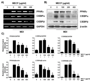
3. 3T3-L1 세포에서 지방 생성 유도 유전자의 발현 에 미치는 MECF의 영향
지방 세포의 분화에 따른 지방 생성은 지방 세포 에서 지방 축적에 중요한 역할을 하는 특정 지방 생 성 유전자의 발현에 의해 엄격하게 조절된다[33,34].
그중에서 PPARγ와 C/EBP은 지방 세포 분화와 지방 산 합성의 핵심 조절자 역할을 한다. 이들의 발현이 증가되면, fatty-acid synthase, adiponectin, leptin 및 adipocyte protein 2와 같은 지방 세포 특이 마커의 발현이 향상되어 지방 생성이 촉진된다[35,36].
Figure 3에 나타낸 RT-PCR 및 Western blot 분석의 결과에 의하면, MDI 처리에 의해 분화가 유도된
3T3-L1 세포에서는 PPARγ, C/EBPα 및 C/EBPβ의 발현이 전사 및 번역 수준에서 모두 현저하게 증가 하였음을 알 수 있다. 따라서 MECF에 의한 지방 세 포 분화 억제가 이들 유전자의 발현 감소와 연관성 이 있는지를 조사하였으며, 증가된 이들 유전자의 발현은 MECF가 존재하는 조건에서는 모두 MECF 처리 농도 의존적으로 감소되었다. 따라서 MECF는 지방 세포 분화와 지방 합성 조절에 관여하는 유전 자들의 발현을 억제함으로서 지방 세포로의 분화를 억제하고 지질의 형성을 차단하였음을 알 수 있다.
Figure 3. Effect of MECF on the expression of transcription factors involved in adipogenesis in 3T3-L1 cells. The cells were treated with the indicated concentrations of MECF alon g with differentiation induction medium and allowed to differ entiatie for 8 days. (A) The cellular RNA was isolated on the day 9, and RT-PCR was performed after measurement and reverse transcription. (B) Whole cell lysate was isolated and analyzed by Western blot analysis with respective antibo dies. GAPDH and β-actin levels are shown as loading control s. (C) Bands were quantified using ImageJ and normalized to GAPDH and β-actin, and the ratio was determined. Data are expressed as the mean ± SD of three independent experim ents (*p < 0.05 indicates a significant difference compared to the control group).
4. B16F10 세포의 증식에 미치는 MECF의 영향 다음은 MECF의 멜라닌 생성 억제 효능 평가를 위한 실험 조건의 설정을 위하여 3T3-L1 세포에서와 같이 MTT 분석에 의한 세포 생존력에 미치는 MECF의 영향을 조사하였다. 동일한 조건에서 배양 된 B16F10 세포에서도 3T3-L1 세포에서처럼 400 μ
g/ml 처리군까지는 세포 생존력에 변화가 없었지만, 600 μg/ml 이상 처리군에서는 유의적인 세포 생존율 의 저하가 관찰되었다(Figure 4). 따라서 B16F10 세 포에서도 MECF의 효능 평가를 위한 처리 농도를 세포 독성이 없는 400 μg/ml 이하로 설정하였다.
Figure 4. Effect of MECF on cell viability of B16F10 cells.
The cells were seeded in 24-well plates at a density of 2x10⁴ cells/well and incubated with the indicated concentrations of MECF. After 72 h, cell viability was analyzed by an MTT assay. Values are expressed as mean ± SD of triplicate experi ments (*p < 0.05 indicates a significant difference compared to the control group).
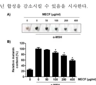
5. B16F10 세포의 멜라닌 합성에 미치는 MECF의 영향
생체 내에서 멜라닌의 합성은 tyrosine으로부터 멜 라닌 생성 효소에 의해 촉매되는 일련의 반응을 통 하여 이루어진다[37,38]. MECF의 미백 효과를 조사 하기 위하여 B16F10 세포에서 α-MSH의 처리에 의 한 멜라닌의 합성에 미치는 MECF의 억제 효과를 조사하였다. Figure 5의 결과에서 알 수 있듯이, MECF가 전치리된 조건에서는 α-MSH 단독 처리된 세포에 비하여 MECF의 처리 농도가 증가할수록 세 포 내 멜라닌의 함량이 유의적으로 억제되었다. 특 히 400 μg/ml의 MECF가 전처리된 경우, α-MSH에 의한 멜라닌의 생성을 40% 정도 억제시켰다.
Tyrosinase는 포유류에서 멜라닌 합성의 핵심 효소 중 하나로서 tyrosine을 quinone과 indolquinone 화합 물 등과 같은 중간체를 거쳐 멜라닌이 생성되게 한 다[16,17,39]. 따라서 MECF에 의한 멜라닌 생성 억 제가 tyrosinase 활성 저해와 연관성이 있는지를 조사 하였다. 이를 위하여 동일 조건에서 배양된 B16F10 세포의 tyrosinase 활성을 비교한 결과, MECF 처리 논도가 증가할수록 α-MSH에 의해 유도된 tyrosinase 의 활성이 유의적으로 억제되었다(Figure 6A). 이러 한 결과는 MECF가 tyrosinase 활성을 억제하여 멜라
닌 합성을 감소시킬 수 있음을 시사한다.
Figure 5. Anti-melanogenesis effect of MECF on B16-F10 melanoma cells. The cells were exposed to various concentrat ions of MECF for 72 h in the presence of 100 nM α-MSH.
At the end of the treatment, the melanin contents were measur ed. (A) Images of pellets of B16F10 cells after harvest. (B) Determination of relative melanin contents. Data are reported as the means ± SD of three independent experiments carried out in triplicate (*p < 0.05 indicates a significant difference compared to α-MSH-treated cells).
6. B16F10 세포에서 멜라닌 합성 관련 유전자의 발 현에 미치는 MECF의 영향
멜라린 합성에 중요하게 관여하는 유전자는 TRP-1 및 TRP-2를 포함하는 tyrosinase 계열 단백질 (family protein)이며, MITF는 이들의 발현을 조절하 는 핵심 전사조절인자이다[39,40]. α-MSH는 MITF 발현을 강력하게 유도하여 tyrosinase, TRP-1 및 TRP-2 발현을 전사적으로 자극하여 멜라닌 생성을 증가시킨다[17,41]. 따라서 이상에서 확인된 MECF 의 멜라닌 생성 억제 기전을 밝히기 α-MSH에 의하 여 유도된 이들 유전자의 발현에 미치는 MECF의 영향을 조사하였다. Figure 6B에 나타난 바와 같이, α-MSH로 자극된 B16F10 세포에서 tyrosinase의 단백 질 수준은 α-MSH 단독 처리된 세포에 비해 MECF의 처리 농도 의존적으로 감소되어 MECF의 tyrosinase 활성 억제 효과는 이 효소의 발현 감소에 의한 것임 을 알 수 있다. 이와 함께 TRP-1 및 TRP-2의 발현 또한 억제되었으며, 이는 MITF 발현 감소와 연관성 이 있음을 보여주었다. 이러한 결과는 MECF에 의한 멜라닌 생성 억제가 MITF의 발현 억제를 통한 ty- rosinase, TRP-1 및 TRP-2의 발현 차단에 의한 결과 임을 시사한다.
Figure 6. Effect of MECF on the activity of tyrosinase and expression of tyrosinase, TRP-1, TRP-2 and MITF in B16F1 0 melanoma cells. The cells were treated with 100 nM α-MS H in presence or absence of MECF at the indicated concentrat ions for 72 h. (A) Tyrosinase activity was measured by absor bance at 490 nm. Data are reported as the means ± SD of three independent experiments carried out in triplicate (*p
< 0.05 indicates a significant difference compared to α-MSH- treated cells). (B) Whole cell lysate was isolated and analyzed by Western blot analysis with respective antibodies. β-actin levels are shown as a loading control. (C) Bands were quantif ied using ImageJ and normalized to β-actin, and the ratio was determined. Data are expressed as the mean ± SD of three independent experiments (*p < 0.05 indicates a signific ant difference compared to the control group).
본 연구에서는 청각의 기능성 평가를 위하여 MECF이 지질 및 멜라닌 합성에 미치는 영향을 조사 하였다. 본 연구의 결과에 의하면 MECF는 3T3-L1 세포에서 MDI의 자극에 의한 지질의 형성을 유의적 으로 억제하였으며, 이는 PPARγ, C/EBPα 및 C/EBP β와 같은 지방 세포의 분화에 관여하는 핵심 유전자 들의 발현 억제와 연관성이 있었다. 아울러 MECF는 B16F10 세포에서 α-MSH 자극에 의한 멜라닌의 생 성을 유의적으로 감소시켰으며, 이는 MITF의 전사 활성 감소에 의한 tyrosinase, TRP-1 및 TRP-2의 발현 차단에 의한 것임을 유추할 수 있었다. 비록 본 연구 의 결과가 청각의 항비만 및 미백 효과에 대한 기초 자료로 활용이 될 수는 있겠지만, 본 연구의 결과를 바탕으로 이러한 효능과 연계된 세포 내 신호 전달 계의 역할과 비만 및 미백 조절 관련 유전자들의 역 할에 대한 추가적인 연구가 수행되어야 할 것이다.
결 론
본 연구에서는 녹조류인 청각 메탄올 추출물 (MECF)의 항비만 및 멜라닌 생성 억제 효과를 평가 하였다. 본 연구의 결과에 의하면 MECF는 3T3-L1 지방 세포에서 TG의 축적을 억제하였으며, 지방 세 포로의 분화 억제는 PPARγ, C/EBPα 및 C/EBPβ와 같은 지방 형성 관련 전사 인자의 발현 억제와 관련 이 있었다. 또한 MECF는 α-MSH 존재하에서 B16F10 세포의 tyrosinase 활성 및 melanin 함량을 감 소시켰을 뿐만 아니라 색소침착에 핵심적인 역할을 하는 tyrosinase, TRP-1, TRP-2 및 MITF의 발현을 감 소시켰다. 따라서 청각 추출물은 비만과 피부 색소 침착의 예방 및 치료를 위한 유망한 기능성 소재로 활용될 수 있음을 알 수 있다.
References
1. Hodson, L., Rosqvist, F., Parry, S. A. 2020. The influence of dietary fatty acids on liver fat content and metabolism.
Proc. Nutr. Soc. 79, 30-41.
2. Westerterp, K. R. 2018. Exercise, energy balance and body composition. Eur. J. Clin. Nutr. 72, 1246-1250.
3. Peyton, K. J., Liu, X .M., Shebib, A. R., Johnson, F. K., Johnson, R. A., Durante, W. 2018. Arginase inhibition prevents the development of hypertension and improves insulin resistance in obese rats. Amino Acids 50, 747– 754.
4. Koenen, M., Hill, M. A., Cohen, P., Sowers, J. R. 2021.
Obesity, adipose tissue and vascular dysfunction. Circ.
Res. 128, 951-968.
5. Dias, S., Paredes, S., Ribeiro, L. 2018. Drugs Involved in dyslipidemia and obesity treatment: Focus on adipose tissue. Int. J. Endocrinol. 2018, 2637418.
6. Rajjo, T., Mohammed, K., Alsawas, M., Ahmed, A. T., Farah, W., Asi, N., Almasri, J., Prokop, L. J., Murad, M. H. 2017. Treatment of pediatric obesity: An umbrella systematic review. J. Clin. Endocrinol. Metab. 102, 763-775.
7. Kumar, A., Chauhan, S. 2021. Pancreatic lipase inhibitors:
The road voyaged and successes. Life Sci. 271, 119115.
8. Narayanaswami, V., Dwoskin, L. P. 2017. Obesity:
Current and potential pharmacotherapeutics and targets.
Pharmacol. Ther. 170, 116-147.
9. Siebenhofer, A., Jeitler, K., Horvath, K., Berghold, A.,
Posch, N., Meschik, J., Semlitsch, T. 2016. Long-term effects of weight-reducing drugs in people with hypertension. Cochrane. Database Syst. Rev. 3, CD007654.
10. Krentz, A. J., Fujioka, K., Hompesch, M. 2016.
Evolution of pharmacological obesity treatments: focus on adverse side-effect profiles. Diabetes Obes. Metab.
18, 558-570.
11. Desmedt, B., Courselle, P., De Beer, J. O., Rogiers, V., Grosber, M., Deconinck, E., De Paepe, K. 2016.
Overview of skin whitening agents with an insight into the illegal cosmetic market in Europe. J. Eur. Acad.
Dermatol. Venereol. 30, 943–950.
12. Qian, W., Liu, W., Zhu, D., Cao, Y., Tang, A., Gong, G., Su, H. 2020. Natural skin-whitening compounds for the treatment of melanogenesis (Review). Exp. Ther.
Med. 20, 173-185.
13. Costin, G. E., Hearing, V. J. 2007. Human skin pigmen- tation: Melanocytes modulate skin color in response to stress. FASEB J. 21, 976–994.
14. Ohbayashi, N., Fukuda, M. 2020. Recent advances in understanding the molecular basis of melanogenesis in melanocytes. F1000Res 9 F1000.
15. Pillaiyar, T., Manickam, M., Jung, S. H. 2017. Recent development of signaling pathways inhibitors of melanogenesis. Cell. Signal. 40, 99-115.
16. Pillaiyar, T., Namasivayam, V., Manickam, M., Jung, S. H. 2018. Inhibitors of melanogenesis: An updated review. J. Med. Chem. 61, 7395-7418.
17. Smit, N., Vicanova, J., Pavel, S. 2009. The hunt for natu- ral skin whitening agents. Int. J. Mol. Sci. 10, 5326– 5349.
18. Draelos, Z. D. 2007. Skin lightening preparations and the hydroquinone controversy. Dermatol. Ther. 20, 308 –313.
19. Ramos-Romero, S., Torrella, J. R., Pagès, T., Viscor, G., Torres, J. L. 2021. Edible microalgae and their bio- active compounds in the prevention and treatment of metabolic alterations. Nutrients 13, 563.
20. Thiyagarasaiyar, K., Goh, B. H., Jeon, Y. J., Yow, Y.
Y. 2020. Algae metabolites in cosmeceutical: An over- view of current applications and challenges. Mar. Drugs 18, 323.
21. Muhamad, I. I., Zulkifli, N., Selvakumaran, S. A., Lazim, N. A. M. 2019. Bioactive algal-derived polysaccharides:
Multi-functionalization, therapeutic potential and bio-
22. Wan-Loy, C., Siew-Moi, P. 2016. Marine algae as a po- tential source for anti-obesity agents. Mar. Drugs 14, 222.
23. Sanjeewa, K. K. A., Lee, W., Jeon, Y.-J. 2018. Nutrients and bioactive potentials of edible green and red seaweed in Korea. Fish. Aquat. Sci. 21, 19.
24. Kim, E., Cui, J., Kang, I., Zhang, G., Lee, Y. 2021.
Potential antidiabetic effects of seaweed extracts by up- regulating glucose utilization and alleviating in- flammation in C2C12 myotubes. Int. J. Environ. Res.
Public Health 18, 1367.
25. Monmai, C., Rod-In, W., Jang, A. Y., Lee, S. M., Jung, S. K., You, S., Park, W. J. 2020. Immune-enhancing ef- fects of anionic macromolecules extracted from Codium fragile coupled with arachidonic acid in RAW264.7 cells. PLoS One 15, e0239422.
26. Lee, C., Park, G. H., Ahn, E. M., Kim, B. A., Park, C. I., Jang, J. H. 2013. Protective effect of Codium frag- ile against UVB-induced pro-inflammatory and oxidative damages in HaCaT cells and BALB/c mice. Fitoterapia 86, 54-63.
27. Lee, S. A., Moon, S. M., Choi, Y. H., Han, S. H., Park, B. R., Choi, M. S., Kim, J. S., Kim, Y. H., Kim, D.
K., Kim, C. S. 2017. Aqueous extract of Codium fragile suppressed inflammatory responses in lip- opolysaccharide-stimulated RAW264.7 cells and carra- geenan-induced rats. Biomed. Pharmacother. 93, 1055-1064.
28. Kang, C. H., Choi, Y. H., Park, S. Y., Kim, G. Y. 2012.
Anti-inflammatory effects of methanol extract of Codium fragile in lipopolysaccharide-stimulated RAW 264.7 cells. J. Med. Food. 15, 44-50.
29. Kolsi, R. B. A., Jardak, N., Hajkacem, F., Chaaben, R., Jribi, I., Feki, A. E., Rebai, T., Jamoussi, K., Fki, L., Belghith, H., Belghith, K. 2017. Anti-obesity effect and protection of liver-kidney functions by Codium fragile sulphated polysaccharide on high fat diet induced obese rats. Int. J. Biol. Macromol. 102, 119-129.
30. Le Lay, S., Dugail, I. 2009. Connecting lipid droplet biology and the metabolic syndrome. Prog. Lipid Res.
31. Padilla-Benavides, T., Velez-delValle, C., Marsch- Moreno, M., Castro-Muñozledo, F., Kuri-Harcuch, W.
2016. Lipogenic enzymes complexes and cytoplasmic lipid droplet formation during adipogenesis. J. Cell Biochem. 117, 2315-2326.
32. Yang, X., Heckmann, B. L., Zhang, X., Smas, C. M., Liu, J. 2013. Distinct mechanisms regulate ATGL-medi- ated adipocyte lipolysis by lipid droplet coat proteins.
Mol. Endocrinol. 27, 116-126.
33. Ali, A. T., Hochfeld, W. E., Myburgh, R., Pepper, M.
S. 2013. Adipocyte and adipogenesis. Eur. J. Cell Biol.
92, 229-236.
34. Spiegelman, B. M., Flier, J. S. 2001. Obesity and the regulation of energy balance. Cell 104, 531–543.
35. Muruganandan, S., Ionescu, A. M., Sinal, C. J. 2020.
At the crossroads of the adipocyte and osteoclast differ- entiation programs: Future therapeutic perspectives. Int.
J. Mol. Sci. 21, 2277.
36. Rosen, E. D., Walkey, C. J., Puigserver, P., Spiegelman, B.M. 2020. Transcriptional regulation of adipogenesis.
Genes Dev. 14, 1293–1307.
37. Serre, C., Busuttil, V., Botto, J. M. 2018. Intrinsic and extrinsic regulation of human skin melanogenesis and pigmentation. Int. J. Cosmet. Sci. 40, 328-347.
38. Rzepka, Z., Buszman, E., Beberok, A., Wrześniok, D.
2016. From tyrosine to melanin: Signaling pathways and factors regulating melanogenesis. Postepy Hig. Med.
Dosw (Online). 70, 695-708.
39. D'Mello, S. A., Finlay, G. J., Baguley, B. C., Askarian-Amiri, M. E. 2016. Signaling pathways in melanogenesis. Int. J. Mol. Sci. 17, 1144.
40. Wan, P., Hu, Y., He, L. 2011. Regulation of melanocyte pivotal transcription factor MITF by some other tran- scription factors. Mol. Cell. Biochem. 354, 241-246.
41. Schallreuter, K. U. 2007. Advances in melanocyte basic science research. Dermatol. Clin. 25, 283-291,