http://e-nrp.org
Peanut sprouts extract (Arachis hypogaea L.) has anti-obesity effects by controlling the protein expressions of PPARγ and adiponectin of adipose tissue in rats fed high-fat diet
Nam E Kang
1, Ae Wha Ha
2, Hye Won Woo
2and Woo Kyoung Kim
2§1Department of Food and Nutrition, Eulji University, 553 Sanseong-Daero,Seongnam-Si, Gyeonggi 461-632, Korea
2Department of Food Science and Nutrition, Dankook University, 152 Jukjeon-Ro, Suji-Gu, Yongin-Si, Gyeonggi 448-701, Korea
BACKGROUD/OBEJECTIVES: This study aims to find out the effects of peanut sprout extracts on weight controls and protein expressions of transcription factors related to adipocyte differentiation and adipocytokine in rats under high-fat diets.
MATERIALS/METHODS: Four week-old Sparague-Dawley (SD) were assigned to 4 groups; normal-fat (NF) diets (7% fat diet), high-fat (HF) diets (20% fat diet), high fat diets with low peanut sprout extract (HF + PSEL) diet (20% fat and 0.025% peanut sprout extract), and high fat diets with high peanut sprout extract (HF + PSEH) diet (20% fat and 0.05% peanut sprout extract).
Body weight changes, lipid profiles in adipose tissue, and the mRNA protein expressions, such as peroxisome proliferator-activated receptor γ (PPARγ), CCAAT element binding protein α (C/EBP α), leptin, and adiponectin, were determined.
RESULTS: After 9 weeks of feeding, the HF + PSEH group had significantly less weight gains than the HF group (P < 0.05).
However, the total dietary intakes or food efficiency ratios among groups were not significantly different. The weight of epididymal fat in HF + PSEH group, 3.61 ± 0.5 g, or HF + PSEL group, 3.80 ± 0.7 g, was significantly lower than the HF group, 4.39 ± 0.4g, (P < 0.05). Total lipids and total cholesterol in adipose tissue were significantly decreased in HF + PSEH group compared to those in the HF group, respectively (P < 0.05). PSEH supplementation caused AST and ALT levels to decrease when it compared to HF group, but it was not statistically significant. The protein expression of PPARγ in HF + PSEH group was significantly lower than the HF group (P < 0.05). Comparing with the HF group, the protein expression of adiponectin in HF + PSEH group was significantly increased (P < 0.05). The protein expressions of C/EBP α and leptin in HF + PSEH group were lower than the HF group, but it was not statistical significant.
CONCLUSIONS: In conclusion, peanut sprout extract has anti-obesity effect by lowering the expressions of PPARγ which regulates the expression of adiponectin.
Nutrition Research and Practice 2014;8(2):158-164; doi:10.4162/nrp.2014.8.2.158; pISSN 1976-1457 eISSN 2005-6168
Keywords: Peanut sprout extract, obesity, PPARγ, adiponectin, high fat diet
INTRODUCTION
6)Adipocytes are known simply as energy reservoirs, but since the 1990s, studies have revealed that adipocytes directly and indirectly regulate body energy metabolisms by secreting a protein called adipocytokine [1,2]. Adipocytokines which includes leptin and adiponectin play a pivotal role in regulating energy metabolisms. Adipocytokine have been noted for causing metabolic disorders like obesity, diabetes, and arteriosclerosis, and thus, regulating the adipocytokine has been considered a novel approach in treating such metabolic disorders [3].
Differentiation of adipocytes is a complicated process affected by various hormones and transcription factors. Adipocyte differ- entiation is primarily regulated by various transcription factors such as peroxisome proliferator activated receptors (PPARs) and
CCAAT/enhancer binding proteins (C/EBPs) [4]. These factors are expressed at different stages of differentiation process. PPAR expression is regulated by C/EBP family. Specifically, the C/EBP induces early expressions of the adipocyte differentiation of PPAR;
and the PPARγ regulates the expressions of several adipocyte- secreted proteins including adiponectin [5].
Researchers have shown interests in developing natural ingredients that can prevent and manage obesity. Resveratrol is a phytochemical produced naturally by several plants when under attack by pathogens such as bacteria or fungi [6].
Resveratrol has been a topic of numerous animal and human studies into anti-aging effects or various chronic diseases preven- tions [7-11]. Resveratrol also triggers mechanism that coun- teracts obesity-related effects like restraining fat synthesis and accumulation, and resveratrol was shown to be more effective
This works was supported by R&D program of MKE/KEIT(10033818).
§Corresponding Author: Woo Kyoung Kim, Tel. 82-31-8005-3172, Fax. 82-31-8005-3170, Email. [email protected] Received: August 12, 2013, Revised: December 27, 2013, Accepted: December 30, 2013
This is an Open Access article distributed under the terms of the Creative Commons Attribution Non-Commercial License (http://creativecommons.org/licenses/by-nc/3.0/) which permits unrestricted non-commercial use, distribution, and reproduction in any medium, provided the original work is properly cited.
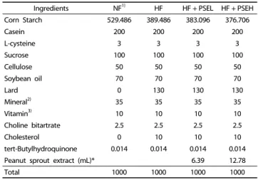
Ingredients NF1) HF HF + PSEL HF + PSEH
Corn Starch 529.486 389.486 383.096 376.706
Casein 200 200 200 200
L-cysteine 3 3 3 3
Sucrose 100 100 100 100
Cellulose 50 50 50 50
Soybean oil 70 70 70 70
Lard 0 130 130 130
Mineral2) 35 35 35 35
Vitamin3) 10 10 10 10
Choline bitartrate 2.5 2.5 2.5 2.5
Cholesterol 0 10 10 10
tert-Butylhydroquinone 0.014 0.014 0.014 0.014
Peanut sprout extract (mL)* 6.39 12.78
Total 1000 1000 1000 1000
1)NF: normal-fat diets (7% fat diet), HF: high-fat diets (20% fat diet), HF + PSEL:
high fat diets with low peanut sprout extract diet (20% fat and 15 mg/kg peanut sprout extract (0.025% resveratrol), and HF + PSEH: high fat with high peanut sprout extract diets (20% fat and 30 mg/kg peanut sprout extract (0.05%
resveratrol).
2)Mineral mixture (per kg): Calcium carbonate anhydrous, 357 g; Potassium phosp- hate monobasic, 196 g; Potassium citrate tripotassium monohydrate, 70.78 g;
Potassium sulfate Sodium chloride, 74 g: Magnesium oxide, 24 g; Ferric citrate, 6.06 g; Zinc carbonate, 1.65 g; Sodium meta-silicate, 1.45 g; Manganous carbonate, 0.63 g; Cupric carbonate, 0.30 g; Chromium potassium sulfate, 0.275 g; Boric acid, 81.5 mg; Sodium fluoride, 63.5 mg; Nickel carbonate, 31.8 mg; Lithium chloride, 17.4 mg; Sodium selenate anhydrous, 10.25 mg; Potassium iodate, 10.0 mg; Ammonium paramolybdate, 6.66 mg; Powdered sucrose, 221.026 g
3)Vitamin mixture (per kg) : Nicotinic acid, 3.0 g; Ca Pantothenate, 1.6 g; Pyridoxine HCl 0.7 g; Thiamin HCl, 0.6 g; Riboflavin 0.6 g; Folic acid, 0.2 g; Biotin, 0.02 g; Vitamin B12, 2.5 g; Vitamin E, 15.0 g; Vitamin A, 0.8 g; Vitamin D3, 0.25 g;
Vitamin K-1, 0.075 g; Powdered sucrose, 974.655 g
* Peanut sprout water extracts: 38.17 mg/mL resveratrol
Table 1. Compositions of experimental diets (g/kg)
in high-fat diets than in normal diets [12,13].
Major food sources of resveratrols are skin of grapes, wines, and peanuts [6]. Studies have shown that the resveratrol content in peanut sprouts is 110.3 μg/g, which is much higher than grapes, 3 μg/g and wine, 0.6 μg/g and peanut, 0.018~1.25 μ g/g [6]. Peanut sprouts refer to germinated peanut seeds.
Peanut sprout is germinated and grown by spraying potable water under humid and warm condition and it can be harvested after 7 days from starting spray [12].
Peanuts itself contains resveratrol as little as 0.15 μg/mg while when peanut is germinated and grown into sprout, resveratrol is significantly increased to 14.2 μg/mg, as much as 90 times of peanuts [14]. Peanut sprout contains isoflavones such as daidzin, genistin, genistein and various types of polypenols [15]. Peanut sprout also includes essential fatty acids and essential amino acids, especially plenty of asparagine, which are far more than peanut itself [15].
Despite of these nutritional values of peanut sprout, the study determining physiological functions of peanut sprout is very limited, and most of them are in vitro studies regarding antioxidant ability of peanut sprouts [16-18]. In Korea, since bean sprouts is habitually consumed as a vegetable, there is a great possibility that peanut sprout can be used as a fresh vegetable as well as functional health foods Therefore, this study aims to find out the effects of peanut sprout extracts that contain large amounts of resveratrol on weight controls and protein expressions of transcription factors related to adipocyte differentiation and adipocytokine for rats under high-fat diets.
MATERIALS AND METHODS
Preparations of peanut sprout extract (PSE)
The peanut sprout extracts were prepared from Chonnam National University, by the modified methods of Wang et al.
[19]. Briefly, fresh peanut sprout, which was germinated for 7 days with mature peanut (Arachis hypogaea L.) kernels, was obtained from a local processing plant (JangSuChae Co, Gyeonggi-do, South Korea). Peanut sprouts were cooked in a boiling water bath. After they were heated, peanut sprouts were cooled with tap water and subjected to lyophilization to prepare dried sprout powders. Dried peanut sprout was extracted with ethanol (1:10 v:v) at 60℃ for 90 min and filtered. The extraction procedure was repeated 3 times. Ethanol in the sample was removed using a rotary vacuum evaporator (N-11 Eyela, Tokyo Rikakikai Co. Tokyo, Japan). Resveratrol contents in PSE were determined by the modification of Wang et al’s study [16]. The mean resveratrol content of PSE used in this study was 38.17 mg/mL.
Animals and Experimental diets
Four week-old Sparague-Dawley (SD) were assigned to two groups; 10 normal-fat (NF) diets, 7% fat diets, and 30 high-fat (HF) diets, 20% fat diets, according to a randomized block design. After 4 weeks, the high-fat diet group was divided into 3 groups; high-fat (HF), high-fat with low levels of peanut sprout extracts (HF + PSEL), and high-fat with high levels of peanut sprout extracts (HF + PSEH), and were all fed for another five weeks. Low peanut sprout extract diets (HF + PSEL) had 15
mg/kg (0.025% resveratrol) and high peanut sprout extract diets (HF + PSEH) had 30 mg/kg (0.05% resveratrol) (Table 1). Food and water were freely accessed to rats in a stainless steel cage, and the temperature of room was maintained at 24 ± 1℃. All experimental diets were stored at 4℃ in the refrigerator. Dietary intake was recorded twice a week and weighed once a week at a fixed time. All experiments were approved by the guidelines of Animal Testing Ethics Committee of Eulji University (EUIACUC 10-09).
Specimen preparation of various organs
After 9 weeks of feeding, the experimental animals were fasted for 12 hours and anesthetized with ethylether. Blood samples were drawn from hearts with a syringe after laparatomy.
The blood sample was centrifuged for 30 minutes at 3,000 rpm, and the plasma was stored at 70℃ before use. Specimens taken from liver, kidney, spleen, epididymal fat, internal fat, kidney fat, and thymus were washed with saline solution, dried in a filter paper, and then weights were measured.
Lipid measurements in adipose tissue
Adipose tissues were collected from various sites such as epididymal fat, kidney fat, and abdominal fat. Total lipids in adipose tissues were extracted with chroloform:methanol (2:1, v/v) using a modified method described by Folch et al. [20].
After extraction, total lipids were determined as described by
Frings and Dunn [21], with slight modifications. Total cholesterol
Parameter NF (n = 10) HF (n = 10) HF + PSEL (n = 10) HF + PSEH (n = 10)
Initial body weight (g) 112.4 ± 1.41)NS2) 112.8 ± 0.9 111.3 ± 1.1 112.5 ± 1.3
Final body weight (g) 423.6 ± 15.7b3) 462.5 ± 11.8a 463.4 ± 10.1a 446.0 ± 8.5ab
Weight gain (g/9weeks) 311.1 ± 14.9b 349.7 ± 11.7a 352.1 ± 10.0a 333.5 ± 9.1ab
Total food intake (g/9weeks) 1290.5 ± 42.8a 1155.8 ± 23.3b 1143.8 ± 18.0b 1128.0 ± 24.6b
FER4) 0.24 ± 0.01b 0.30 ± 0.01a 0.30 ± 0.01a 0.30 ± 0.01a
1)Mean ± SE
2)NS: not significant
3)The significant was determined by Duncan's multiple range test at α = 0.05
4)FER (food efficiency ratio) = Body weight gain for experimental period / Food intake for experimental period Table 2. Body weight, body weight gain, total food intakes and food efficiency ratio of rats
Parameter NF
(n = 10) HF
(n = 10) HF + PSEL
(n = 10) HF + PSEH (n = 10) Internal fat 9.51 ± 1.21)NS2) 10.97 ± 0.7 9.77 ± 0.8 9.32 ± 0.8 Kidney fat 8.66 ± 1.2NS 10.62 ± 0.8 10.07 ± 0.8 9.05 ± 0.9 Epididymal fat 4.05 ± 1.0b3) 4.39 ± 0.4a 3.80 ± 0.7c 3.61 ± 0.5c Total fat 22.5 ± 2.7b 26.0 ± 2.0a 24.9 ± 1.7a 23.8 ± 1.7a
1)Mean ± SE
2)NS: not significant
3)The significant was determined by Duncan's multiple range test at α = 0.05
Table 4. Adipose tissues weight of rats (g)
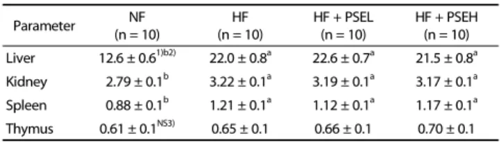
Parameter NF
(n = 10) HF
(n = 10) HF + PSEL
(n = 10) HF + PSEH (n = 10) Liver 12.6 ± 0.61)b2) 22.0 ± 0.8a 22.6 ± 0.7a 21.5 ± 0.8a Kidney 2.79 ± 0.1b 3.22 ± 0.1a 3.19 ± 0.1a 3.17 ± 0.1a Spleen 0.88 ± 0.1b 1.21 ± 0.1a 1.12 ± 0.1a 1.17 ± 0.1a Thymus 0.61 ± 0.1NS3) 0.65 ± 0.1 0.66 ± 0.1 0.70 ± 0.1
1)Mean ± SE
2)The significant was determined by Duncan's multiple range test at α = 0.05
3)NS: not significant
Table 3. Organ weights of the rats fed the experimental diets (g)
and triacylglycerol were measured enzymatically using a comm- ercial kit (Asan Pharmaceutical Co., Seoul, Korea).
Plasma AST (aspartate aminotransferase) and ALT (alanine amino- transferase)
Plamsa AST and ALT activity was determined by commercial assay kit (Asan Pharmaceutical Co., Seoul, Korea) and determined absorbance at 505 nm with spectrophotometer.
Western blot analysis for PPAR γ , C/EBP α , adiponectin, and leptin The protein expressions of PPARγ and C/EBPα, and adipocy- tokines including adiponectin and leptin were determined [13].
Adipose tissue previously stored at -70℃ was finely chopped, mixed with 1 mL of cold phosphate buffered saline (PBS), and centrifuged at 3,000 rpm for 10 minutes. The 400 μL of lysis buffer (Cell Signaling Technology, Boston MA, USA) was added and mixed for 30 minutes, and the mixture was centrifuged at 4℃ at 13,000 rpm for 15-30 minutes. Protein concentration was measured at 595nm by Bio-red method.
After protein was quantified, the sample was dissolved in SDS-PAGE, and transferred to immobilon
™-p membrane (Millipore, Bedford, MA, USA). The membrane was 5% milk/TBST (20 mM Tris-HCl, 137 mM Nacl, 0.1% Tween 20, pH 7.4) and incubated at room temperature for 1 hour. Antibody of the protein was prepared by diluting PPARγ, C/EBPα, leptin (Santacruz, CA, USA), and adiponectin (Cell Signaling Technology, Boston, MA, USA) with 5% milk/TBST and then incubated. After being washed with TBST, the membrane was incubated with Peroxidase- Affinipure Sheep Anti-Mouse IgG (Jackson immunoresearch, West Grove, PA, USA) or Perox-Affinipure Donkey Anti-Rabbit IgG (Jackson immunoresearch, West Grove, PA, USA). Its color was developed by WEST-ZOL® Plus Western Blot Detection System (iNtRON Biotechnology, Korea) and X-Omat Film (Kodak, Tokyo, Japan) and the molecular weight was compared and analyzed with High-Range Rainbow
™Molecular Weight Marker (GE Healthcare, Uppsala, Sweden). Each band was measured for its density by an imaging program called image J Launcher (provided by NCBI).
Statistical analysis
The statistical analysis was performed using Statistical Analysis System (SAS Institute, Cary, NC, USA). The results were reported as mean with standard error, and statistically significant differences among groups were determined by using one way-ANOVA (analysis of variance). Statistically significant differences among the means of groups were tested at α = 0.05 using Duncan's multiple range tests.
RESULTS
The changes of weight and dietary intake
After 9 weeks of feeding, the NF group had an average weight gain of 311.1 ± 14.9 g in NF group, 349.7 ± 11.7 g in HF group, 352.1 ± 10.0 g in HF + PSEL group, and 333.5 ± 9.1 g in HF + PSEH group, showing HF + PSEH group have less weight gains than the HF group. The total dietary intake after 9 weeks was 1,290.5 ± 42.8 g for NF group, 1,155.8 ± 23.3 g for HF group, 1,143.8 ± 18.0 g for HF + PSEL group, and 1,128.0 ± 24.6 g for HF + PSEH group. No significant differences among the HF group, HF + PSEL group, and the HF + PSEH group were shown.
The changes of organ weights
The liver weight of the HF group (22.0 ± 0.8 g), HF + PSEL group (22.6 ± 0.7 g), or HF + PSEH group (21.5 ± 0.8 g) were significantly heavier than the NF group (12.6 ± 0.6 g), but there was no significant difference between HF group, and HF + PSEH group (Table 3). Likewise, the kidney, and thymus weights of the HF group, HF + PSEL group, and HF + PSEH group were not significantly different.
The weight change of adipose tissues
Although HF + PSEH group showed the lower internal fat
weight, 9.32 ± 0.8 g, than that the HF group, 10.97 ± 0.7 g, but
Parameter NF
(n = 10) HF
(n = 10) HF + PSEL
(n = 10) HF + PSEH (n = 10) Total lipid
(μmol/g) 196.0 ± 12.91)b2) 240.5 ± 13.7a 228.1 ± 13.7c 227.0 ± 9.8c Cholesterol
(μmol/g) 6.3 ± 0.3b 7.5 ± 0.3a 6.6 ± 0.4b 6.5 ± 0.3b Triglyceride
(μmol/g)
80.3 ± 4.6NS3) 96.8 ± 7.6 86.8 ± 5.2 84.0 ± 5.9
1)Mean ± SE
2)The significant was determined by Duncan's multiple range test at α = 0.05
3)NS: not significant
Table 5. Concentration of adipose tissue lipids
Parameter NF
(n = 10) HF
(n = 10) HF + PSEL
(n = 10) HF + PSEH (n = 10) AST (IU/ℓ) 89.5 ± 5.01)NS2) 99.0 ± 3.4 98.5 ± 2.7 95.1 ± 3.1 ALT (IU/ℓ) 61.4 ± 1.3NS 70.5 ± 4.3 70.3 ± 3.6 67.2 ± 4.2
1)Mean ± SE
2)NS: not significant
Table 6. The plasma concentration of AST and ALT
Fig. 1. Effects of peanut sprout extracts on PPARγ expression in adipose tissues of rats. Frozen adipose tissue was homogenized in lysis buffer. Protein concentration was determined by using a Bio-Rad method. Equal amounts of proteins (30 μg) were resolved by SDS-PAGE, transferred to the membranes and probed with PPAR γ. Above photographs of chemiliuminiscent detection of the western blots, are shown and under the graph of quantitative analysis which were representatives of three independent experiments. Each bar represents the mean ± SE (n = 10).
Comparison among different concentrations of peanut sprout extracts that yielded significant differences (P< 0.05) are indicated by the different letters above each bar.
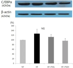
Fig. 2. Effect of peanut sprout extracts on C/EBPα expression in adipose tissue of rats. Frozen adipose tissue was homogenized in lysis buffer. Protein concentration was determined by using a Bio-Rad method. Equal amounts of proteins (30 μg) were resolved by SDS-PAGE, transferred to the membranes and probed with C/EBP α. Above photographs of chemiliuminiscent detection of the western blots, are shown and under the graph of quantitative analysis which were representatives of three independent experiments. Each bar represents the mean ± SE
there were no significant difference among groups (Table 4).
HF + PSEH group had lowest epididymal fat weight, and the statistical difference between HF group and HF + PSEH group were shown (P < 0.05). The total fat weight between HF group and HF + PSEL or HF + PSEH was not statistically significant.
The lipid concentrations of adipose tissue
In Table 5, total lipids in the HF + PSEL group (228.1 ± 13.7 μ mol/g) and HF + PSEH group (227.0 ± 9.8 μmol/g) decreased significantly compared to those in the HF group (240.5 ± 13.7 μ mol/g) group (P < 0.05). Total cholesterol levels also decreased significantly in HF + PSEL group (6.6 ± 0.4 μmol/g or HF + PSEH group (6.5 ± 0.3 μmol/g) compared to those in the HF group (7.5 ± 0.3 μmol/g) (P < 0.05). No significant differences were observed for triglyceride levels among groups.
Plasma AST and ALT concentration
To determine the safety of peanut sprout extract as a supplementation, the plasma concentration of ALT and AST, as index of liver damage, were determined. The plasma concen- tration of AST and ALT were increased when rats fed with high fat diet. PSEH supplementation caused AST and ALT levels to decrease, when it compared to HF group, but it was not statistically significant.
Effects of peanut sprout extracts on protein expressions of PPAR γ and C/EBP α
By setting the NF at 100%, the effects of sprout peanut extracts on the protein expressions of PPARγ and C/EBPα in HF Groups were determined (Fig. 1, 2). The protein expression of PPARγ in HF + PSEH group was significantly lower than the HF group (P < 0.05, Fig.1). The protein expressions of C/EBPα in HF + PSEL group and HF + PSEH group were lower than that of the HF group, although the differences were not statistically significant (Fig. 2).
Effect of peanut sprout extracts on adiponectin and leptin The protein expression level of adiponectin was significantly
increased following the treatment with peanut sprout ethanol
extracts at high levels (HF + PSEH group) when it compared with
HF group (P < 0.05, Fig. 3). The HF group had 58.9%, HF + PSEL
had 67.2%, and HF + PSEH had 76.2%. The protein expression
of leptin in HF group was higher than that in NF group without
Fig. 3. Effect of peanut sprout extracts on adiponectin expression in adipose tissue of rats. Frozen adipose tissue was homogenized in lysis buffer. Protein concentration was determined by using a Bio-Rad method. Equal amounts of proteins (30 μg) were resolved by SDS-PAGE, transferred to the membranes and probed with adiponectin. Above photographs of chemiliuminiscent detection of the western blots, are shown and under the graph of quantitative analysis which were representatives of three independent experiments. Each bar represents the mean
± SE Comparison among different concentrations of peanut sprout extracts that yielded significant differences (P< 0.05) are indicated by the different letters above each bar.
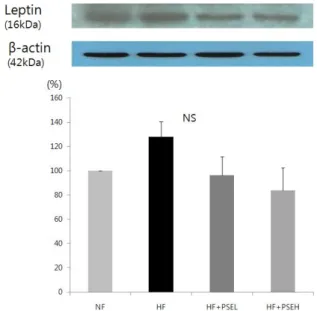
Fig. 4. Effect of peanut sprout extracts on leptin protein expression in adipose tissue of rats. Frozen adipose tissue was homogenized in lysis buffer. Protein concentration was determined by using a Bio-Rad method. Equal amounts of proteins (30 μg) were resolved by SDS-PAGE, transferred to the membranes and probed with leptin. Above photographs of chemiliuminiscent detection of the western blots, are shown and under the graph of quantitative analysis which were represen- tatives of three independent experiments. Each bar represents the mean ± SE
statistical significances (Fig. 4). Although there was a much lower leptin level in the peanut sprout treated group than the HF group, the difference was not statistically significant.
DISCUSSION
World Health Organization (WHO) has announced obesity as a disease in need of treatment. Peanut sprouts refer to germinated peanut seeds and produce lots of resveratrol during germination [22]. In addition to abundant resveratrol contents in peanut sprouts, it also contains various bio-functional components such as pholypherols, isoflavones, and essential amino acids [15]. Hence, to evaluate peanut sprouts as a functional food material, anti-obesity effects of peanut sprout extract were examined by providing rats with high fat diet supplemented with low peanut sprout extract (0.025%
resveratol) or high peanut sprout extract (0.05% resveratrol).
In this study, the body weight gain in high peanut sprout extract group was significantly suppressed, as compared with high fat group. However, the total food intakes were similar for the high fat groups and the peanut sprout extract groups.
Likewise, Studies reported that hamsters provided with high fat diets added with resveratrol showed no significant differences in dietary intake and food efficiency ratio, but a significant decrease in weight [23,24].
In addition to decreased weight gains, there were also significant decreases in epididymal fat weights (Table 4) and low total lipids and cholesterol contents in adipose tissue of peanut sprout extract treated rats (Table 5), compared high fat only diet, which demonstrated clearly that peanut sprout has some anti-obesity effects. Many in vivo resveratol studies reported that 0.04% pure resveratrol administered into a high fat diet was effective in reducing body weight or decreasing adipose tissue weights while 0.01% resveratrol was not very effective [7-12].
In this study, the PSE supplementation (containing 0.05%
resveratrol) showed significantly decreased visceral adipose fat weights, even much higher effects than pure 0.05 % resveratrol supplementation reported by previous studies [9-12]. Two previous in vitro studies reported that combination treatment with resveratrol and genistein or other natural polypherols suppressed lipid accumulation significantly more than the responses to resveratrol alone [25,26]. Those studies suggested that resveratrols’s anti-adipogenic activity was substantially enhanced by the other natural isoflavornes or pholypherols compound in foods. Therefore, we believe that several bio- functional components of peanut sprout, in addition to resve- ratrol, allowed obtaining much stronger effects at reducing adipose body fat weights and body weight gain. This study has limitation that other physiological bio-components of peanut sprouts were not analyzed, and thus more studies regarding various components of peanut sprout on anti-obesity effects are needed.
In this study, the addition of the PSE supplementation in high
fat diet lowered the epididymal fat weight even more so than
the normal diet. A research found that the epididymal fat cell
size of rats fed a normal diet with natural plants supple-
mentation was significantly less than that of rats fed normal
diet [27]. Based on our data of AST and ALT concentration, and
the fact that adiponectin expression in adipose tissue signi-
ficantly increased with the PSE supplementation, it can be
concluded that the PSE supplement does not incur any side
effects. However, further research may be needed to explore the causes for this decrease in epididymal fat weight.
Peanut sprout extract in this study also significantly dec- reased the expression of PPARγ (Fig. 1.), adipogenic transcription factor, and the target gene, adiponectin (Fig. 4.). These changes of PPARγ and adiponectin may contribute to the lower weight gain and epidermal fat weights in rats fed with high fat with peanut sprout extracts. In research on adipocyte differentiation in human and rats, the transcription factors such as PPARs and C/EBPs are reported to play an important role in adipocyte- specific gene expressions and adipocyte differentiations [4].
Vadal-Puig et al. [28] observed decrease in PPARγ expression due to restriated food intakes and increases in high-fat diets.
Also, in a study of obese patients, it was found that PPARγ expression occurs mainly in human adipose tissues and is positively correlated with body mass index [29]. Our previous study which uses 3T3-L1 adipocytes [13] showed that resveratrol was down-regulated with adipocyte differentiation in a dose dependent manner.
Adiponectin is a direct target of regulation by PPARγ, and Tsuchida et al. reported that activation of PPAR increased adiponectin receptor 1 and 2 in both white adipose tissues (WAT) and brown adipose tissues (BAT) in vivo [30]. When treated with Pioglitazone, a PPARγ agonist, 3T3-L1 adipocytes presented enhanced insulin sensitivity through an upregulation of adiponectin receptor 2 (AdipoR2) and adiponectin [31,32].
In this study, high-fat diet groups (HF) showed significant decreases in protein expression of adiponectin, while peanut sprout extract diet groups showed significant increases in adiponectin. Adiponectin is a peptide hormone that is inversely related with abdominal visceral fat area, and diminishes the concentration of blood fatty acids and level of triglycerides [33,34]. Barnea et al. [35] concluded that adiponectin was decreased for mice fed with high fat diets and Rogers et al.
[36] reported that rats fed with resveratrol showed increases in the concentrations of adiponectin in serum and suppressions of fat accumulation. Thus this can suggest that peanut sprout extract supplementation activates the protein expressions of PPARγ which inhibits adipocyte differentiation and improves adiponectin secretion in adipose tissues which has been shown to play important roles in lipolysis and thermogenesis.
Blood leptin concentration is also known to be closely related with body mass index and amount of body fat; an obese person tends to have higher blood leptin concentrations than a normal individual [37]. Szkudelska et al. [38] noted that resveratrol directly restrains leptin secretion by observing the effects of resveratrol in restraining leptin secretion in mice adipocytes. In this study, we observed the tendency of the peanut sprout extract groups to have lower leptin expression levels than the high-fat diet groups, though the difference was not significant.
In conclusion, the peanut sprout extract demonstrated decreasing weight gain and reducing body fat contents in adipose tissues of rats fed with high fat diets. Peanut sprout extracts can restrain adipocyte differentiation by lowering the expressions of PPARγ and adiponectin which are necessary for the adipocyte differentiation process.
REFERENCES
1. Coelho M, Oliveira T, Fernandes R. Biochemistry of adipose tissue:
an endocrine organ. Arch Med Sci 2013;9:191-200.
2. Fonseca-Alaniz MH, Takada J, Alonso-Vale MI, Lima FB. Adipose tissue as an endocrine organ: from theory to practice. J Pediatr (Rio J) 2007;83:S192-203.
3. Matsuzawa Y. The metabolic syndrome and adipocytokines. FEBS Lett 2006;580:2917-21.
4. Fajas L, Fruchart JC, Auwerx J. Transcriptional control of adipoge- nesis. Curr Opin Cell Biol 1998;10:165-73.
5. Grimaldi PA. The roles of PPARs in adipocyte differentiation. Prog Lipid Res 2001;40:269-81.
6. Kopp P. Resveratrol, a phytoestrogen found in red wine. A possible explanation for the conundrum of the 'French paradox'? Eur J Endocrinol 1998;138:619-20.
7. Rocha KK, Souza GA, Ebaid GX, Seiva FR, Cataneo AC, Novelli EL.
Resveratrol toxicity: effects on risk factors for atherosclerosis and hepatic oxidative stress in standard and high-fat diets. Food Chem Toxicol 2009;47:1362-7.
8. Shang J, Chen LL, Xiao FX, Sun H, Ding HC, Xiao H. Resveratrol improves non-alcoholic fatty liver disease by activating AMP- activated protein kinase. Acta Pharmacol Sin 2008;29:698-706.
9. Aubin MC, Lajoie C, Clément R, Gosselin H, Calderone A, Perrault LP. Female rats fed a high-fat diet were associated with vascular dysfunction and cardiac fibrosis in the absence of overt obesity and hyperlipidemia: therapeutic potential of resveratrol. J Pharmacol Exp Ther 2008;325:961-8.
10. Rivera L, Morón R, Zarzuelo A, Galisteo M. Long-term resveratrol administration reduces metabolic disturbances and lowers blood pressure in obese Zucker rats. Biochem Pharmacol 2009;77:1053-63.
11. Pearson KJ, Baur JA, Lewis KN, Peshkin L, Price NL, Labinskyy N, Swindell WR, Kamara D, Minor RK, Perez E, Jamieson HA, Zhang Y, Dunn SR, Sharma K, Pleshko N, Woollett LA, Csiszar A, Ikeno Y, Le Couteur D, Elliott PJ, Becker KG, Navas P, Ingram DK, Wolf NS, Ungvari Z, Sinclair DA, de Cabo R. Resveratrol delays age-related deterioration and mimics transcriptional aspects of dietary restric- tion without extending life span. Cell Metab 2008;8:157-68.
12. Szkudelska K, Nogowski L, Szkudelski T. Resveratrol, a naturally occurring diphenolic compound, affects lipogenesis, lipolysis and the antilipolytic action of insulin in isolated rat adipocytes. J Steroid Biochem Mol Biol 2009;113:17-24.
13. Kang NE, Ha AW, Kim JY, Kim WK. Resveratrol inhibits the protein expression of transcription factors related adipocyte differentiation and the activity of matrix metalloproteinase in mouse fibroblast 3T3-L1 preadipocytes. Nutr Res Pract 2012;6:499-504.
14. Rural Development Administration, National Institute of Crop Science (KR). Rural Development Administration News [Internet].
Suwon: Rural Development Administration; 2010 [cited 2010 Oct 14]. Available from: http://rda.korea.kr/gonews.
15. Kang HI, Kim JY, Park KW, Kang JS, Choi MR, Moon KD, Seo KI.
Resveratrol content and nutritional components in peanut sprouts.
Korean J Food Preserv 2010;17:384-90.
16. Kang HI, Kim JY, Kwon SJ, Park KW, Kang JS, Seo KI. Antioxidative effects of peanut sprout extracts. J Korean Soc Food Sci Nutr 2010;
39:941-6.
17. Kim HJ, Kang JS, Park HR, Hwang YI. Neuroprotective effects of methanolic extracts from peanut sprouts. J Life Sci 2010;20;253-9.
18. Lee SE, Park CH, Bang JK, Seong NS, Chung TY. Comparison on antioxidant potential of several peanut varieties. J Korean Soc Food Sci Nutr 2004;33:941-5.
19. Wang KH, Lai YH, Chang JC, Ko TF, Shyu SL, Chiou RY. Germination of peanut kernels to enhance resveratrol biosynthesis and prepare sprouts as a functional vegetable. J Agric Food Chem 2005;53:242-6.
20. Folch J, Lees M, Sloane Stanley GH. A simple method for the isolation and purification of total lipides from animal tissues. J Biol Chem 1957;226:497-509.
21. Frings CS, Dunn RT. A colorimetric method for determination of total serum lipids based on the sulfo-phospho-vanillin reaction. Am J Clin Pathol 1970;53:89-91.
22. Lin BS, Lien TF, Chao MR, Lai TY, Chang JC, Chou SJ, Liao HF, Chiou RY. Toxicological and nutraceutical assessments of peanut sprouts as daily supplements to feed Sprague-Dawley rats for 18 weeks.
J Sci Food Agric 2008;88:2201-7.
23. Cho IJ, Ahn JY, Kim S, Choi MS, Ha TY. Resveratrol attenuates the expression of HMG-CoA reductase mRNA in hamsters. Biochem Biophys Res Commun 2008;367:190-4.
24. Park SH, Park TS, Cha YS. Grape seed extract (Vitis vinifera) partially reverses high fat diet-induced obesity in C57BL/6J mice. Nutr Res Pract 2008;2:227-33.
25. Rayalam S, Della-Fera MA, Yang JY, Park HJ, Ambati S, Baile CA.
Resveratrol potentiates genistein's antiadipogenic and proapoptotic effects in 3T3-L1 adipocytes. J Nutr 2007;137:2668-73.
26. Park HJ, Yang JY, Ambati S, Della-Fera MA, Hausman DB, Rayalam S, Baile CA. Combined effects of genistein, quercetin, and resveratrol in human and 3T3-L1 adipocytes. J Med Food 2008;11:773-83.
27. Kim HS, Kim TW, Kim DJ, Hwang HJ, Lee HJ, Choe M. Effects of natural plants supplementation on adipocyte size of the epididymal fat pads in rats. J Korean Soc Food Sci Nutr 2007;36:419-23.
28. Vidal-Puig A, Jimenez-Liñan M, Lowell BB, Hamann A, Hu E, Spiegelman B, Flier JS, Moller DE. Regulation of PPARγ gene exp- ression by nutrition and obesity in rodents. J Clin Invest 1996;97:
2553-61.
29. Vidal-Puig AJ, Considine RV, Jimenez-Liñan M, Werman A, Pories
WJ, Caro JF, Flier JS. Peroxisome proliferator-activated receptor gene expression in human tissues. Effects of obesity, weight loss, and regulation by insulin and glucocorticoids. J Clin Invest 1997;
99:2416-22.
30. White UA, Stephens JM. Transcriptional factors that promote forma- tion of white adipose tissue. Mol Cell Endocrinol 2010;318:10-4.
31. Tsuchida A, Yamauchi T, Takekawa S, Hada Y, Ito Y, Maki T, Kadowaki T. Peroxisome proliferator-activated receptor (PPAR)alpha activation increases adiponectin receptors and reduces obesity-related inflam- mation in adipose tissue: comparison of activation of PPARalpha, PPARgamma, and their combination. Diabetes 2005;54:3358-70.
32. Chinetti G, Zawadski C, Fruchart JC, Staels B. Expression of adipone- ctin receptors in human macrophages and regulation by agonists of the nuclear receptors PPARalpha, PPARgamma, and LXR. Biochem Biophys Res Commun 2004;314:151-8.
33. Kazumi T, Kawaguchi A, Sakai K, Hirano T, Yoshino G. Young men with high-normal blood pressure have lower serum adiponectin, smaller LDL size, and higher elevated heart rate than those with optimal blood pressure. Diabetes Care 2002;25:971-6.
34. Yamamoto Y, Hirose H, Saito I, Tomita M, Taniyama M, Matsubara K, Okazaki Y, Ishii T, Nishikai K, Saruta T. Correlation of the adipocyte-derived protein adiponectin with insulin resistance index and serum high-density lipoprotein-cholesterol, independent of body mass index, in the Japanese population. Clin Sci (Lond) 2002;
103:137-42.
35. Barnea M, Shamay A, Stark AH, Madar Z. A high-fat diet has a tissue-specific effect on adiponectin and related enzyme expression.
Obesity (Silver Spring) 2006;14:2145-53.
36. Rogers CQ, Ajmo JM, You M. Adiponectin and alcoholic fatty liver disease. IUBMB Life 2008;60:790-7.
37. Considine RV, Caro JF. Leptin: genes, concepts and clinical pers- pective. Horm Res 1996;46:249-56.
38. Szkudelska K, Nogowski L, Szkudelski T. The inhibitory effect of resveratrol on leptin secretion from rat adipocytes. Eur J Clin Invest 2009;39:899-905.