INTRODUCTION
Atherosclerosis (AS) is a chronic inflammatory disease in arte-
rial walls, which is characterized by lipids deposition in the ar- terial vessel wall.1,2 AS causes various acute cardiovascular events, including myocardial infarction and stroke, and severely influences the patients’ quality of life.3,4 Due to the change in di- etary structure, AS incidence has been increasing worldwide and may reach epidemic proportions in the next few decades.5 Therefore, it is urgent to find new therapeutic methods and targets for the treatment of AS.
Mesenchymal stem cells (MSCs) have the functions of pro- moting healing and immunomodulatory,6 which provide an opportunity of using autologous MSC transplantation to pro- mote tissue repair and regeneration in clinical practice.7 Adi- pose-derived stem cells (ADSCs) are isolated from adipose tis- sue, and are ubiquitous in all kinds of MSCs.8 Recently, MSC
Adipose-Derived Stem Cell Transplantation Inhibits Vascular Inflammatory Responses and
Endothelial Dysfunction in Rats with Atherosclerosis
Mingqiang Fan1*, Jing Bai1*, Tao Ding1, Xiangxiang Yang1, Qiaoke Si1, and Dengmei Nie2
1Department of Dardiovascular, Pingliang People’s Hospital, Pingliang;
2Department of Pathology, Second Provincial People’s Hospital, Lanzhou, China.
Purpose: This study aimed to investigate the effect of adipose-derived stem cell (ADSC) transplantation on atherosclerosis (AS) and its underlying mechanisms.
Materials and Methods: In our study, rat AS model was established, and ADSCs were isolated and cultured. Atherosclerotic plaque and pathological symptoms of thoracic aorta were measured by Oil Red O staining and Hematoxylin-Eosin staining, re- spectively. Total cholesterol (TC), triglyceride (TG), high-density lipoprotein cholesterol (HDL-C), and low-density lipoprotein cholesterol (LDL-C) levels were measured by an automatic biochemical analyzer. Expressions of vascular endothelial growth fac- tor (VEGF), vascular cell adhesion molecule-1 (VCAM-1), intercellular adhesion molecule-1 (ICAM-1), aortic endothelin-1 (ET- 1), interleukin-6 (IL-6), c-reactive protein (CRP), and tumor necrosis factor α (TNF-α) were measured by enzyme linked immuno- sorbent assay, VEGF, VCAM-1, ICAM-1, ET-1, respectively, and NF-κB p65 mRNA expressions were detected by quantitative real- time polymerase chain reaction. Protein expressions of VEGF, VCAM-1, ICAM-1, ET-1, NF-κB p65, p-NF-κB p65, and IκBα were measured by western blot. Moreover, NF-κB p65 expression was measured by immunofluorescence staining.
Results: ADSC transplantation alleviated the pathological symptoms of aortic AS. ADSC transplantation decreased the levels of TC, TG, and LDL-C and increased serum HDL-C level. Meanwhile, ADSC transplantation decreased the levels of IL-6, CRP, and TNF-α in AS rats. Moreover, the expressions of VEGF, ET-1, VCAM-1, and ICAM-1 were decreased by ADSC transplantation.
ADSC transplantation inhibited phosphorylation of NF-κB p65 and promoted IκBα expression in AS rats.
Conclusion: Our study demonstrated that ADSC transplantation could inhibit vascular inflammatory responses and endothelial dysfunction by suppressing NF-κB pathway in AS rats.
Key Words: ADSC, atherosclerosis, endothelial dysfunction, inflammatory responses, NF-κB pathway
pISSN: 0513-5796 · eISSN: 1976-2437
Received: May 14, 2019 Revised: August 12, 2019 Accepted: September 11, 2019
Corresponding author: Dengmei Nie, MD, Department of Pathology, Second Pro- vincial People’s Hospital, 1 Hezheng West Road, Chengguan District, Lanzhou, Gan- su 730000, China.
Tel: 86-0933-8639060, Fax: 86-0933-8639060, E-mail: [email protected]
*Mingqiang Fan and Jing Bai contributed equally to this work.
•The authors have no potential conflicts of interest to disclose.
© Copyright: Yonsei University College of Medicine 2019
This is an Open Access article distributed under the terms of the Creative Com- mons Attribution Non-Commercial License (https://creativecommons.org/licenses/
by-nc/4.0) which permits unrestricted non-commercial use, distribution, and repro- duction in any medium, provided the original work is properly cited.
Yonsei Med J 2019 Nov;60(11):1036-1044 https://doi.org/10.3349/ymj.2019.60.11.1036
transplantation has been recognized as a new technique for treating various diseases. Previously, researchers have report- ed that ADSCs could enhance liver regeneration in acute liver injury.9 Zhang, et al.10 have indicated that ADSC transplanta- tion alleviates brain edema from intracerebral hemorrhage.
However, precise roles of ADSCs in AS and their potential mo- lecular mechanisms remain to be further investigated.
Nowadays, it is well recognized that endothelial dysfunc- tion is the initial step in the pathogenesis of AS.11 The abnor- mal endothelial function further promotes a series of inflam- matory response.12 Nuclear factor-κB (NF-κB) has long been recognized as a key component of signaling mechanisms in- volved in the pathogenesis of a number of inflammatory re- sponses.13 Under normal physiological condition, NF-κB stays in the cytoplasm by interacting with the inhibitor of κB (IκB) proteins, of which the prototypical member is IκBα. Inflam- matory factors, including interleukin-6 (IL-6), c-reactive pro- tein (CRP), and tumor necrosis factor α (TNF-α), could acti- vate IκBα.13
In our study, we investigated the effects of ADSC transplan- tation on AS and its related molecular mechanisms. Our re- sults suggested that ADSC transplantation could inhibit vas- cular inflammatory responses and endothelial dysfunction by suppressing NF-κB signaling pathway in AS rats. The results of our study may provide new theoretical foundation for deeply exploring the treatment of AS.
MATERIALS AND METHODS
Animal model of AS
Forty-eight male Sprague-Dawley (SD) rats (200–220 g) were supplied by Dashuo Co., LTD. (Chengdu, China). The rats were housed at room temperature with 45–60% humidity for at least a week to adapt to the environment. The rats were ran- domly assigned to two groups (24 rats in each group): normal group and AS group. Rats in AS group were intraperitoneally injected with vitamin D3 at a dose of 700000 IU/kg over 3 d.
Subsequently, the rats were fed a high-fat vitamin D3 diet (2%
cholesterol, 0.5% sodium cholate, 0.2% propyl thiouracil, 3%
lard, 5% sugar, 0.0125% vitamin D3 powder, and 82.3% basic diet) every day for 3 weeks. Meanwhile, rats in normal group were fed a normal diet and injected with physiological saline under the same conditions. All animal experiments were con- ducted strictly in accordance with the guideline for the care and use of laboratory animals, and approved by the ethics commit- tee of Second Provincial People’s Hospital.
Determination of serum lipids
After 3 weeks of modeling, 6 mL of blood was collected. Total cholesterol (TC), triglyceride (TG), high-density lipoprotein cholesterol (HDL-C), and low-density lipoprotein cholesterol (LDL-C) levels were then measured using an Olympus AU2700
automatic biochemical analyzer (Interscience, Saint Nom La Breteche, France).
Oil Red O staining assay
The aorta, which was cut from the root to the bifurcation of tho- racic arteries, was fixed in 10% formalin. Aortic roots were sectioned to 5-μm thick sections and then stained with Oil Red O for 10 min. Subsequently, the sections were counter- stained with hematoxylin to visualize the nuclei. Finally, the plaques were visualized with an optical microscope (Olympus, Tokyo, Japan).
Isolation and culture of ADSCs
ADSCs were isolated from the subcutaneous adipose tissue of 8–10 week old male C57BL/6J mice. In brief, the subcutane- ous adipose tissue was chopped and digested with 0.1% colla- genase type I (Gibco, Grand Island, NY, USA)/1% BSA solution at 37°C for 30 min. Following filtration, centrifugation, and re- suspension, ADSCs were cultivated in Dulbecco’s Modified Eagle Medium (DMEM) (Gibco, USA) supplemented with 10% fetal bovine serum (Beckman Coulter Inc., Miami, FL, USA) and 1% penicillin/streptomycin (Gibco). After 48 h, the adherent cells were washed with phosphate-buffered saline (Sigma-Aldrich Chemie, Steinheim, Germany) to remove loose cellular debris, and the medium was then changed every 3 days. ADSCs at three passage were stained for 15 min at room temperature with monoclonal PE-conjugated antibodies for CD34 and CD45 (BD Biosciences, San Diego, CA, USA) or FITC- conjugated antibodies for CD44 and CD90 (BD Biosciences), and then incubated with the appropriate fluorochrome-con- jugated secondary antibody. Isotype control IgG was used to stain the cells as control. Finally, samples were analyzed by flow cytometer (Beckman Coulter, Brea, CA, USA).
ADSC transplantation
After modeling, normal and AS rats were randomly divided into four groups (eight rats in each group): normal group, normal+
ADSCs group, AS group, and AS+ADSCs group. ADSCs cell suspension, labeled by PKH26 dye (Sigma-Aldrich Chemie) with a concentration of 1×10¹⁰ L-1 (0.5 mL), was slowly injected into the rats of normal+ADSCs group and AS+ADSCs group through their tail vein. Furthermore, rats of normal group and AS group were injected with equal volume of DMEM medium.
Enzyme linked immunosorbent assay (ELISA)
After 3 weeks of ADSC transplantation, the rats were decapi- tated and their blood was rapidly collected. The levels of vas- cular endothelial growth factor (VEGF), vascular cell adhesion molecule-1 (VCAM-1), intercellular adhesion molecule-1 (ICAM-1), and aortic endothelin-1 (ET-1) were measured us- ing ELISA kits (R&D Systems Inc., Minnesota, MN, USA) ac- cording to the methods provided by the manufacturer. Mean- while, inflammatory factors including IL-6, CRP, and TNF-α
were also detected.
Real-time fluorogenic PCR assay
Thoracic aortas were collected and the total RNA was extract- ed using TRIZOL (Invitrogen, Carlsbad, CA, USA) after 3 weeks of ADSCs transplantation. Then, total 500-ng RNA was re- verse-transcribed into cDNA by Revert Aid First Strand cDNA Synthesis Kit (Thermo Scientific, Fremont, CA, USA), and measured using quantitative real-time polymerase chain re- action (qRT-PCR) (Bio-Rad Laboratories, Hercules, CA, USA) with SYBR green qPCR Master Mix (Thermo Scientific). Prim- ers used for qRT-PCR analysis were as follows: VEGF (for- ward): 5'-ATGTGTGTCCGTCTAGATGT-3', (reverse): 5'- GGAAGTGTTGATTGGAAAACTGA-3'; ET-1 (forward):
5'-ACCCAGCCTATCCAGAATCC-3', (reverse): 5'-AT- GAAGCTGGGCTCTGAGAA-3'; VCAM-1 (forward): 5'- G C A G A A G T G G A AT TA G T T G - 3 ' , ( r e v e r s e ) : 5 ' - CTGGGTTCTCCAAGAAAA-3'; ICAM-1 (forward): 5'- CACAGGTGGTGCTICTGAAC- 3', (reverse): 5'- CTCCT- G A G C C T T C T G TA AC T TG - 3 ' ; G A P D H ( f o r w a rd ) : 5'-ATTGTCAGCAATGCATCCTG-3', (reverse): 5'-GTAGGCCAT- GAGGTCCACCA-3'.
Western blot analysis
Total proteins were extracted from the thoracic aortas by lysis buffer, and protein concentration was measured by BCA kit (TaKaRa, Kyoto, Japan). Protein samples (30 μg) were subject- ed to 10% SDS-PAGE and then transferred onto nitrocellulose membrane. Following blocking with 5% skim milk, the mem- branes were incubated with the primary antibody (ICAM-1, 1:1000; VCAM-1, 1:1000; ET-1, 1:1000; NF-κB p65, 1:1000;
p-NF-κB p65, 1:1000, Abcam, Cambridge, MA, USA; VEGF, 1:1000; IκBα, 1:1000, Cell Signaling Technology, Beverly, MA, USA; GAPDH, 1:1000, Sino Biologial, Beijing, China) at 4°C overnight. Afterwards, peroxidase-labeled secondary anti- body (anti-rabbit IgG, 1:5000, Abcam) was used for incuba- tion for 1 h at room temperature. Protein blots were visualized with an enhanced chemiluminescence kit. Finally, density of western blot bands was analyzed using Quantity One 1-D Anal- ysis Software (Bio-Rad Laboratories).
Hematoxylin-Eosin (HE) staining
The aorta, which was cut from the root to the bifurcation of tho- racic arteries, was fixed in 10% formalin for 24 h. Afterwards, paraffin-embedded tissues were sectioned to 5-μm thick aor- ta sections after dehydration and vitrification. The aorta sec- tions were deparaffined with xylene, rehydrated with gradient ethanol, and stained with HE dye in proper order. Histopatho- logical changes were visualized with an optical microscope (Olympus) at ×400 magnification. Moreover, we also mea- sured the thickness of the intima (I) and media of the aorta (M), and calculated the ratio of I/(I+M).
Immunofluorescence staining
Immunofluorescence staining for thoracic aorta tissue was performed on paraffin sections, which were prepared using the same method as in HE staining. After being blocked with 3% BSA to avoid nonspecific immunoreactions, the sections were incubated overnight at 4°C with primary antibody of NF- κB p65 (1:100, Abcam). The sections were washed three times and incubated at 37°C for 2 h with FITC-labeled goat anti-rab- bit IgG (1:500, Abcam). Afterwards, the sections were stained by 4', 6-diamidino-2-phenylindole (DAPI) for 5 min at room temperature. Fluorescence images were randomly scanned by a single investigator, who was blind to sample identity, using a confocal laser scanning microscope at ×400 magnification.
Statistical analysis
All statistical analyses were performed using SPSS ver. 22.0 Statistical Software (IBM Corp., Armonk, NY, USA). The results were presented in the form of mean±standard deviation. Dif- ferences between various groups were analyzed by one-way ANOVA followed by Tukey’s post hoc test, and data of the two groups were assessed using Student’s t test. All experiments were repeated three times. p<0.05 was considered statistically significant.
RESULTS
Rats AS model is successfully constructed
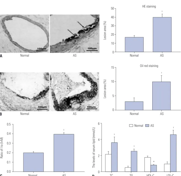
The results of HE staining (Fig. 1A) showed that the aorta in normal group was normal and did not show hyperplasia.
However, the aorta had disorganized structures and visible atherosclerotic plaques in AS group. Meanwhile, the intima and media thickness of aorta increased significantly in AS group (p<0.001). The atherosclerotic plaque in aorta was de- termined by Oil Red O staining (Fig. 1B). Compared to normal group, the number of Oil Red O positive lipid droplets in AS group significantly increased (p<0.01). Furthermore, the ratio of I/(I+M) remarkedly increased in AS group compared to normal group (p<0.001) (Fig. 1C). As shown in Fig. 1D, the se- rum levels of TC, TG, and LDL-C in AS group were significantly higher than those in normal group (p<0.001). In contrast, the serum levels of HDL-C in AS group were significantly lower than those in normal group (p<0.05). All of the results above revealed that AS model of rats was successfully constructed in our research.
Identification of ADSCs
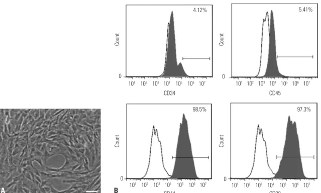
As shown in Fig. 2A, P3 ADSCs had adherent growth ability, morphology of fibroblast-like cells, and radial shape appear- ance. The results of flow cytometry confirmed that ADSCs ex- pressed CD44 (98.5%) and CD90 (97.3%), but not CD34 (4.12%) and CD45 (5.41%), which was consistent with the surface mo- lecular characteristics of ADSCs (Fig. 2B).
ADSCs transplantation alleviates the pathological symptoms of aortic AS in rats
As shown in Fig. 3A, after 3 weeks of ADSCs transplantation, red positive cells were found in normal+ADSCs group and AS+ADSCs group, while no fluorescent cells were found in normal group and AS group, indicating that ADSC transplan- tation was successful. In accordance with the aortic morphol- ogy observed via HE (Fig. 3B), atherosclerotic plaques were formed in the vascular tissue of AS group (p<0.001), and the thickness of whole vascular wall increased (p<0.001). The visi- ble atherosclerotic plaques and increased vascular wall thick- ness were alleviated in AS+ADSCs group (p<0.01). Consistent with the results of HE staining, the ratio of I/(I+M) significantly increased in AS group compared to normal and normal+ADSCs groups (p<0.001) (Fig. 3C). Compared to AS group, the ratio of
I/(I+M) markedly decreased in AS+ADSCs group (p<0.01) (Fig.
3C). All of these results suggest that ADSC transplantation could alleviate the symptoms of aortic AS.
ADSC transplantation decreases the levels of lood lipids and inflammatory cytokines in AS rats
Compared to the values in normal and normal+ADSCs group, the serum levels of TC, TG, and LDL-C significantly increased (p<0.001) and serum HDL-C levels markedly decreased in AS group (p<0.01) (Fig. 4A). Compared to AS group, the serum lev- els of TC, TG, and LDL-C significantly decreased (p<0.001) and serum HDL-C levels markedly increased in AS+ADSCs group (p<0.05) (Fig. 4A). Furthermore, the levels of IL-6, CRP, and TNF-α in AS group significantly increased compared to those in normal and normal+ADSCs groups (p<0.001) (Fig. 4B-D).
50 40 30 20 10 0
15
10
5
0
6
4
2
0 0.5
0.4 0.3 0.2 0.1 0.0
Normal AS
Normal AS
HE staining
Oil red staining
TC TG HDL-C LDL-C Normal AS
Lesion area (%)Lesion area (%)
The levels of serum lipid (mmol/L)
Ratio of I to (I+M)
A
B
C D
‡
Normal AS
Normal AS
†
Normal AS
‡
‡
‡
‡
*
Fig. 1. Successful construction of rat atherosclerosis (AS) model (n=8 in each group). (A) Hematoxylin-Eosin (HE) staining of aortic cross-sections. Arrows indicate atheromatous plaque. Magnification: ×200. (B) Oil Red O staining of aortic sections. Magnification: ×400. (C) Ratio of I/(I+M) (I=thickness of aortic intima, M=thickness of aortic media). (D) The levels of total cholesterol (TC), triglyceride (TG), high-density lipoprotein cholesterol (HDL-C), and low-densi- ty lipoprotein cholesterol (LDL-C) were measured by automatic biochemical analyzer. Data are presented as mean±standard deviation, with repetition of three times. Bar=100 μm. *p<0.05, †p<0.01, ‡p<0.001, vs. Normal group.
101 102 103 104 105 106 107 CD34
CD44
CD45
CD90
97.3%
98.5%
4.12% 5.41%
0
0 0
0 101 102 103 104 105 106 107
101 102 103 104 105 106 107
101 102 103 104 105 106 107
CountCount CountCount
A B
Fig. 2. Identification of adipose-derived stem cells (ADSCs). (A) Morphological characteristics of ADSCs at three passages. Magnification: ×200.
Bar=100 μm. (B) Expressions of ADSC surface antigens (CD34, CD44, CD45, and CD90) were detected by flow cytometry.
A
B Normal Normal+ADSCs AS AS+ADSCs Normal Normal+ADSCs AS AS+ADSCs
50 40 30 20 10 0
0.5 0.4 0.3 0.2 0.1 0.0
Lesion area (%) Ratio of I to (I+M)
C Normal Normal+ADSCs AS AS+ADSCs Normal Normal+ADSCs AS AS+ADSCs
‡ ‡
\\
\\
HE staining
Fig. 3. Adipose-derived stem cells (ADSC) transplantation alleviated the pathological symptoms of aortic atherosclerosis (AS) (n=8 in each group). (A) Expression of PKH26-labeled ADSCs in aortic tissues of rats. Bar=100 μm. (B) Hematoxylin-Eosin (HE) staining of aortic sections. Magnification: ×200.
(C) Ratio of I/(I+M) (I=thickness of aortic intima, M=thickness of aortic media). Data are presented as mean±standard deviation, with repetition of three times. ‡p<0.001, vs. Normal group and Normal+ADSCs group; \\p<0.01, vs. AS group.
The levels of IL-6 (p<0.001), CRP (p<0.001), and TNF-α (p<0.01) in AS+ADSCs group were lower than those in AS group (Fig.
4B-D).
5 4 3 2
1 0
80
60
40
20
0 TC TG HDL-C LDL-C
The levels of serum lipid (mmol/L) The level of IL-6 (ng/mL)
A B
‡
‡
†
‡
§
¶
¶
¶
Normal Normal+ADSCs AS
AS+ADSCs
Normal Normal+ADSCs AS AS+ADSCs
‡
¶
50
40 30 20 10 0
10
8 6 4 2 0
The level of TNT-α (pg/mL) The level of CRP (pg/mL)
C Normal Normal+ADSCs AS AS+ADSCs D Normal Normal+ADSCs AS AS+ADSCs
‡ ‡
\\
¶
Fig. 4. Adipose-derived stem cell (ADSC) transplantation decreased the levels of lood lipids and inflammatory cytokines in atherosclerosis (AS) rats (n=8 in each group). (A) The levels of total cholesterol (TC), triglyceride (TG), high-density lipoprotein cholesterol (HDL-C), and low-density lipoprotein cholesterol (LDL-C) were measured by automatic biochemical analyzer. (B-D) Expression of interleukin-6 (IL-6), tumor necrosis factor α (TNF-α), and c-reactive protein (CRP) was detected by Enzyme linked immunosorbent assay. Data are presented as mean±standard deviation, with repetition of three times. †p<0.01, ‡p<0.001, vs. Normal group and Normal+ADSCs group; §p<0.05, \\p<0.01, ¶p<0.001, vs. AS group.
60 40 20 0
120 90 60 30 0
8 6 4 2 0 8
6 4 2
The level of VEGF (pg/mL) The level of ET-1 (ng/L) 0 The level of ICAM-1 (pg/mL)
The level of VCAM-1 (pg/mL)
A NormalNormal+ ADSCs B NormalNormal+ C Normal D Normal
ADSCs Normal+
ADSCs Normal+
ADSCs
ADSCsAS+ AS+
ADSCs AS+
ADSCs ADSCsAS+
AS AS AS AS
† ‡ ‡ ‡
¶
¶
¶ ¶
3
2
1
0
2.5 2.0 1.5 1.0 0.5
VEGF ET-1 VCAM-1 ICAM-1 0.0 VEGF ET-1 VCAM-1 ICAM-1
Relative mRNA expression Relative protein expression
E F
Normal Normal+
ADSCs AS+
ADSCs AS VEGF
ET-1 VCAM-1 ICAM-1 GAPDH
151 KD 24 KD 110 KD 140 KD 37 KD
‡
‡
‡ ‡ ‡ ‡
‡ ‡
¶
§
¶
¶ § \\
¶
§
Normal Normal+ADSCs
AS AS+ADSCs Normal Normal+ADSCs
AS AS+ADSCs
Fig. 5. Adipose-derived stem cell (ADSC) transplantation inhibited endothelial dysfunction in atherosclerosis (AS) rats (n=8 in each group). (A-D) The levels of vascular endothelial growth factor (VEGF), endothelin-1 (ET-1), vascular cell adhesion molecule-1 (VCAM-1), and intercellular adhesion mol- ecule-1 (ICAM-1) were detected by Enzyme linked immunosorbent assay. (E) The mRNA expressions of VEGF, ET-1, VCAM-1, and ICAM-1 were mea- sured by quantitative real-time polymerase chain reaction (qRT-PCR). (F) Protein expressions of VEGF, ET-1, VCAM-1, and ICAM-1 were measured by western blot. Data are presented as mean±standard deviation, with repetition of three times. †p<0.01, ‡p<0.001, vs. Normal group and Normal+ADSCs group; §p<0.05, \\p<0.01, ¶p<0.001, vs. AS group. GAPDH, glyceraldehyde 3-phosphate dehydrogenase.
ADSCs transplantation inhibits endothelial dysfunction in AS rats
The results of ELISA revealed that the levels of VEGF, ET-1,
VCAM-1, and ICAM-1 in AS group were remarkedly higher than those in normal and normal+ADSCs groups (p<0.001) (Fig. 5A-D). Compared to AS group, the levels of VEGF, ET-1, VCAM-1, and ICAM-1 decreased in AS+ADSCs group (p<0.001) (Fig. 5A-D). Meanwhile, the mRNA and protein expressions of VEGF, ET-1, VCAM-1, and ICAM-1 were measured by qRT- PCR (Fig. 5E) and western blot (Fig. 5F). The results also con- firmed that both mRNA and protein expressions of VEGF, ET- 1, VCAM-1, and ICAM-1 in AS group significantly increased compared to those in normal and normal+ADSCs groups (p<
0.001). The mRNA expressions of VEGF, ET-1, VCAM-1, and ICAM-1 in AS+ADSCs group dramatically decreased com- pared to AS group (p<0.001). Similarly, the protein expressions of VEGF (p<0.05), ET-1 (p<0.05), VCAM-1 (p<0.01), and ICAM- 1 (p<0.05) in AS+ADSCs group were significantly lower than those in AS group. All of these results suggest that ADSC trans- plantation could inhibit endothelial dysfunction in AS rats.
ADSCs transplantation inhibits NF-κB signaling pathway in AS rats
NF-κB is essential in regulating inflammatory gene expres- sion. Activation of NF-κB was presented as the ratio of NF-κB p65 in cytoplasmic to nuclear localization. To confirm the ac- tivation of NF-κB in thoracic aortas, immunofluorescence staining was performed to detect the translocation of NF-κB p65 in four different groups (Fig. 6A). Immunofluorescence
staining results showed that the number of NF-κB p65-posi- tive cells markedly increased in AS group compared to normal and normal+ADSCs groups (p<0.001). Compared to AS group, the number of NF-κB p65-positive cells decreased in AS+ADSCs group (p<0.01). The results were also confirmed by qRT-PCR (Fig. 6B). The mRNA expression of NF-κB p65 markedly in- creased in AS group compared to normal and normal+ADSCs groups (p<0.01). Compared to AS group, the mRNA expres- sion of NF-κB p65 decreased in AS+ADSCs group (p<0.05). In addition, western blot results revealed that p-NF-κB p65 ex- pression in AS group significantly increased compared to nor- mal and normal+ADSCs groups (p<0.001) (Fig. 6C). However, the expression of IκBα in AS group was markedly lower com- pared to normal and normal+ADSCs groups (p<0.01) (Fig.
6C). Compared to AS group, p-NF-κB p65 expression de- creased (p<0.01) and IκBα expression increased (p<0.05) in AS+ADSCs group (Fig. 6C). All of these results suggest that ADSC transplantation could inhibit NF-κB signaling pathway in AS rats.
DISCUSSION
AS is a chronic disease of the arterial wall, which is the main reason for death and loss of life expectancy of patients all over the world.14,15 Therefore, it is urgent to explore and discover in-
60 40 20 0
2.5 2.0 1.5 1.0 0.5
NF-κB positive cells (%) Relative mRNA expression of NF-κB0.0
A B
C
2.5 2.0 1.5 1.0 0.5
0.0 p-NF-κB/NF-κB IκBα
Relative protein expression
Normal Normal+ADSCs AS AS+ADSCs
Normal
Normal
Normal Normal
+ADSCs
Normal+
ADSCs
Normal +ADSCs ADSCsAS+
ADSCsAS+
ADSCsAS+
AS
AS
AS
65 KD 65 KD 39 KD 37 KD NF-κB
p-NF-κB IκBα GAPDH DAPI
FITC-NF-κB
Merge
‡ †
\\
§
†
§
‡
\\
Normal Normal+ADSCs AS
AS+ADSCs
Fig. 6. Adipose-derived stem cell (ADSC) transplantation inhibited NF-κB signaling pathway in atherosclerosis (AS) rats (n=8 in each group). (A) Ex- pression of NF-κB p65 was measured by immunofluorescence staining. Scale bar=20 μm. (B) Expression of NF-κB p65 was detected by quantitative real-time polymerase chain reaction (qRT-PCR). (C) Expressions of NF-κB p65, p-NF-κB p65, and IκBα were measured by western blot. Data are pre- sented as mean±standard deviation, with repetition of three times. †p<0.01, ‡p<0.001, vs. Normal group and Normal+ADSCs group; §p<0.05, \\p<0.01, vs.
AS group. DAPI, 4', 6-diamidino-2-phenylindole; GAPDH, glyceraldehyde 3-phosphate dehydrogenase.
novative and effective treatment strategies to better treat this disease. In the present study, we demonstrated that ADSC transplantation could inhibit vascular inflammatory responses and endothelial dysfunction by suppressing NF-κB signaling pathway in AS rats.
MSCs could be isolated from various tissues including bones, embryos, fat, muscles, and bone marrow.16 Previous re- search have confirmed that MSCs play important roles in tis- sue healing and regeneration.17,18 ADSCs, which are a group of multipotent stem cells isolated from adipose tissue, are ubiq- uitous and can be easily harvested.19 Recently, transplantation of MSCs was shown to improve the recovery of patients in vari- ous diseases.20 Therefore, ADSCs could be considered as ideal seed cells in regenerative medicine therapies and translation- al medicine research. Previously, researchers have reported that ADSCs could enhance liver regeneration in acute liver in- jury.9 In our study, we investigated the effects of ADSC trans- plantation in AS, and found that ADSCs transplantation could alleviate the pathological symptoms of aortic AS. Moreover, some studies have demonstrated that the levels of TC and LDL- C are higher in AS, while HDL-C level is lower.21 Our results confirmed that ADSC transplantation could decrease the se- rum levels of TC, TG, and LDL-C and increase HDL-C level, suggesting that ADSC transplantation could be beneficial for the treatment of AS.
Accumulating evidence have demonstrated that endotheli- al dysfunction is the initial step in the pathogenesis of AS.11 Endothelial dysfunction is an imbalance of vascular regulator produced by vascular endothelium, such as nitric oxide and ET-1.22 It is worth noting that ET-1 is an indicator molecules in the formation of AS23 which also controls the effects of vaso- constrictors.24 Adhesion molecules (ICAM-1 and VCAM-1) re- leased by vascular endothelium are considered as other activa- tors of early AS.25 VEGF is a chemotactic factor during vascular inflammation, which is correlated to the progression of AS.26,27 In the present study, we found that ADSC transplantation could decrease the expressions of VEGF, ET-1, VCAM-1, and ICAM- 1 in AS rats. Subsequently, the abnormal endothelial function further promotes a series of inflammatory responses.12 Some research have shown that inflammatory factors could pro- mote the development of AS.28 Our results have confirmed that ADSC transplantation could decrease the levels of IL-6, CRP, and TNF-α in AS rats. NF-κB is a pleiotropic regulator of nu- merous genes involved in innate immunity and inflammatory response, and it also regulates the secretion of inflammatory mediators, such TNF-α IL-6, and IL-1β.29 In the canonical NF- κB signaling pathway, the phosphorylation and degradation of IκBα could result in the release of NF-κB, followed by its translocation to the nucleus where it regulates proinflammato- ry gene expression.30,31 Accumulating evidence has demon- strated that NF-κB activation is an crucial pathway in the de- velopment of AS.32 Furthermore, NF-κB signaling pathway contributes to the upregulation of ICAM-1 and VCAM-1.33
Zhou, et al.34 have reported that human ADSCs play an impor- tant role in immunoregulation by inhibiting NF-κB activation in T cells via PD-L1/PD-1 and Gal-9/TIM-3 pathways. In the present study, we investigated whether ADSC transplantation could play a role in AS through NF-κB signaling pathway, and found that ADSC transplantation could decrease the phos- phorylation of NF-κB p65 and increase the expression of IκBα.
Our results suggested that ADSC transplantation could inhibit NF-κB signaling pathway in AS rats.
In conclusion, ADSC transplantation could inhibit vascular inflammatory responses and endothelial dysfunction through the inhibition of NF-κB signaling pathway in AS rats. Our re- search provides an innovatively regulatory mechanism re- garding ADSC transplantation in AS, and suggests a new way to treat AS. Further studies are needed to identify the mecha- nisms by which ADSC transplantation regulate other inflam- matory signaling pathways, including mitogen-activated pro- tein kinase family pathways and extracellular signal-regulated kinase.
AUTHOR CONTRIBUTIONS
Conceptualization: Mingqiang Fan. Data curation: Jing Bai. Formal analysis: Mingqiang Fan and Tao Ding. Investigation: Mingqiang Fan.
Methodology: Xiangxiang Yang. Project administration: Mingqiang Fan. Resources: Qiaoke Si. Software: Qiaoke Si. Supervision: Dengmei Nie. Validation: Dengmei Nie. Visualization: Dengmei Nie. Writing—
original draft: All authors. Writing—review & editing: All authors.
ORCID iDs
Mingqiang Fan https://orcid.org/0000-0002-3795-054X Jing Bai https://orcid.org/0000-0003-2629-4097 Tao Ding https://orcid.org/0000-0002-7042-375X Xiangxiang Yang https://orcid.org/0000-0002-0974-5952 Qiaoke Si https://orcid.org/0000-0002-4295-0915 Dengmei Nie https://orcid.org/0000-0001-9091-6052
REFERENCES
1. Emini Veseli B, Perrotta P, De Meyer GRA, Roth L, Van der Donckt C, Martinet W, et al. Animal models of atherosclerosis. Eur J Phar- macol 2017;816:3-13.
2. Ross R. Atherosclerosis is an inflammatory disease. Am Heart J 1999;138(5 Pt 2):S419-20.
3. Libby P, Ridker PM, Hansson GK. Progress and challenges in translating the biology of atherosclerosis. Nature 2011;473:317-25.
4. Weber C, Noels H. Atherosclerosis: current pathogenesis and ther- apeutic options. Nat Med 2011;17:1410-22.
5. Hansson GK. Inflammation, atherosclerosis, and coronary artery disease. N Engl J Med 2005;352:1685-95.
6. Schlosser S, Dennler C, Schweizer R, Eberli D, Stein JV, Enzmann V, et al. Paracrine effects of mesenchymal stem cells enhance vascu- lar regeneration in ischemic murine skin. Microvasc Res 2012;83:
267-75.
7. Mimeault M, Hauke R, Batra SK. Stem cells: a revolution in thera- peutics-recent advances in stem cell biology and their therapeu-
tic applications in regenerative medicine and cancer therapies.
Clin Pharmacol Ther 2007;82:252-64.
8. Zuk PA, Zhu M, Ashjian P, De Ugarte DA, Huang JI, Mizuno H, et al. Human adipose tissue is a source of multipotent stem cells.
Mol Biol Cell 2002;13:4279-95.
9. Choi JS, Ryu HA, Cheon SH, Kim SW. Human adipose derived stem cells exhibit enhanced liver regeneration in acute liver inju- ry by controlled releasing hepatocyte growth factor. Cell Physiol Biochem 2019;52:935-50.
10. Zhang Y, Deng H, Hu Y, Pan C, Wu G, Li Q, et al. Adipose-derived mesenchymal stem cells stereotactic transplantation alleviate brain edema from intracerebral hemorrhage. J Cell Biochem 2019;120:14372-82.
11. Onat D, Brillon D, Colombo PC, Schmidt AM. Human vascular endothelial cells: a model system for studying vascular inflamma- tion in diabetes and atherosclerosis. Curr Diab Rep 2011;11:193- 202.
12. Libby P. Inflammation in atherosclerosis. Arterioscler Thromb Vasc Biol 2012;32:2045-51.
13. Negi G, Sharma SS. Inhibition of IκB kinase (IKK) protects against peripheral nerve dysfunction of experimental diabetes. Mol Neu- robiol 2015;51:591-8.
14. Charo IF, Taub R. Anti-inflammatory therapeutics for the treat- ment of atherosclerosis. Nat Rev Drug Discov 2011;10:365-76.
15. Taleb S. Inflammation in atherosclerosis. Arch Cardiovasc Dis 2016;109:708-15.
16. Baranowska A, Skowron B, Nowak B, Ciesielczyk K, Guzdek P, Gil K, et al. Changes in viability of rat adipose-derived stem cells iso- lated from abdominal/perinuclear adipose tissue stimulated with pulsed electromagnetic field. J Physiol Pharmacol 2017;68:253-64.
17. Maziarz A, Kocan B, Bester M, Budzik S, Cholewa M, Ochiya T, et al. How electromagnetic fields can influence adult stem cells: pos- itive and negative impacts. Stem Cell Res Ther 2016;7:54.
18. Pittenger MF, Mackay AM, Beck SC, Jaiswal RK, Douglas R, Mosca JD, et al. Multilineage potential of adult human mesenchymal stem cells. Science 1999;284:143-7.
19. Liu MH, Li Y, Han L, Zhang YY, Wang D, Wang ZH, et al. Adipose- derived stem cells were impaired in restricting CD4(+)T cell pro- liferation and polarization in type 2 diabetic ApoE(-/-) mouse.
Mol Immunol 2017;87:152-60.
20. Lee JS, Hong JM, Moon GJ, Lee PH, Ahn YH, Bang OY, et al. A long- term follow-up study of intravenous autologous mesenchymal stem cell transplantation in patients with ischemic stroke. Stem Cells 2010;28:1099-106.
21. Skrzep-Poloczek B, Tomasik A, Tarnawski R, Hyla-Klekot L, Dy- duch A, Wojciechowska C, et al. Nephrotic origin hyperlipidemia, relative reduction of vitamin E level and subsequent oxidative
stress may promote atherosclerosis. Nephron 2001;89:68-72.
22. Giannotti G, Landmesser U. Endothelial dysfunction as an early sign of atherosclerosis. Herz 2007;32:568-72.
23. Fang Y, Sang H, Yuan N, Sun H, Yao S, Wang J, et al. Ethanolic ex- tract of propolis inhibits atherosclerosis in ApoE-knockout mice.
Lipids Health Dis 2013;12:123.
24. Hou HF, Yuan N, Guo Q, Sun T, Li C, Liu JB, et al. Citreoviridin en- hances atherogenesis in hypercholesterolemic ApoE-deficient mice via upregulating inflammation and endothelial dysfunction.
PLoS One 2015;10:e0125956.
25. Krieglstein CF, Granger DN. Adhesion molecules and their role in vascular disease. Am J Hypertens 2001;14(6 Pt 2):44S-54S.
26. Kim I, Moon SO, Kim SH, Kim HJ, Koh YS, Koh GY. Vascular en- dothelial growth factor expression of intercellular adhesion mole- cule 1 (ICAM-1), vascular cell adhesion molecule 1 (VCAM-1), and E-selectin through nuclear factor-kappa B activation in endotheli- al cells. J Biol Chem 2001;276:7614-20.
27. Wang HW, Lo HH, Chiu YL, Chang SJ, Huang PH, Liao KH, et al.
Dysregulated miR-361-5p/VEGF axis in the plasma and endothe- lial progenitor cells of patients with coronary artery disease. PLoS One 2014;9:e98070.
28. Alexander MR, Moehle CW, Johnson JL, Yang Z, Lee JK, Jackson CL, et al. Genetic inactivation of IL-1 signaling enhances athero- sclerotic plaque instability and reduces outward vessel remodeling in advanced atherosclerosis in mice. J Clin Invest 2012;122:70-9.
29. Gao Y, Jiang W, Dong C, Li C, Fu X, Min L, et al. Anti-inflammatory effects of sophocarpine in LPS-induced RAW 264.7 cells via NF-κB and MAPKs signaling pathways. Toxicol In Vitro 2012;26:1-6.
30. Israf DA, Khaizurin TA, Syahida A, Lajis NH, Khozirah S. Carda- monin inhibits COX and iNOS expression via inhibition of p65NF- kappaB nuclear translocation and Ikappa-B phosphorylation in RAW 264.7 macrophage cells. Mol Immunol 2007;44:673-9.
31. Pateras I, Giaginis C, Tsigris C, Patsouris E, Theocharis S. NF-κB signaling at the crossroads of inflammation and atherogenesis:
searching for new therapeutic links. Expert Opin Ther Targets 2014;
18:1089-101.
32. Brånén L, Hovgaard L, Nitulescu M, Bengtsson E, Nilsson J, Jo- vinge S. Inhibition of tumor necrosis factor-alpha reduces athero- sclerosis in apolipoprotein E knockout mice. Arterioscler Thromb Vasc Biol 2004;24:2137-42.
33. Cao C, Zhu Y, Chen W, Li L, Qi Y, Wang X, et al. IKKα knockout prevents high fat diet induced arterial atherosclerosis and NF-κB signaling in mice. PLoS One 2013;8:e64930.
34. Zhou K, Guo S, Tong S, Sun Q, Li F, Zhang X, et al. Immunosup- pression of human adipose-derived stem cells on T cell subsets via the reduction of NF-kappaB activation mediated by PD-L1/PD-1 and Gal-9/TIM-3 pathways. Stem Cells Dev 2018;27:1191-202.