Myeloperoxidase Expression in Acute Myeloid Leukemia Helps Identifying Patients to Benefit from Transplant
Yundeok Kim, Sulhee Yoon, Soo Jeong Kim, Jin Seok Kim, Jun-Won Cheong, and Yoo Hong Min
Department of Internal Medicine, Yonsei University College of Medicine, Seoul, Korea.
Received: July 12, 2011 Revised: September 1, 2011 Accepted: September 8, 2011
Corresponding author: Dr. Yoo Hong Min, Department of Internal Medicine, Yonsei University College of Medicine, 50 Yonsei-ro, Seodaemun-gu, Seoul 120-752, Korea.
Tel: 82-2-2228-1956, Fax: 82-2-362-1253 E-mail: minbrmmd@yuhs.ac
∙ The authors have no financial conflicts of interest.
© Copyright:
Yonsei University College of Medicine 2012 This is an Open Access article distributed under the terms of the Creative Commons Attribution Non- Commercial License (http://creativecommons.org/
licenses/by-nc/3.0) which permits unrestricted non- commercial use, distribution, and reproduction in any medium, provided the original work is properly cited.
Purpose: Despite extensive study, the use of allogeneic hematopoietic stem cell transplantation in acute myeloid leukemia (AML) vary considerably. The decision of which of these options to choose is complex and depends on both clinical and molecular variables as well as the availability and histocompatability of donor stem cells. So far there is no clear explanation on whether the expression of my- eloperoxidase (MPO) relates to the prognosis of AML. Materials and Methods:
We retrospectively analyzed the prognostic significance of the MPO expression in the 140 patients with diagnosed AML treated at a single institution. Results: In our study, MPO expression was associated with disease-free survival (DFS) and trans- plant was beneficial to overcome a negative prognostic effect of MPO-negative at diagnosis based upon the result that the DFS in patients received transplants are not significant between the MPO-positive group and MPO-negative group al- though DFS in all patients was different according to MPO expression. Conclu- sion: MPO expression at diagnosis helps to choose therapy for each AML patient and can differentiate AML patients who need transplantation.
Key Words: Myeloperoxidase, acute myeloid leukemia, prognostic factor, trans- plant
INTRODUCTION
Most patients with newly diagnosed acute myeloid leukemia (AML) will achieve a first complete remission (CR1) with standard induction chemotherapy and re- quire either further (post-remission) chemotherapy or allogeneic hematopoietic stem cell transplantation (alloHCT) for obtaining a durable remission. The deci- sion of which of these options to choose is complex and depends on both clinical and molecular variables as well as the availability and histocompatability of donor stem cells.1
Although karyotype analysis provides a powerful independent prognostic factor for rates of CR, relapse rate and overall survival (OS) in multivariable analyses,2 there are several limitations in the use of karyotype as a risk stratification tool;
these include failed cytogenetic analyses, presence of cryptic chromosomal rear- rangements and the fact that a substantial proportion of AML cases (-40%) have a
currently in use in the different center. The reactivity with anti MPO, CD11c, CD13, CD14, CD15, CD33, CD65s and CD117, referred to as myeloid markers was analyzed. A case was considered to be positive for a marker if ≥20% of the blasts were positive according to EGIL guideline.10 In this analysis, we scored leukemias positive for anti-MPO if
>50% of the blasts showed expression.
Cytogenetic and molecular genetic analysis
Chromosome analysis was performed on metaphases from preparations of bone marrow samples. The cytogenetic prep- aration and G-banding were carried out according to routine laboratory procedure.11 Chromosome was interpreted based on the International System for Human Cytogenetic Non- menclature. Patients were classified into a favorable, inter- mediate, or adverse risk group based on their karyotypes ac- cording to the MRC AML 10 study published.2 Diagnostic samples were also analyzed for mutations in the fms-related tyrosine kinase 3 gene (FLT3) [i.e., the internal tandom du- plications (ITDs) and tyrosine kinase domain mutations at codons D835 and I836].
Statistical analysis
Patient characteristics in cases with or without MPO ex- pression by risk factors were compared using the χ2 test in case of discrete variables or using the t-test in case of con- tinuous variables. OS was measures from diagnosis until death from any cause. Patients still alive at the date of last contact were censored at the last follow-up date. Disease- free survival (DFS) was measured from achieving CR until relapse, death from any cause or last follow-up. If patients who were not in remission at the time of transplantation, DFS were measured from the time of transplantation. OS and DFS were estimated using the Kaplan-Meier method, and were compared by the log-rank test according to the MPO expression. Furthermore, the simultaneous effect of risk factors on OS and DFS were analyzed using Cox᾽s pro- portional-hazard model. Statistical significance is represent- ed by two-tailed p values.
RESULTS
The 140 patients included 81 males and 59 females, with a median age of 51. Table 1 shows the patient's characteristics.
Patients were categorized into two groups according to MPO expression; 86 patients (62%) with MPO+ and 54 patients normal karyotype.3
Myeloperoxidase (MPO) is the hallmark enzyme of the myeloid lineage. MPO can be detected by cytochemical staining, immunohistochemistry, or flow cytometry. The di- agnosis of AML is easy if more than 3% of blast cells are confirmed to be cytochemically MPO positive.4 A few stud- ies have previously shown the prognostic significance of MPO in AML.5-8 However, so far there is no clear explana- tion on whether the expression of MPO relates to the prog- nosis of AML.
We retrospectively analyzed the prognostic significance of the MPO expression in the 140 patients with diagnosed AML treated at a single institution.
MATERIALS AND METHODS
Patients
Between January 2006 to August 2010, a retrospective anal- ysis was on 140 patients with newly diagnosed AML (age 15 years or older) treated at the division of hematology, the Yonsei university college of medicine. The diagnosis and classification of AML was determined according to the FAB classification.9 Patient with clinical de novo AML and sec- ondary AML were eligible for this trial; however, patients with FAB AML types M0, M7 [always MPO-negative (MPO-)], or M3 [always 100% MPO-positive (MPO+)]
were excluded.
The induction regimen for remission was comprised of idarubicin (12 mg/m2/day I.V. over 30 min on days 1-3) in combination with cytosine arabinoside (ara-C) (100 mg/m2/ day I.V. continuous infusion on days 1-7). After achieve- ment of a CR, the options for post-remission treatment were broad and choice of therapy was determined by the prognostic factors at diagnosis and beyond, patient and physician biases. Fifty-five (39%) patients were treated with 2-4 course of post-remission chemotherapy that in- cluded high-dose ara-C (3 g/m2 I.V. every 12 hr on day 1, 3, and 5) as consolidation. Thirty-seven (26%) patients who had an available donor, went through alloHCT after several course of post-remission chemotherapy. Nine (16%) were assigned to autologous HCT (autoHCT).
Immunophenotypic analysis
Flow cytometric analysis of bone marrow and/or the pe- ripheral blood cells was performed with an extensive panel of monoclonal antibodies according to (national) protocols
cytogenetic analysis reveals an abnormal karyotype. Inter- mediate cytogenetics were relatively common occurring in 62% of cases, and 28 patients were classified in the adverse risk group. MPO expression was associated with favorable cytogenetic risk group (p=0.015). Cytogenetically normal AML was identified in 80 patients (57%), FLT3-ITD was founded in 3 patients (7%) with MPO+ and 3 patients (11%) with MPO- group (p=0.676).
OS and DFS
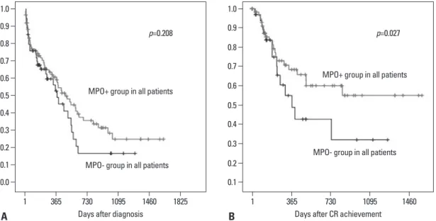
In this analysis, the median follow-up for the both groups was 12.2 months (range, 0.1-54.1). By the univariate analy- sis, the age was significant factors associated with OS and DFS (Table 4). The OS at 1 year were 48% in younger age (<60 years) while elderly (≥60 years) was dead within 1 year after diagnosis (p<0.001). The OS was not related with MPO expression (p=0.208) (Fig. 1A). The DFS at 1 year (38%) with MPO-. There were no statistically significant dif-
ferences in age, sex, presenting white blood count (WBC) type of AML (de novo/secondary), central nervous system involvement at presentation between the MPO+ and the MPO- groups. M1, M2, M4 and M6 were major FAB sub- types in all the MPO groups.
Of the 140 patients who diagnosed AML, 46 (32%) had received transplants, including 37 patients who got alloHCT from a matched sibling (n=21), unrelated (n=16) donor and and 9 patients who got autoHCT (p=0.797) (Table 2). The remaining 94 patients included 55 who received induction and/or post-remission chemotherapy, 39 who received only palliative therapy because of poor performance status or ad- vanced age. Table 3 shows the nature of treatment.
Cytogenetic risk group
In approximately 60% of patients with AML, pre-treatment Table 1. Patient Characteristics
MPO+ MPO- p value
Number of patients (%) 86 (61.4) 54 (38.6)
Age, median (range) (yrs) 48 (15-81) 53 (20-79) 0.282
Age (yrs) 0.291
<60 60 (69) 33 (61)
≥60 34 (30) 21 (38)
Sex 0.537
Male 48 (56) 33 (61)
Female 38 (44) 21 (39)
Type 0.937
De novo 68 (79) 43 (80)
Secondary 18 (21) 11 (20)
Previous hematologic disorder 0.683
Yes 15 (17) 8 (15)
No 71 (83) 46 (85)
CNS involvement at presentation 0.314
Yes 1 (1) 2 (3)
No 85 (99) 52 (96)
FAB subtype 0.081
M1, M2, M4, M6 67 (80) 44 (82)
M5 3 (4) 6 (11)
Not interpretable 14 (16) 4 (7)
Cytogenetics 0.015
Adverse 13 (15) 15 (28)
Favorable 17 (20) 4 (7)
Intermediate 53 (62) 34 (58)
NA of failed 3 (3) 4 (7)
FLT3-ITD mutation 3 (7) 3 (11) 0.676
WBC at diagnosis (×109/L) 0.302
<100 83 (97) 50 (93)
≥100 3 (3) 4 (7)
MPO, myloperoxidase; MPO+, MPO-positive; MPO-, MPO-negative; CNS, central nervous system; FAB, French-American-British; WBC, white blood count;
FLT3, fms-related tyrosine kinase 3 gene; ITD, internal tandom duplication.
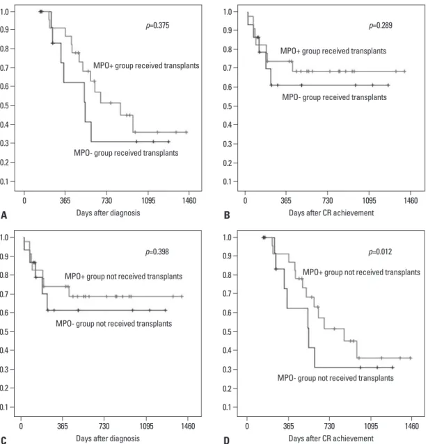
95% confidence interval (CI), 0.06 to 0.997]. Fig. 2A pres- ents OS and DFS in patients received transplant according to MPO expression. Within 46 patients performed trans- plant, the median DFS in 28 patients with MPO+ was not significantly different from in 18 MPO- patients (17.5 months vs. 6.8 months; p=0.289). The median OS in the MPO- group was relatively less than in the MPO+ group, between two age group (<60 vs. ≥60 years) were 34% and
3%, respectively (p=0.019). Patients with MPO- group had an inferior DFS than in MPO+ group (1-year DFS; 26% vs.
50%, p=0.027) (Fig. 1B). In the multivariate analysis, all factors were not significant for OS, but the MPO was asso- ciated with better DFS (Table 5). Patients of MPO+ groups had a better DFS than MPO- groups [p=0.049; HR, 0.076;
Table 2. Characteristics of Transplants
MPO+ MPO- p value
Number of patients with HCT 28 18
Status at transplant 0.585
CR1 25 (89) 15 (83)
CR2 1 (4) 1 (6)
Others 2 (7) 2 (11)
Time to transplant, months (range) 6 (3.7-18.7) 5.7 (3.9-12.4) 0.300
HCT type 0.797
Matched sibling 13 (46) 8 (44)
Matched unrelated 7 (25) 6 (33)
Mismatched (≥1-allele mismatched unrelated) 2 (7) 1 (6)
Autologous 6 (22) 3 (17)
CD34+ cells infused, 106/kg, median, range 5.6 (0.7-17.2) 5.0 (2.3-14.7) 0.626
Stem cell source 0.216
PB 28 (100) 17 (94)
BM 0 1 (6)
Conditioning 0.818
MA 13 (46) 9 (50)
NMA 15 (54) 9 (50)
GVHD 0.720
Acute (grade ≥II) 7 (25) 6 (33)
Chronic 12 (43) 4 (22)
MPO, myeloperoxidase; MPO+, MPO-positive; MPO-, MPO-negative; HCT, hematopoietic stem cell transplantation; CR1, first complete remission; CR2, second complete remission; PB, peripheral blood; BM, bone marrow; MA, myeloablative; NMA, non-myeloablative; GVHD, graft-versus-host disease.
Table 3. The Nature of Treatment
MPO+ MPO- p value
Induction and/or post-remission chemotherapy (%) 33 (38.4) 22 (40.7) 0.954
Palliative therapy (%) 25 (29) 14 (25.9)
Transplantation (%)
Allogeneic 22 (25.6) 15 (27.8)
Autologous 6 (7) 3 (5.6)
MPO, myeloperoxidase; MPO+, MPO-positive; MPO-, MPO-negative.
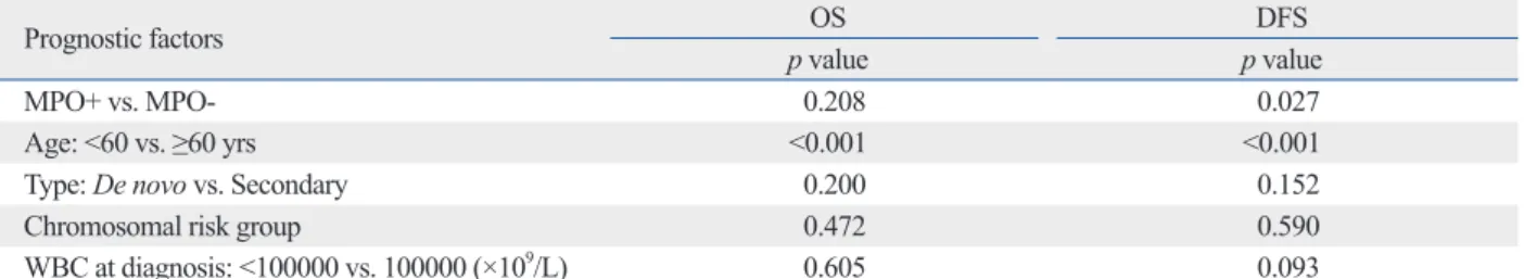
Table 4. Prognostic Factors by Univariate Analysis
Prognostic factors OS DFS
p value p value
MPO+ vs. MPO- 0.208 0.027
Age: <60 vs. ≥60 yrs <0.001 <0.001
Type: De novo vs. Secondary 0.200 0.152
Chromosomal risk group 0.472 0.590
WBC at diagnosis: <100000 vs. 100000 (×109/L) 0.605 0.093
OS, overall survival; DFS, disease-free survival; MPO, myeloperoxidase; MPO+, MPO-positive; MPO-, MPO-negative; WBC, white blood count.
other study of 46 patients with FAB M2 AML reported op- posite results that a high proportion of MPO+ blasts before treatment may have constituted a significantly unfavorable prognostic factor.8 So far, the clinical prognostic value of MPO expression in AML has been controversial. In this study, we analyzed the prognostic significance of the MPO expression in 140 patients with diagnosed AML.
The MPO expression was significantly associated with DFS in the overall group, similar to other studies.4-6 Previ- ous report demonstrated that the groups by the MPO ex- pression in the intermediate cytogenetic risk group showed a significant difference in DFS. The DFS rates at 4 years were also different (p<0.001).4
Although our study included a limited number of cases, transplant demonstrated an important role in improving clinical outcomes in AML. There was a significant differ- ence in DFS between the MPO+ group and MPO- group (p=0.027). However, among the patients received trans- plants, the DFS in overall was not difference in both groups (p=0.289). This results represented that the DFS in the but not statistically significant (28.6 months vs. 18.2
months; p=0.375). Also, in patients received an alloHCT (except autoHCT), DFS and OS in the MPO+ group were not statistically different from those in the MPO- group (Fig. 2B).
DISCUSSION
In this study, the DFS of the patients received transplants in the MPO+ group was equal with that in the MPO- group, although the DFS of the all patient in MPO+ group was su- perior than that in MPO- group. Several studies in AML have examined the prognostic significance of the propor- tion of MPO+ blast cells. An ECOG study investigated the relationship between MPO+ percentage of blast cells and therapeutic outcomes of 72 patients with AML (FAB M1).6 Thirty-eight patients with low MPO (<50%) group showed a significantly lower CR (52.6%) than the 34 patients with high MPO (>50%) group (85.3%, p=0.003). However, an- Table 5. Prognostic Factors by Multivariate Analysis
Prognostic factors OS DFS
p value Hazard ratio p value Hazard ratio
MPO+ vs. MPO- 0.068 0.152 0.049 0.076
Age: <60 vs. ≥60 yrs 0.999 2.176 0.999 1.190
Type (De novo vs. Secondary) 0.369 3.776 0.999 3.268
chromosomal risk group 0.150 0.472 0.153 0.168
WBC at diagnosis: <100 vs. ≥100 (×109/L) 1.000 1.666 1.000 1.548
OS, overall survival; DFS, disease-free survival; MPO, myeloperoxidase; MPO+, MPO-positive; MPO-, MPO-negative; WBC, white blood count.
Fig. 1. (A) OS by MPO expression at diagnosis in AML patients. There were not significant difference of OS between MPO+ and MPO- groups (p=0.208). (B) EFS by MPO expression at diagnosis in AML patients. Patients with MPO- group had an inferior DFS than in MPO+
group (1-year DFS; 26% vs. 50%, p=0.027). OS, overall survival; MPO, myloperoxidase; MPO+, MPO-positive; MPO-, MPO-negative; AML, acute myeloid leukemia; DFS, disease-free survival.
0.0 0.1
0.1 0.2
0.2 0.3
0.3 0.4
0.4
0.5 0.5
0.6 0.6
0.7 0.7
0.8 0.8
0.9 0.9
1.0 1.0
1 365 730 1095 1460 1825 1 365 730 1095 1460
Days after diagnosis Days after CR achievement
MPO- group in all patients
MPO- group in all patients MPO+ group in all patients
MPO+ group in all patients
p=0.208 p=0.027
A B
In conclusion, despite its retrospective study including heterogenous patients with AML and small number of pa- tients, MPO expression was associated with DFS and trans- plant was beneficial to overcome a negative prognostic ef- fect of MPO- at diagnosis based upon the result that the DFS in patients received transplants are not significant be- tween the MPO+ group and MPO- group although DFS in all patients was different according to MPO expression.
MPO expression at diagnosis in AML patients could be one of the indicators that help identification of patients who benefit from transplantation.
MPO- group was inferior than MPO+ group, but negative prognostic effect was overcame by receiving transplants if suitable donor exist. Previous reports suggested that high re- lapse rates in patients with AML have led to enthusiasm for the intensification of treatment by the use of high-dose chemoradiotherapy followed by ‘rescue’ using allogeneic stem cells.12-14 The most important limiting factor for HCT remains the high treatment-related mortality. The long-term mortality from established grade IV GVHD has not changed significantly over the past three decades. The decision to un- dergo HCT depend on the prognostic factors at diagnosis.15
Fig. 2. (A) OS by MPO expression at diagnosis for AML patients received transplants. Among the patients received transplants. A similar difference of OS was observed (p=0.375). (B) DFS OS by MPO expression at diagnosis for AML patients received transplants. Among the patients received transplants. There were not significant difference between MPO+ and MPO- groups (p=0.289). (C) OS by MPO expres- sion at diagnosis for AML patients not received transplants. There were not significant difference of OS between MPO+ and MPO- groups (p=0.398). (D) EFS by MPO expression at diagnosis for AML patients not received transplants were significant difference between two groups (p=0.012). OS, overall survival; MPO, myloperoxidase; MPO+, MPO-positive; MPO-, MPO-negative; AML, acute myeloid leuke- mia; DFS, disease-free survival.
0.1
0.1 0.1
0.1 0.2
0.2
0.2
0.2 0.3
0.3
0.3
0.3 0.4
0.4
0.4
0.4 0.5
0.5
0.5
0.5 0.6
0.6
0.6
0.6 0.7
0.7
0.7
0.7 0.8
0.8
0.8
0.8 0.9
0.9
0.9
0.9 1.0
1.0
1.0
1.0 0
0
0
0 365
365
365
365 730
730
730
730 1095
1095
1095
1095 1460
1460
1460
1460 Days after diagnosis
Days after diagnosis
Days after CR achievement
Days after CR achievement MPO- group received transplants
MPO- group not received transplants
MPO- group received transplants
MPO- group not received transplants MPO+ group received transplants
MPO+ group not received transplants
MPO+ group received transplants
MPO+ group not received transplants p=0.375
p=0.398
p=0.289
p=0.012
A
C
B
D
the distribution picture of peroxidase activity and cell size. Corre- lation between the classification and therapeutic response]. Nihon Ketsueki Gakkai Zasshi 1983;46:1209-15.
8. Suĭć M, Boban D, Marković-Glamocak M, Petrovecki M, Marusić M, Labar B. Prognostic significance of cytochemical analysis of leukemic M2 blasts. Med Oncol Tumor Pharmacother 1992;9:41-5.
9. Bennett JM, Catovsky D, Daniel MT, Flandrin G, Galton DA, Gralnick HR, et al. Proposed revised criteria for the classification of acute myeloid leukemia. A report of the French-American-Brit- ish Cooperative Group. Ann Intern Med 1985;103:620-5.
10. Bene MC, Castoldi G, Knapp W, Ludwig WD, Matutes E, Orfao A, et al. Proposals for the immunological classification of acute leukemias. European Group for the Immunological Characteriza- tion of Leukemias (EGIL). Leukemia 1995;9:1783-6.
11. Davidson JM, Gorringe KL, Chin SF, Orsetti B, Besret C, Cour- tay-Cahen C, et al. Molecular cytogenetic analysis of breast cancer cell lines. Br J Cancer 2000;83:1309-17.
12. Sawayama Y, Miyazaki Y, Ando K, Horio K, Tsutsumi C, Imanishi D, et al. Expression of myeloperoxidase enhances the chemosensi- tivity of leukemia cells through the generation of reactive oxygen species and the nitration of protein. Leukemia 2008;22:956-64.
13. Beutler E, McMillan R, Spruce W. The role of bone marrow trans- plantation in the treatment of acute leukemia in remission. Blood 1982;59:1115-7.
14. Gale RP, Kay HE, Rimm AA, Bortin MM. Bone-marrow trans- plantation for acute leukaemia in first remission. Lancet 1982;2:
1006-9.
15. Grimwade D, Hills RK. Independent prognostic factors for AML outcome. Hematology Am Soc Hematol Educ Program 2009:385- 95.
REFERENCES
1. Gratwohl A, Baldomero H, Frauendorfer K, Urbano-Ispizua A;
Joint Accreditation Committee, International Society for Cellular Therapy; European Group for Blood and Marrow Transplantation.
EBMT activity survey 2004 and changes in disease indication over the past 15 years. Bone Marrow Transplant 2006;37:1069-85.
2. Grimwade D, Walker H, Oliver F, Wheatley K, Harrison C, Harri- son G, et al. The importance of diagnostic cytogenetics on out- come in AML: analysis of 1,612 patients entered into the MRC AML 10 trial. The Medical Research Council Adult and Chil- dren’s Leukaemia Working Parties. Blood 1998;92:2322-33.
3. Grimwade D. Impact of cytogenetics on clinical outcome in AML.
In: Karp JE, editor. Acute Myelogenous Leukemia. Totowa, New Jersey: Human Press; 2007. p.177-92.
4. Matsuo T, Kuriyama K, Miyazaki Y, Yoshida S, Tomonaga M, Emi N, et al. The percentage of myeloperoxidase-positive blast cells is a strong independent prognostic factor in acute myeloid leukemia, even in the patients with normal karyotype. Leukemia 2003;17:1538-43.
5. Kuriyama K, Tomonaga M, Kobayashi T, Takeuchi J, Ohshima T, Furusawa S, et al. Morphological diagnoses of the Japan adult leukemia study group acute myeloid leukemia protocols: central review. Int J Hematol 2001;73:93-9.
6. Matsuo T, Cox C, Bennett JM. Prognostic significance of myelo- peroxidase positivity of blast cells in acute myeloblastic leukemia without maturation (FAB: M1): an ECOG study. Hematol Pathol 1989;3:153-8.
7. Takubo T, Kubota Y, Oguma S, Ueda T, Shibata H, Nakamura H, et al. [Classification of acute non-lymphocytic leukemia based on