Original Article
Copyright © 2017 Korean Society of Medical Physics
CCThis is an Open-Access article distributed under the terms of the Creative Commons Attribution Non-Commercial License (http://creativecommons.org/licenses/by- nc/4.0) which permits unrestricted non-commercial use, distribution, and reproduction in any medium, provided the original work is properly cited.
Progress in Medical Physics 28(4), December 2017 https://doi.org/10.14316/pmp.2017.28.4.218 pISSN 2508-4445, eISSN 2508-4453
Evaluations and Comparisons of Body Surface Doses during Breast Cancer Treatment by Tomotherapy and LINAC Radiotherapy Devices
Hyun-Jik Lee*,†, Sun-Hyun Bae*, Kwang Hwan Cho*, Jae-Hong Jeong*, Su-Il Kwon†, Kil-Dong Lee†
*Department of Radiation Oncology, Soonchunhyang University Bucheon Hospital, Bucheon, †Department of Medical Physics, General Graduate School, Kyonggi University, Seoul, Korea
Received 21 November 2017 Revised 19 December 2017 Accepted 20 December 2017
Corresponding author Kil-Dong Lee ([email protected]) Tel: 82-31-249-9621 Fax: 82-31-253-1165
Effects on skin caused by the dose from linear accelerator (LINAC) opposing portal irradiation and TomoDirect 3-D modeling treatment according to the radiation devices and treatment methods were measured, and a comparative analysis was performed. Two groups of 10 patients each were created and measurements were carried out using an optically stimulated luminescence dosimeter. These patients were already receiving radiation treatment in the hospital. Using the SPSS statistical program, the minimum and maximum average standard deviations of the measured skin dose data were obtained. Two types of treatment method were selected as independent variables; the measured points and total average were the dependent variables. An independent sample T-test was used, and it was checked whether there was a significance probability between the two groups. The average of the measured results for the LINAC opposing portal radiation was 117.7 cGy and PDD 65.39% for the inner breast, 144.7 cGy and PDD 80.39% for the outer breast, 143.2 cGy and PDD 79.56% for the upper breast, 151.4 cGy and PDD 84.11% for the lower breast, 149.6 cGy and PDD 83.11% for the axilla, and 141.32 cGy and PDD 78.51% for the total average. In contrast, for TomoDirect 3-D conformal radiotherapy, the corresponding measurement values were 137.6 cGy and PDD 76.44%, 152.3 cGy and PDD 84.61%, 148.6 cGy and PDD 82.56%, 159.7 cGy and PDD 88.72%, and 148.6 cGy PDD 82.56%, respectively, and the total average was 149.36 cGy and PDD 82.98%. To determine if the difference between the total averages was statistically significant, the independent sample T-test of the SPSS statistical program was used, which indicated that the P-value was P=0.024, which was 0.05 lower than the significance level. Thus, it can be understood that the null hypothesis can be dismissed, and that there was a difference in the averages. In conclusion, even though the treatment dose was similar, there could be a difference in the dose entering the body surface from the radiation treatment plan;
however, depending on the properties of the treatment devices, there is a difference in the dose affecting the body surface. Thus, the absorbed dose entering the body surface can be high. During breast cancer radiotherapy, radiation dermatitis occurs in almost all patients. Most patients have a difficult time while undergoing treatment, and therefore, when choosing a radiotherapy treatment method, minimizing radiation dermatitis is an important consideration.
Keywords: TomoDirect, LINAC, Breast cancer skin dose, OSL dosimetry
Introduction
If statistics from recent years are examined, breast cancer
cases comprise the second-largest percentage of the female cancer incidence rate, and they are increasing every year.1) Majority of breast cancer patients undergo surgery,
and according to the clinical stage, total mastectomy or partial mastectomy, which preserves the breasts, is performed. As radiation therapy progresses, in the case of patients where radiotherapy accompanies the radiotherapy treatment as it progresses as well as after performing a partial mastectomy, it is reported that a portion show a decrease in relapses and an increase in survival.2,3) Breast cancer survivors who receive radiotherapy after a partial mastectomy show a survival rate of over 95%.4) There is a trend that the number of patients receiving radiotherapy is increasing.5–7)
International standards for breast cancer treatment recommend 50 Gy as the dosage value, with the tumor part receiving an additional 10 Gy dose.3) With a 60-Gy distributed dose, even though disturbances to normal tissues are minimized, in the majority of patients, acute radiation dermatitis greater than stage 1 occurs.8) In domestic research on radiation dermatitis that follows the 2010 Radiation Therapy Oncology Group’s (RTOG’s) classification of side-effect levels, of a total of 284 patients, 207 patients had 0th and 1st stage minor radiation dermatitis, and 77 patients had 2nd or greater stage serious radiation dermatitis.9) The main symptom of radiation dermatitis is red spots, and oily or dry skin accompanied by severe itching and pain in a portion of the patients.10,11)
The reason why each patient has different degrees of symptoms, from the patient perspective, is the cancer size or skin type and the degree of skin moisture; when looking at it from the treatment perspective, it is due to the treatment dose and energy and the radiation therapy method.12) Among various factors, changes in the skin dose depending on the radiotherapy method are the largest.
Related recent reports indicate that, in three-dimensional radiation therapy or intensity modulated radiation therapy, more than in general radiotherapy, the measured breast cancer skin dose is lower; thus, it is reported that the degree of radiation dermatitis is low.13,14)
However, in the authors’ recent experiences, in the case of breast cancer treatment therapy, the degree of radiation therapy from tomotherapy, which is an intensity- modulated radiotherapy, or TomoDirect, which is a three- dimensional radiation therapy, is greater than in the opposing portal irradiation-type LINAC radiation therapy.
It is thought that this difference is because the radiotherapy devices, measurement devices, radiotherapy treatment plans, etc. in each report are slightly different. Therefore, this report aims to measure and compare the skin dose treatment from LINAC opposing portal irradiation and the TomoDirect three-dimensional radiation therapy. To measure the actual dose received by the body surface, 20 patients who were currently receiving radiotherapy were divided into two groups of 10 people each, and the skin dose was measured. There are many measuring devices to measure the skin dose, but since they have to adhere to the actual skin, an optically stimulated luminescence dosimeter, which is a small, flat probe that can adhere to the skin curves, was selected.15)
Materials and Methods
1. Properties of the optically stimulated luminescence dosimeter
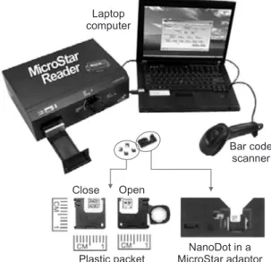
The optically stimulated luminescence dosimeter system is composed of a reader (Microstar reader, LANDAUER, USA) and Al2O3:C device built in a 9×9×1.5-mm3 plastic packet (Nano DOT, LANDAUER Inc., USA) (Fig. 1).
Until a few years ago, a thermoluminescent dosimeter (TLD) that had the advantages that it was small and could measure skin dose and other similar doses;
however, an optically stimulated luminescence dosimeter available today can perform faster and more accurate measurements, and it is being used frequently in place of the TLD. For the optically stimulated luminescence dosimeter, the element carbon is placed inside Al2O3, and on a support that is like a film, a coating is applied. It is made similar to a 3D gel. Through the absolute dose and counter value, which indicates the type of medium and the value of the absorbed dose, the relative dose measurement can be obtained. Therefore, two types of measurements are possible.
The advantage of the optically stimulated luminescence dosimeter is that it is a small device and very easy to use.
Further, compared to film or glass dosimeters that must be read after roughly 1 h, it is possible to read the optically stimulated luminescence dosimeters aafter 10 min.
Compared to count rest analog glass dosimeters, which, after using heat for annealing, return to room temperature after 1 h, and therefore take a significant amount time, the optically stimulated luminescence dosimeter uses light and can be count reset after 30 min. Thus, it is possible to reuse it easily and quickly.16,17)
2. Calibrating an optically stimulated luminescence dosimeter
In this research, to calibrate the optically stimulated luminescence dosimeter, the LINAC and tomotherapy were calibrated. For setting the LINAC (Primus, Siemens, Germany), when the source-to-axis distance (SAD) was 100 cm and the dose area was 10×10 cm2 at a 6-MV reference depth of 1.5 cm to ensure the delivery of a 100-cGy dose, the number of monitor units was 100 MU. The geometrical structure for the setting the optically stimulated lumi ne- scence dosimeter was as follows: a solid phantom was positioned, an optically stimulated luminescence do- simeter was placed on top of it, and with a height corres- ponding to 1.5 cm, a 0.5-cm bolus and 1 cm solid phantom were placed on top of the dosimeter (Fig. 2). The bolus was placed in the center to decrease inaccuracy due to the influence of the air gap on the measuring device.18) For calibrating the tomotherapy (Accuray Inc., Sunnyvale, CA, USA) at the reference depth, a fixed quantity could not be
dosed; therefore, the absorbed quantity during a 10-s dose was set as the reference. With a SAD 80 cm and a dose size of 5×10 cm2, at a 6-MV reference depth of 1.5 cm, there was a 10-s dose, and it was checked that the absorbed quantity received was 141.66 cGy. The geometrical structure for the setting of the optically sti mula ted luminescence dosimeter was that a solid phan tom was put in place, and an optically stimulated lumi nescence dosimeter was placed on top of it, and with a height corresponding to 1.5 cm, a 0.5- cm bolus and 1-cm solid phantom were placed on top of the dosimeter. When the dose was on for 10 s, to measure the absorbed dose at 1 cm point, a farmer chamber was placed 1 cm below the optically stimulated luminescence dosimeter inside the solid phantom (Fig. 3). The settings according to the serial number of each optically stimulated luminescence dosimeter were entered. Subtracting the background count from the readout counter value, the LINAC 100-cGy absorption count was LINAC 100 cGy, and the TomoDirect count was 141.66 cGy. With these counter values used as a reference, the skin dose measured counter values were calculated in proportion to these counter values, and the skin dose was calculated.
3. Measurement of cancer patient skin dose
The target of this research was 20 breast cancer patients divided into two groups of 10 people each who received radiation treatment through LINAC and TomoDirect.
Five positions inside the patient’s breast (based on the standard of the center of the dose field, at a point 3 cm
Fig. 1. InLight OSL dosimetry system.
Laptop computer
Bar code scanner Close Open
Plastic packet NanoDot in a MicroStar adaptor
Fig. 2. Setup for calibration of OSL dosimeter — LINAC.
1 cm 0.5 cm
10 cm Solid phantom
Bolus
OSL Dosimeter
Solid phantom
from the outside, at a point 3 cm from the inside, at a point 5 cm above the surface, at a point 5 cm below the surface, and in the center of the axilla) were selected, the optically stimulated luminescence dosimeter was put in position, and the skin dose was measured (Fig. 4). The prescribed dose amount for one treatment in a day was such that a 180-cGy absorption amount would be delivered. The MU value from the treatment plan was recorded, and both the LINAC and TomoDirect used 6-MV X-rays. To minimize errors according to breast size, large and small sizes were excluded, and measurements were performed on a comparatively midrange-size breast, and measurements were done once on each patient. A record of schematized measurements was made for each patient, and after the dose treatment, the serial number of the optically stimulated luminescence dosimeter was entered into the chart, and the counter value in the reader was measured.
Each counter value was calculated in proportion to the appropriate absorption counter value. For the LINAC, it was 100 cGy, and for the TomoDirect, it was 141.66 cGy.
Finally, the absorption dose amount for each point was obtained.
4. Statistical analysis methods of the skin dose measurement instrument
Statistical analysis was performed using the SPSS statis- tical program. For the measured skin dose in the two treatment devices (LINAC and TomoDirect), for each point, the minimum and maximum average standard deviation was obtained. To compare the dose differences based on treatment method, the averages compared were the same point from the two treatment devices. Further,
with the LINAC and TomoDirect treatment methods as independent variables, and with each treatment point as well as all the treatment points as a dependent variable, the independent sample T-test was used to determine if there was a significance probability in the average difference between the two groups.
Results
The difference in the dose that affected the skin of the patients who were divided into two groups of 10 each and received radiation treatment from either the LINAC or the TomoDirect was based on the results of the treatment method of the radiation treatment device. Based on the
Field margin
3 cm
Field margin Isocenter 5 cm
5 cm 3 cm
Fig. 4. Locations of dose measurement where the OSL dosimeters were attached: ① Upper Breast, ② Inner Breast, ③ Lower Breast,
④ Outer Breast, and ⑤ Axilla.
1 cm 0.5 cm
Solid phantom
OSL Dosimeter Farmer Chamber
Bolus 1 cm Solid phantom
Fig. 3. Setup for calibration of OSL dosimeter — Tomotherapy.
skin dose measurement results from the LINAC treatment opposing portal irradiation method, the lowest measured point was the inner breast with an average value of 117.7 cGy and PDD 65.39%, and the highest measured point was the axilla, which had an average value of 149.6 cGy and PDD 83.11%. The measurements of the other points were 143.2 cGy and PDD 79.56% for the upper breast, 151.4 cGy and PDD 84.11% for the lowest breast, and 144.7 cGy and PDD 80.39% for the outer breast (Table 1). Summing up the points for the entire breast, the result for the average skin dose was 141.32 cGy and PDD 78.51%.
Based on the skin dose measurement results from the TomoDirect 3-D radiation method, of the five points, the lowest measured point was the inner breast with an average of 137.6 cGy and PDD 76.44%, whereas the highest measured point was the lower breast with an average of 159.7 cGy and PDD 88.72%. The measurements of the other points were 148.6 cGy and PDD 82.56% for the upper breast, 152.3 cGy and PDD 84.61% for the outer breast, and 148.6 cGy and PDD 82.56% for the axilla (Table 2).
Summing up the points for the entire breast, the result for the average skin dose was 149.36 cGy and PDD 82.98%. On comparing the LINAC and TomoDirect skin dose point- by-point, there are four points in the TomoDirect that had values higher than that in the LINAC. Values in the TomoDirect were higher in the upper breast by 5.4 cGy and
PDD 3.0%, in the lower breast by 8.3 cGy and PDD 4.6%, in the inner breast by 19.9 cGy and PDD 11.1%, and in the outer breast by 7.6 cGy and PDD 4.2% than in the LINAC (Table 3). The difference between the lowest skin dose and highest skin dose for each point had low points and high points. This can be confirmed in the average and standard deviations. In the LINAC, the standard deviation for the upper breast was 9.4, for the lower breast 10.0, for the inner breast 6.0, for the outer breast 9.2, and for the axilla 11.4. In the TomoDirect the standard deviation for the upper breast was 16.1, for the lower breast 11.3, for the inner breast 13.4, for the outer breast 16.6, and for the axilla 30.9, and as these values were higher than those in the LINAC, the standard deviations were relatively large. The minimum skin dose in the axilla was 106 cGy and PDD 58.7%, and the maximum skin dose was 216 cGy and PDD 119.7%.
In addition, three measurements above the prescribed dose of 180 cGy occurred only in the TomoDirect, where the values measured in the lower breast were 184 cGy and PDD 102.3%, in the outer breast 182 cGy and PDD 100.9%, and in the axilla 216 cGy and PDD 119.7%. Combining all the points, results depending on the treatment device were that, in the LINAC, the total average was 141.32 cGy and PDD 78.51%, and in the TomoDirect, the total average was 149.36 cGy and PDD 82.98%. The measurements in the TomoDirect were shown to be about 8 cGy higher (Table 4).
Table 1. Skin dose of LINAC patient.
Measurement point
Minimum dose (cGy)
Minimum PDD
Maximum dose (cGy)
Maximum PDD
Average dose (cGy)
Average PDD
Standard deviation
Upper breast 130 71.96% 154 85.40% 143.2 79.56% 9.4
Lower breast 134 74.52% 163 90.68% 151.4 84.11% 10.0
Inner breast 110 61.34% 127 70.60% 117.7 65.39% 6.0
Outer breast 131 72.62% 158 87.54% 144.7 80.39% 9.2
Axilla 128 71.36% 163 90.36% 149.6 83.11% 11.4
Table 2. Skin dose of TomoDirect patient.
Measurement point
Minimum dose (cGy)
Minimum PDD
Maximum dose (cGy)
Maximum PDD
Average dose (cGy)
Average PDD
Standard deviation
Upper breast 113 62.76% 169 93.97% 148.6 82.56% 16.1
Lower breast 143 79.45% 184 102.26% 159.7 88.72% 11.3
Inner breast 114 63.33% 157 87.19% 137.6 76.44% 13.4
Outer breast 133 73.82% 182 100.86% 152.3 84.61% 16.6
Axilla 106 58.70% 216 119.73% 148.6 82.56% 30.9
Taking the LINAC and the TomoDirect treatment variables as independent variables, and the measured points as dependent variables, the SPSS statistical program independent sample T-test was used to determine whether there was a significance probability between the averages of the two groups. The null hypothesis “There was no difference between the two groups” was used, and with a significance level set at 0.05, the P-value was obtained. For the upper breast, P=0.372, for the lower breast, P=0.099, for the inner breast, P=0.000, for the outer breast, P=0.222, and for the axilla, P=0.925. The P-value for the inner breast was P=0.000, which falls under the condition P<0.05 according to which the null hypothesis should be rejected.
Therefore, it can be understood that there was a difference in the averages of the two groups. Further, excluding the axilla, close results were obtained for all the other points.
In addition, combining all the points, the results from the independent sample T-test was that the P-value, P=0.024, was less than the 0.05 significance level, and therefore, the null hypothesis could be rejected; thus, the result was that there was a difference in the LINAC and TomoDirect averages.
Discussion and Conclusion
In this research, we analyzed and compared the influence on the skin from breast cancer treatment with the LINAC and TomoDirect treatment devices. The results measuring skin dose were that when comparing measurements from all points, for four out of five points, measurements from the TomoDirect were higher, and in all the total point averages, it was about 8 cGy higher.
In terms of statistics, to determine whether there was a
difference in the averages with LINAC and the TomoDirect the independent variable, the independent sample T-test was used, and results from a comparative analysis showed that with a P-value of 0.024, the difference was significant.
Further, as shown in the measurement results, the LINAC opposing portal irradiation was distributed comparatively close to the average; in other words, the standard deviation was small, but for the TomoDirect 3D radiation treatment, the standard deviation was comparatively high. This means that, compared with the LINAC, the dose’s influence on the skin by the tomotherapy treatment machine had a large range between the minimum and maximum values.
With a 70% to 80% dose compared with the prescribed dose, deviation from the treatment dose could be seen.
When looking from the side of the treatment plan, in the LINAC opposing portal irradiation, the iso-center is taken as the normalization point, and 180 cGy is the prescribed dose in the treatment plan. In contrast, in the TomoDirect 3D radiation treatment, the treatment plan is that the 180-cGy dose enters 3~5 mm below the body surface.
Considering this point, from the level of the treatment plan, there is a small difference in the dose entering the surface; however, according to the treatment devices, there is a clear difference in the dose entering the body surface.
Tomotherapy and similar intensity-modulated radiation therapy treatments have the advantage that they deliver a large dose to the target, and a small dose to normal tissues;
however, compared to the 2D and 3D conformal radiation treatments of the LINAC treatment devices, they use more field and higher MU. Thus, the absorbed amount on the body surface can be large. Currently, related research is comparing the normal tissue dose based on breast cancer Table 3. Comparison of skin dose measurement LINAC patient
and TomoDirect patient.
Measurement point
LINAC TomoDirect
Average dose (cGy)
Average PDD
Average dose (cGy)
Average PDD Upper breast 143.2 79.56% 148.6 82.56%
Lower breast 151.4 84.11% 159.7 88.72%
Inner breast 117.7 65.39% 137.6 76.44%
Outer breast 144.7 80.39% 152.3 84.61%
Axilla 149.6 83.11% 148.6 82.56%
Table 4. Comparison of skin dose measurement LINAC patient and TomoDirect patient.
Measurement point
LINAC TomoDirect
Average dose (cGy)
Standard deviation
Average dose (cGy)
Standard deviation
Upper breast 143.2 9.4 148.6 16.1
Lower breast 151.4 10.0 159.7 11.3
Inner breast 117.7 6.0 137.6 13.4
Outer breast 144.7 9.2 152.3 16.6
Axilla 149.6 11.4 148.6 30.9
Total sum 141.32 15.2 149.36 19.5
radiation treatment. The intensity-modulated radiation therapy and volumetric-modulated arc therapy use a minimum of two times more output than 3D conformal radiation therapy, and they cause scattered rays and leaked rays. Therefore, by a large secondary dose, it is reported that the dose to normal tissues cannot help but be large.
Among the currently developed radiation treatment methods, intensity-modulated radiation therapy and volumetric-modulated arc therapy in the case of a deep target, the protection of normal tissue is large, but in the case of breast cancer radiotherapy, it is confirmed that a higher dose is measured in the normal tissue than in radiation treatment that uses tangential beam irradiation.19) As seen in the skin dose measurement results in this paper, measurements in the TomoDirect were generally higher than in the LINAC. Further, a special property was that, in some parts, the surface skin dose in the TomoDirect was higher than the prescribed treatment dose of 180 cGy. Various variables were in effect, but among those, it is thought that the largest factor originated from the treatment method and properties of the treatment devices.
Compared with the LINAC opposing portal irradiation, the TomoDirect 3-D radiation as well as intensity- modulated radiation therapy has the advantage of uniformly and accurately delivering a dose to the target.
The dose going into the normal lung tissue can be reduced;
however, it was found that the surface body skin dose was a little high. The goal of radiation therapy is to deliver a uniform, maximum dose to tumor tissues, and to deliver a minimum dose to normal tissues. When deciding on the radiotherapy treatment method, these two points should be appropriately considered. Among them, the protection of normal tissue is very important in breast cancer radiotherapy. Decreasing the dose delivered to the lungs and thick skin and epidermal skin at the same time is good, but if that cannot be done, an approach is needed that probabilistically affects the patient the least.
Although there are differences in the degree of radiation dermatitis, it occurs in almost all patients. Various things must be considered in the choice of breast cancer radiotherapy treatment methods and treatment devices, but it is necessary to seriously consider a method that minimizes radiation dermatitis. In that regard, it is hoped
that these skin dose measurements will be of at least some help in setting up radiation treatment plans and choosing treatment methods and treatment devices.
Acknowledgements
This work was supported by the Nuclear Safety Research Program through the Korea Radiation Safety Foundation (KORSAFe) and the Nuclear Safety and Security Commission (NSSC), Republic of Korea (Grant No. 1305033).
Conflicts of Interest
The authors have nothing to disclose.
Availability of Data and Materials
All relevant data are within the paper and its Supporting Information files.
Ethics Approval and Consent to Participate
The study was approved by the institutional review board (IRB approval number; 2017-07-024).
References
1. National Cancer Registry Annual Report. Annual report of cancer statistics in Korea in 2014 Korea Central Cancer Registry Cancer Registration & Statistics Branch, Division of Cancer Registration & Surveillance, National Cancer Center, Korea. 2016.
2. Veronesi U, Salvadori B, Luini A, et al. Breast conservation is a safe method in patients with small cancer of the breast long-term results of three randomized trials on 1,973 patients. Eur J Cancer. 1995;31A:1574-1579.
3. Clarke M, Collins R, Darby S, et al. Effects of radiotherapy and of differences in the extent of surgery for early breast cancer on local recurrence and 15-year survival: An over- view of the randomised trials. Lancet. 2005;366:2087-2106.
4. Kim JH, Byun SJ. Long-term results of breast conserving surgery and radiation therapy in early breast cancer. J Korean Soc Ther Radiol Oncol. 2009;27:153-162.
5. Jung YS, Na KY, Kim KS, et al. Nation-wide Korean breast cancer data from 2008 using the breast cancer registration program. J Breast Cancer. 2011;14:229-236.
6. Ahn SH, Yoo KY. Chronological changes of clinical cha- racteristics in 31115 new breast cancer patients among Koreans during 1996-2004. Breast Cancer Res Treat.
2006;99:209-214.
7. Kim JH, Kim OB, Kim YS. Breast conserving operation and radiation therapy in early breast cancer: interim analysis.
J Korean Soc Ther Radiol Oncol. 2001;19:27-33.
8. Salvo N, Barnes E, van Draanen J, et al. Prophylaxis and management of acute radiation-induced skin reactions: a systematic review of the literature. Curr Oncol. 2010;17(4):
94-112.
9. Lee SY, Kwon HC, Kim JS, Lee HK. An analysis of the incidence and related factors for radiation dermatitis in breast cancer patients who received radiation therapy. J Korean Soc Ther Radiol Oncol. 2010;28:16-22.
10. Hymes SR, Strom EA, Fife C. Radiation dermatitis: clinical presentation, pathophysiology, and treatment 2006. J Am Acad Dermatol. 2006;54:28-46.
11. Harper JL, Franklin LE, Jenrette JM, Aguero EG. Skin toxi city during breast irradiation: pathophysiology and management. South Med J. 2004;97:989-993.
12. Kraus-Tiefenbacher U, Sfintizky A, Welzel G, et al. Factors of influence on acute skin toxicity of breast cancer patients treated with standard three-dimensional conformal ra-
diotherapy (3D-CRT) after breast conserving surgery (BCS). Radiat Oncol. 2012;7:217.
13. Rudat V, Nour A, Alaradi1 AA, et al. In vivo surface dose mea surement using GafChromic film dosimetry in breast cancer radiotherapy: comparison of 7-field IMRT, tan- gential IMRT and tangential 3D-CRT. Radiat Oncol. 2014;9:
156.
14. Pignol JP, Olivotto I, Rakovitch E, et al. A multicenter randomized trial of breast intensity-modulated radiation therapy to reduce acute radiation dermatitis. J Clin Oncol.
2008;26:2085-2092.
15. Im IC, Yu YS, Lee JS. Measurement of skin dose for rectal cancer patients in radiotherapy using optically stimulated luminescence detectors (OSLDs). J Radiat Protect. 2011;2:
36.
16. Jursinic PA. Characterization of optically stimulated luminescent dosimeters, OSLDs, for clinical dosimetric measurements. Med Phys. 2007;34(12):4594-4604.
17. Viamonte A, da Rosa LA, Buckey LA, Cherpak A, Cygler JE.
Radiotherapy dosimetry using a commercial OSL system.
Med Phys. 2008;35(4):1261-1266.
18. Charles PH, Crowe SB, Kairn T, et al. The effect of very small air gaps on small field dosimetry. Phys Med Biol.
2012;57(21):6947-60.
19. Lee BR, Lee SY, Yoon MG. Dosimetric comparison of radi- ation treatment techniques for breast cancer: 3D-CRT, IMRT and VMAT. J Radiol Sci Technol. 2013;36:237-244.