• 검색 결과가 없습니다.

A case of pulmonic stenosis in a Shihtzu dog

N/A
N/A
Protected

Academic year: 2021

Share "A case of pulmonic stenosis in a Shihtzu dog"

Copied!
4
0
0

로드 중.... (전체 텍스트 보기)

전체 글

(1)

大韓獸醫學會誌(2007) 第47卷 第1號

Korean J Vet Res

(2007) 47(1) : 99~102

99

A case of pulmonic stenosis in a Shihtzu dog

Chul Park, Jong-Hyun Yoo

1

, Dong-In Jung, Ju-Won Kim, Byeong-Teck Kang, Hee-Myung Park*

College of Veterinary Medicine, Konkuk University, Seoul 143-701, Korea

1

BK21 Program of Integrative Network Systems for Veterinarians in Basic Science, Industrial Animals and Preventive Medicines, Konkuk University, Seoul 143-701, Korea

(Accepted: January 16, 2007)

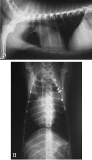
Abstract : A 3-year-old, intact female, Shih-tzu dog was presented with a 15-day history of vomiting, depression, and anorexia. On physical examination, systolic ejection murmurs with precordial thrill at the left heart base were detected. A diagnosis of congenital pulmonic stenosis (PS) was made mainly from the thoracic radiography, electrocardiography, and echocardiography. On complete blood counts and serum biochemistry profiles, the dog had already chronic renal failure (CRF). Thoracic radiography confirmed that main pulmonary artery was tremendously buldged and electrocardiography was suggestive of severe right ventricular hypertrophy. Echocardiographic findings revealed the pulmonic valve stenosis containing valvular dysplasia and poststenotic dilation. On Doppler echocardiography, ejection velocity of the lesion accounted for 3.38 m/sec, meaning mild velocity through the stenotic area. The dog’s condition was deteriorated with the complication of CRF. In this case, CRF was the most complicated problem and resulted in death. However, there has been no reliable relation between PS and CRF. Primary malformation of pulmonic valve was confirmed at necropsy after death.

Key words : congenital heart disease, dog, pulmonic stenosis

Introduction

Pulmonic stenosis (PS) is a common congenital heart defect in dogs and is occasionally recognized in cats [2, 7]. Congenital obstruction of the right ventricular outflow tract (RVOT) can develop in the infundibulum, subvalvular region, and above the pulmonic valve, but primary malformation of the pulmonary valve is the most frequently observed defects in dogs [4]. PS results in obstruction to right ventricular emptying due, in most cases, to partial fusion and dysplasia of the pulmonic valve cusps. Obstruction to RVOT causes an increase in right ventricular systolic pressure, leading to right ventricular hypertrophy, left-ward septal deviation or flattening, and a systolic pressure gradient across the pulmonary valve [6]. High velocity and turbulent flow about the stenosis is associated with systolic ejection murmurs and poststenotic dilation of the main pulmonary artery (MPA). Even though some dogs affected by congenital PS live normal, complicated dogs develop signs of right-sided congestive heart

failure, cardiac arrhythmias, exertional syncope, and sudden death.

The most appropriate form of treatment for severe congenital PS in dogs has not yet been determined.

Recently, the less invasive technique of percutaneous valvuloplasty has been used with increasing frequency [8]. Usually, PS concurrent with chronic renal failure (CRF) is very rare. However, there have been no evident relations between PS and CRF yet.

Case Report

A 3-year-old, intact female, Shih-tzu dog was evaluated because of 15-day history of vomiting, depression, and anorexia at the Veterinary Medical Teaching Hospital of Konkuk University. The dog had been treated with cardiac medication and had been CRF status for about 1 year at the local animal hospital. On physical examination, findings included mild depression, dry muzzle, and retained deciduous teeth. Grade IV/VI of systolic murmurs also was

*Corresponding author: Hee-Myung Park

College of Veterinary Medicine, Konkuk University, Seoul 143-701, Korea

[Tel: +82-2-450-4140, Fax: +82-2-450-3037, E-mail: [email protected]]

(2)

100 Chul Park, Jong-Hyun Yoo, Dong-In Jung, Ju-Won Kim, Byeong-Teck Kang, Hee-Myung Park

ascultated on pulmonic area with precordial thrill.

Complete blood counts (CBC) showed mild thrombo- cytosis and heartworm negative. Results of serum biochemical analysis revealed severe azotemia (BUN, 105.5 mg

/

dl; reference range, 4.8 to 31.4 and CRSC, 5.0 mg

/

dl; reference range, 1.5 to 1.5), hypercholestero- lemia (405 mg

/

dl; reference range, 70 to 303), hyperca- lcemia (13.4 mg

/

dl; reference range, 7.9 to 12.2), and hyperphosphatemia (7.8 mg

/

dl; reference range, 2.2 to 7.9). Thoracic radiographs demonstrated marked bulging of MPA, enlarged right ventricle (RV), and enlarged right atrium (Fig. 1). Based on electrocardiography (ECG) check-up, signs of right ventricular hypertrophy were evident (Fig. 2). There were deep S waves in leads I, II, and III, and right shift of the mean electrical axis (MEA = 237

o

). Echocardiographic examination showed stenosis of pulmonic valvular level, dilation of MPA (post-stenotic dilation), right atrial dilation and mild ejection velocity (3.38 m/sec) through the pulmonic valve (Fig. 3). The pressure gradient was 45.7 mmHg by the modified Bernoulli equation (

P = 4V

2

), meaning mild stenotic condition. The pulmonic valvular performance was tethered and limited with abnormally short valvular leaflets. Tricuspid valve regurgitation was not detected and tricuspid valvular structure was intact. After initial diagnosis, the patient was treated at the local animal hospital. The patient’s condition became worse with deterioration of CRF and died of complication of CRF after 7 days of initial diagnosis.

Finally necropsy was performed and pulmonic valve dysplasia (primary malformation of pulmonic valve) with dilated MPA was confirmed (Fig. 4). The pulmonic valve leaflets were thickened and fused. Post-stenotic dilation of MPA was severe and typical. RV was also Fig. 1. Pulmonic stenosis in a 3-year-old Shihtzu dog.

There are right ventricular enlargement and bulging of the main pulmonary artery in both the left lateral [A] and the ventrodorsal projection [B]. As shown here, right-heart enlargement and prominence of the main pulmonary artery (arrows) are evident.

Fig. 2. Electrocardiography from the patient dog with pulmonic stenosis. Right ventricular hypertrophy is significant, with

axis deviation to the right (MEA = 237

o

) and prominent deep S waves.

(3)

A case of pulmonic stenosis in a Shihtzu dog 101

hypertrophied due to PS. Rumpy-bumpy small kidneys containing green colored materials were observed at the same time.

Discussion

Numerous indices could be chosen to measure and define stenosis severity, including physical stenotic area, effective area determined from the Gorlin formula, and pressure gradient, Doppler jet velocity, blood energy dissipation, and myocardial wall stress [1]. We used Doppler jet velocity to evaluate severity of pulmonary stenotic change in this case. In reality, Doppler

examination revealed mild ejection velocity (3.38 m/sec) through the pulmonic valve, showing mildly increased pressure gradient (45.7 mmHg). Hemodynamically, valvular PS results in a pressure gradient across the stenotic valve because of resistance to right ventricular outflow tract. Therefore, the severity of the lesion is directly related to this pressure gradient. The most common form of PS reported in people [12] and dogs [11] is valvular, in which the semilunar valve leaflets either fused or dysplastic. In this case, the patient showed pulmonic valvular dysplasia with mildly increased velocity through the stenotic valvular lesion.

Also, valvular dysplasia was confirmed by necropsy.

Increased resistance to systolic ejection results in concentric right ventricular hypertrophy, which generally develops in proportion to the severity of the obstructing defect [4]. The echocardiographic examination, electro- cardiographic records, thoracic radiographic images, and pressure gradient of stenotic area were almost in agreement with reasonable results. Many animals may live apparently normal lives with mild degrees of PS.

However, in case of symptomatic animals with PS show dyspnea, exertional fatigue, exercise-induced syncope, right-sided heart failure. The physiologic mechanisms responsible for syncope in dogs with PS have not been evaluated extensively. Hypotension is presumed to develop as a consequence of reduced cardiac output (bradycardia, or worsening of a dynamic obstruction) in combination with peripheral arteriolar vasodilatation (with or in anticipation of exercise) [4].

Although pressure gradient through the stenotic valve is dramatically elevated, post stenotic blood velocity may diminish extensively.

The goal of treatment for dogs with PS is to abolish or to reduce the systolic pressure gradient to the mild range in moderate to severe patients. Surgical therapy had been only useful option until last decade in twentith century. A number of surgical procedures have been advocated for the treatment of severe PS [2, 3, 5]. Valvulotomy, partial valvulectomy, patch-grafting over the outflow tract, and conduits are most popular [6]. Balloon valvuloplasty, an alternative to surgery, has been increasingly utilized over the last decade in twentith century. This technique is successful for some cases of valvular stenosis, especially when the valves are thin and fused and the annulus is not hypoplastic [9, 10]. Generally, balloon valvuloplasty results in a significant decrease (40 to 60%) in the pressure gradient.

Fig. 3. Pulsed wave Doppler tracing the pulmonary outflow tract where pulmonic stenosis exists records mild-velocity flow, documenting velocities of 3.38 m/sec (Pressure gradient is 45.7 mmHg by the modified Bernoulli equation). Post- stenotic dilation is present.

Fig. 4. Pulmonic valve dysplasia (arrow) is confirmed

during necropsy and dilated main pulmonary artery (PA)

aever, ratedt that PS and CRFwall is observed.

(4)

102 Chul Park, Jong-Hyun Yoo, Dong-In Jung, Ju-Won Kim, Byeong-Teck Kang, Hee-Myung Park In this case, pressure gradient through the stenotic area

was mild status. Therefore, there was no indication for surgical correction or balloon valvuloplasty. The limita- tion of this case report was that we were not able to record the RV hypertrophy and interventricular wall thickness by echocardiographic measurement. RV hypertrophy was confirmed at necropsy. It was thought that PS and CRF were separated problems. However, PS might accelerate the renal failure in this case.

Conclusion

In this case, congenital PS was complicated with CRF. Due to lack of reference about relation between PS and CRF, we can not accurately explain the complication of this patient’s condition. We diagnosed this dog as congenital PS based on history taking, clinical signs, physical examination, CBC, serum biochemistry profiles, radiography, ECG, and especially echocardiography with necropsy findings. First of all, the medication was focused on cardiac problem and CRF. Because the cardiac medication was able to influence on the worsening of chronic kidney disease, management was very careful. The dog died of severe complication of CRF. Pulmonic valve dysplasia with right ventricular hypertrophy was confirmed at necropsy.

Acknowledgement

This study was supported by Konkuk University in 2006.

References

1. Brown DJ, Smith FW Jr. Stenosis hemodynamics:

from physical principles to clinical indices. J Vet Intern Med 2002, 16 , 650-657.

2. Buchanan JW. Pulmonic stenosis caused by single coronary artery in dogs: four cases (1965-1984). J Am Vet Med Assoc 1990, 196 , 115-120.

3. Custer MA, Kantor AF, Gilman RA, DeRiemer RH.

Correction of pulmonic stenosis. J Am Vet Med Assoc 1961, 139 , 565-566.

4. Ettinger SJ, Feldman EC. Textbook of Veterinary Internal Medicine. 5th ed. pp. 759-767, Saunders, Philadelphia, 2000.

5. Fingland RB, Bonagura JD, Myer CW. Pulmonic stenosis in the dog: 29 cases (1975-1984). J Am Vet Med Assoc 1986, 189 , 218-226.

6. Fox PR, Sisson D, Moïse NS. Textbook of Canine and Feline Cardiology: Principles and Clinical Practice. 2nd ed. pp. 478-485, Saunders, Philadelphia, 1999.

7. Hunt GB, Church DB, Malik R, Bellenger CR. A retrospective analysis of congenital cardiac anomalies (1977-1989). Aust Vet Pract 1990, 20 , 70-75.

8. Hunt GB, Pearson MR, Bellenger CR, Malik R. Use of a modified open patch-graft technique and valvulectomy for correction of severe pulmonic stenosis in dogs: eight consecutive cases. Aust Vet J 1993, 70 , 244-248.

9. Martin MWS, Godman M, Luis Fuentes V, Clutton RE, Haigh A, Darke PGG. Assessment of balloon pulmonary valvuloplasty in six dogs. J Small Anim Pract 1992, 33 , 443-449.

10. Sisson D, MacCoy DM. Treatment of congenital pulmonic stenosis in two dogs by balloon valvuloplasty.

J Vet Intern Med 1988, 2 , 92-99.

11. Tidholm A. Retrospective study of congenital heart

defects in 151 dogs. J Small Anim Pract 1997, 38 , 94-

12. 98. Weyman AE. Principles and Practice of Echocardio-

graphy. 2nd ed. pp. 869-873, Lea & Febiger,

Philadelphia, 1994.

수치

Fig. 2.  Electrocardiography from the patient dog with pulmonic stenosis. Right ventricular hypertrophy is significant, with axis deviation to the right (MEA = 237 o ) and prominent deep S waves.
Fig. 4.  Pulmonic valve dysplasia (arrow) is confirmed during necropsy and dilated main pulmonary artery (PA) aever, ratedt that PS and CRFwall is observed

참조

관련 문서