비장과 흉선의 림프세포와 LPS에 의해 유도된 사이토카인의 발현에 대한 수은의 영향
김 상 현 *,** . 최 철 희 ** • 임 종 필 *** . 신 태 용 ***,#
*조지아 주립대학, **조선대학교 의과대학, ***우석대학교 약학대학 (Received July 21,2004; Revised August 12,2004)
Oral Exposure to Mercury Alters T Lymphocyte Phenotypes and Augments LPS-induced Cytokine Expressions in Spleen and Thymus
Sang-H yun K im * '* * , C heol-H ee C h o i**, Jong-Pil L im * * * and Tae-Yong S h in * * * ’#
^Interdisciplinary Program of Toxicology, University of Georgia, Athens, Georgia 30602, USA
**Research Center for Resistant Cells, College of Medicine, Chosun University, Gwangju 501-759, Korea College of Pharmacy, Woosuk University, Jeonju 565-701’ Korea
Abstract — Mercury is a widespread metal and consequently there are large populations that currently exposed to low lev
els of mercury. Endotoxin is a component of the gram-negative bacteria and promotes inflammatory responses. The present study was designed to determine the impact of mercury on lymphocytes phenotype populations and endotoxin-induced inflammatory cytokine expressions in immune organ, spleen and thymus. Male BALB/c mice were exposed continuously to 0, 0.3, 1.5, 7.5, or 37.5 ppm of mercuric chloride in drinking water for 14 days and at the end of the treatment period, lipopolysaccharide (LPS, 0.5 mg/kg) was injected intraperitoneally 2 h prior to euthanasia. The dose-range of mercury used did not cause hepatotoxicity. Mercury at 7.5 and 37.5 ppm dose-dependently decreased CD3 + T lymphocytes in spleen; both CD4+ and CD8+ single positive lymphocyte populations were decreased. Exposure to 7.5 and 37.5 ppm of mercury decreased the CD8+ T lymphocyte population in the thymus, whereas double positive CD4 +/CD8+ and CD4+ thymocytes were not altered. Mercury altered LPS-induced inflammatory cytokine gene expressions such as, tumor necrosis factor a, interferon y, and interleukin-12 in spleen and thymus. Results indicated that decreases in T lymphocyte populations in immune organs and altered cytokine gene expression may contribute to the immune-modulative effects of inorganic mer-
Keywords □ mercury, LPS, T lymphocyte, drinking water, inflammatory cytokine, immune-modulation
수은은널리알려진독성물질중의하나이며독성을나타내지 않는저용량의수은이라도만성적으로흡수되면신경독성
,
신장 독성그리고면역계의교란을가져오며특히바이러스감염시숙 주의 대응력을약화시킨다)
ᅵ3)
최근인류건강에 대한경각심과 대체물질사용에의해수은의사용은감소되었으나토양과물에 의한저용량의 만성적인수은중독은여전히일어나고있다.
세계보건기구에서 정한안정농도이하의수은에 노출된사람 을대상으로한실험에서이러한저농도의수은은신경계질환 을일으키지는않지만면역계의이상을가져오는것으로나타났
#본 논문에 관한 문의는 저자에게로
(
전화
) 063-290-1572 (팩스
) 063-290-1567 (E-mail) [email protected]다.4〕면역계는수은에의해독성이나타나는표적기관중의 하나 이고
,
특히저농도수은의 생체에대한영향을실험하는데좋은 연구분야임을알수 있다.
수은에대한만성적인 노출은비록 저농도라할지라도숙주의 면역력을약화시키고또한자가항체 를생성시켜자가면역질환을유발한다.5®
저자등은7J
독성을유발하지 않는범위의 저용량의 수은이
LPS
에의해증가되며 생체방어반응에서 중요한 역할을하는
N O
의생성을 저해하고tum or necrosis factor(TNF)-a, interleukin(IL)-l(3, IL-6
등과 같은 염증유발사이토카인의 생성에 여러 가지transcription
factor
들을조절함으로서 영향을미친다는것을보고한바있다.
지금까지수은에 의해사이토카인의 발현이변화된다는것은알 려져있으나
85
비장과흉선같은면역기관에서 림프세포들의 변화 와사이토카인의발현에대한수은의영향에대해서는연구된바241
가없다
.
따라서본연구에서는저용량의수은이비장과흉선의 림프세 포에미치는영향과그조직에서 나성에의한사이토카인의 발 현에미치는영향을검토하였다
.
또한음식물이나식수가수은 의주노출경로이고경구로흡수된수은의양이농도의존적으 로면역기관으로흡수된다는사실을9>
감만하여수은을:
음용수에 섞어투여하였다.
실 험 방 법
시약및기기
Mercuric chloride, lipopolysaccharide(LPS) 및 tryphan blue 는 Sigma사로부터 구입하여 시용하였다. Hitachi 912 automatic analyzer•는 Roche사(USA), Stomacher blender는 STOM사 (UK), EPICS XL-MCL flow cytometer는 Coulter사(USA), UN-SCAN-IT software는 Silk Scientific사(USA)의 제품을 사용 하였다
.
실험동물
실험에入!용한
BALB/c
계수컷 생쥐(6
주령)는Harlan
사(USA)
에서 구입하여 온도
210C,
상대습도50%
및12
시간마다낮과 밤이반복되도록빛을조절한동물사육실에서1
주일이상순화시켜체중
25-30 g
범위의것을사용하였다.
약물투여
수은
(mercuric chloride
)을0
,0.3, 1.5, 7.5, 37.5 ppm
의농도 로음용수에 섞어2
주동안투여하였다.
대조군동물은증류수 를투여하였다.
매번새롭게만든수은용액을이틀간격으로교 체하였으며,
매일생쥐의 체중과섭취한음용수그리고사료의 양을기록하였다.
수은투여의 마지막날생쥐를하룻동안금식 시키고LPS(0.5 m g lig
)를복강내로투여하고2
시간후에마취 시킨후생쥐의 혈액과비장,
그리고흉선조직을채취하였으며 각각의장기의무게를기록하였다.
간독성 측정
^여한 약물에의한간독성은혈징중
alanine aminotransferase (ALT)
와aspartate aminotransferase(AST)
를H itachi 912 automatic analyzer
를이용하여측정하였다.
비장과 흉선 세포의 분리와 유세포 분석
Johnson
과Sharma
의방법10>에의하여분리하였다.
즉RPM I
배지에보관시킨조직을
Stomacher blender
를이용해단일세포상태로만들고결합조직들은
120 |im
스크린을이용하여 제거하 였다.
분리한세포는R P M I
배지에 현탁시켰으며tryphan blue
염색으로생존율검사시
95%
이상의 생존율을나타내었다.
분리된세포들은
PAB(PBS w ith 1% bovine serum album in and
0.1% NaN3)
용액으로세번세척하고0.5%
포르말린이 함유된PBS
용액에재부유시켰다.
고정된세포들은세포수용체특이적항체를이용하여
EPICS XL-M CL flow cytometer
로유세포 분석하였다.
조직내 人 토카인 발현 분석
조직내
RNA
분리는Tsunoda
와Sharma
의방법11볼 이용하여 분리하였다. cDNA
는Superscript H(Life Technologies
사, USA)
를이용하여 만들고
RT-PCR
를이용하여TNF-a, IFN-y, IL-1(3, IL-12,
그리고p-actin
의발현량을분석하였다. RT-PCR
의조건은 김등의방법7)에준하여실험하였고각각의PCR cycle
은예비실 험을통해최적화하였다. PCR
산물25 n
/을2% agarose
젤로전 기영동하여분리하고ethidium bromide
용액으로염색한후UN- SCAN-IT software
를이용하여수치화하였다.
각사이토?K1
의밴 드는같은샘플에서의p-actin
의양으로정량화하였다.
통계처리
각실험 결과의 통계학적 분석은
SAS statistical software (SAS
사, USA
)를이용하였다.
수치의분석은one way analysis of variance(ANOVA
)와post-hoc Duncan's M ultiple Range
Table
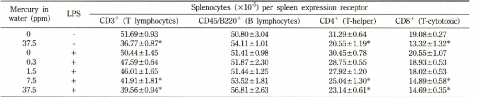
I - Effects of oral exposure to mercury on spleen lymphocyte population Mercury inwater (ppm) LPS Splenocytes ( x 10") per spleen expression receptor
CD3+ (T lymphocytes) CD45/B220+ (B lymphocytes) CD4+ (T-helper) CD8+ (T-cytotoxic)
0 - 51.69±0.93 50.80 ±3.04 31.29±0.64 19.08±0.27
37.5 - 36.77±0.87* 54.11 ±1.01 20.55±1.19* 13.32 ±1.32*
0 + 50.44±1.45 51.41 ±0.98 30.45±0.78 20.55 ±1.07
0.3 + 47.59±0.64 51.87±2.30 28.75±0.55 18.93±0.53
1.5 + 46.01 ±1.65 51.44±1.25 27.92 ±1.20 18.02 ±0.53
7.5 + 41.91±1.81* 53.52 ±1.81 25.04±1.30* 14.89±0.58*
37.5 + 39.56±0.94* 56.81 ±2.63 23.14±0.61* 14.69±0.35*
Populations are represented as absolute cell numbers expression a given receptor.
Mean±SE (n=4).
^Significantly different from the control group (no treatment) at p<0.05.
/.
Pharm. Soc. Korea
LPS LPS
Mercury concentration (ppm)
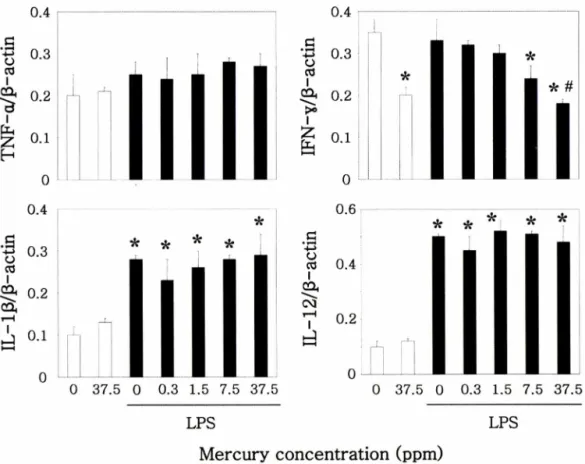
Fig. 1 - The effect of inorganic mercury on the LPS-induced expression of various cytokines in spleen. Male BALB/c mice were treated with 0.3, 1.5, 7.5, 37.5 ppm of mercury in the drinking water for 14 days. Animals were sacrificed 2 h after LPS injection. Extraction and analysis of mRNA performed as described under Materials and methods. Each gene expression was analyzed by RT-PCR. Results are expressed as mean±SE (n=4). *Significantly different than the control group at p<0.05. #Significantly different from the LPS alone group at p<0.05.
test
법을이용하였고p< 0.05
수준에서 유의성을검정하였다.
실 험 결 과
소하였으나유의성을나타내지는않았다
(data not shown).
생쥐 가섭취한사료와음용수의양은가장높은농도의 수은투여군(37.5 ppm
)에서만감소되었다.
간독성과 사료, 음료 섭취량
수은을
0.3, 1.5, 7.5
,37.5 ppm
으로음용수에섞어2
주간투여 한군,
그리고L P S (0 .5 m ^k g
)를복강주사로2
시간투여한군에대해
ALT
와AST
를측정하였을때수은의농도에따라약간감생쥐의 체중과 장기무게의 변화
수은의 투여가생쥐의체중증가와상대적인장기의무게에대
한영향은
2
주간의 실험동안대조군은11.3
%의체중증가를보였으나
37.5 ppm
의수은을투여한군은2.1
%의체중증가에 그Table
II - Effects of oral exposure to mercury on thymus lymphocyte population Mercury inwater (ppm) LPS Thymocytes ( x 10"5) per thymus expression receptor
CD4+/CD8+ (double-positive) CD4+/CD8* (helper) CD47CD8+ (cytotoxic)
0 - 70.35 ±9.94 9.58±0.82 3.99±0.19
37.5 - 67.88±1.41 7.41 ±1.10 2.31 ±0.22*
0 + 66.23±8.88 8.99±3.21 3.21 ±0.55
0.3 + 63.54±6.71 8.06±1.06 3.18±0.19
1.5 + 61.08±7.21 7.63 ±0.82 2.91 ±0.32
7.5 + 70.02 ±2.92 7.56±0.43 2.49±0.31*
37.5 + 66.23±6.21 7.05 ±0.69 2.38±0.16*
Populations are represented as absolute cell numbers expression a given receptor.
Mean±SE (n=4).
*Significantly different from the control group (no treatment) at p<0.05.
o .
o .o.
o.
.sP B - ca/ r
z fe
.sP B -
oa/ OTJ - d .s:
P E - ca/
D - fc^
x
o .o . o .o .
U P O E- ca / ca I— l - d
Vol. 48,No. 4’ 2004
LPS LPS
Mercury concentration (ppm)
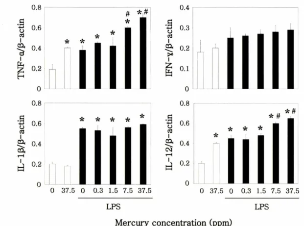
Fig. 2 - The effect of inorganic mercury on the LPS-induced expression of various cytokines in thymus. Male BALB/c mice were treated with 0.3, 1.5, 7.5, 37.5 ppm of mercury in the drinking water for 14 days. Animals were sacrificed 2 h after LPS injection. Extraction and analysis of mRNA performed as described under Materials and methods. Each gene expression was analyzed by RT-PCR. Results are expressed as mean±SE (n=4). *Significantly different than the control group at p<0.05. #Significantly different from the LPS alone group at p<0.05.
쳤다
.
이에반해비장의상대적무게는가장고농도의수은투여 군에서중가하였다.
그러나흉선에서는변화가없었다.
비장과흉선에서 림프세포의변화
수은의투여는비장과흉선에서의 림프세포수를농도의존적 으로감소시켰다
. LPS
의처리는2
시간으로한정되었.0-
口■로림 프세포의수에영향을미치지는않았다.
수은은7.5
와37.5 ppm
의농도로투여시총
C D 3+ T
림프세포를감소시켰으나CD45+
B
림프세포에는영향이 없었다. C D 4
+와CD8+ T
림프세포도 농도의존적으로감소되었고CD3+ T
림프세포의수감소와일 치하였다.
수은7.5
와37.5 ppm
투여군은흉선에서C D 8+ T
림 프세포의감소를나타내었다.
그러나CD4+/CD8
+와C D 4+ T
림 프세포에는영향을나타내지않았다(Table I, Table II).
비장과흉선에서사이토카인발현의 변화
수은을
37.5 ppm
으로투여한군은비장에서IFN-y
의발현을억제하였다
.
그러나TNF-oc, IL -lfi
및IL-12
의발현에는영향이 없었다. LPS
의투여는IFN-y, IL-1(3
및IL-12
의발현을중가시 켰고37.5 ppm
수은투^군은LPS
에의한IFN-
7의발현을억제하였다
.
흉선에서의반응은비장과다른양상으로나타났다.
수 은37.5 ppm
투겨군은TNF-oc
와IL-12
의발현을증가시켰다.
또 한7.5
와37.5 ppm
수은투겨군에서는LPS
에의해중가된TNF-
a 와IL-12
의발현을더욱증가시켰다(Fig. 1, Fig. 2).
고 찰
2
주간수은을Brown Norway
랫트에투여하였을때수은이림프노드에서
CD45+ B
림프세포와CD4+/CD8+ T
림프세포수 의변화없이C D 4
+와CD 8
+의수를감소시킨다고보고되어있다
.12>
이보고의 내용은본실험결과와일치하며이는수은에의한
T
림프세포의 변화가단순히 수은의독성에 의한T
세포들 의감소가아니라또다른특이적인메카니즘에의한것임을암 시한다.
수은은면역억제에서 자가면역까지 넓은범위의면역반응을유발한다
.13)
수은을B6C3F
생쥐에2
주간투여했을때흉선의무게가감소되며 면역저하작용이나타난다는보고가있 다
.14)
또한수은은CD4+
림프노드T
세포의증식을 억제하며IL-3
와IFN-y
의분비를억제하여apoptosis
에이르게한다는보고도있다
.15)
그러나본실험의 결과에서나타난수은에의한일U P O B -
ca /A - z fe
•k
0 37
00.8
0.6
0.4
UP UB - ca/ CNv
lI
o.
o.
o.
o.
UPOB-
ca/D-
fcNX
u }P B -
oa / oa I- T I
J. Pharm. Soc. Korea
부
T
림프세포의 감소는수은에의해유도된apoptosis
에의한 것임이아닌것으로추정된다.
왜냐하면실험결과에서수은의투여가흉선의무게에영향을미치지않았고
apoptosis
에민감하게반응•하는
CD4+/CD8+ T
림프세포의수에변화가없기때문이다.
고용량의
LPS
투여는생체에서 염증반응을유빌하며그결과로다양한면역반응을유발하는
2
차전달물질을발생시키며 염 중을더욱악화시키고결국장기들을손상시킨다.1®
이와는달리저용량의
LPS
를투여하면조직을손상시키지는않지만호중구의축적이나
TNF-ot, IL -ip, IL-12
같은염증유발사미토카인 을유리하며염증반응을촉매한다.1^
나성에의해유도되는조직 의손상은여러가지사이토카인들의발현과밀접한관계가있 다.18)
본실험에서LPS
의투여는2
시간으로한정되어서 림프세 포의수에는영향을미치지않았다.
그러나각면역장기에서염 증유발사이토카인의 발현을중가시켜 염증반응을유발하였고
,
또한수은은LPS
에의해유발되는사이토카인의 발현에영향을나타내었다
.
IFN-y
는major histocompatibility complex
와함께T
림프세포 에의해유발되는면역반응,
특히대식세포의활성화께중요한역 할을차지한다. 37.5 ppm
수은투여시나타난비장IFN-t
의발현 량의감소는비장T
림프세포의감소와상관관계가있는것으로 추정된다.
사이토카인의 발현은다른사이토키인들의발현과의상 호관계에의해결정된다.
면역유발사이토카인의하나인IL-12
는IFN-y
의발현을돕고T-helper l(T h l
)의분화를야기한다.19)
생 쥐의흉선에IL-12
를처리하면CD8+ T
림프세포수가급격히증 가한다.20)
본실험결과에서수은투여에의해흉선CD8+ T
림 프세포수가현저히감소되었기때문에아마도IL-12
가CD8+ T
림프세포로의분화를위해다량으로발현된것으로추측된다
.
본실험에서보여준여러가지사이토카인의 발현결과는수 은을
2
주동안투여한후측정되었다.
사이토카인의 발현은시 간에따라변화됨으로시간의존적으로실험을하는것이이상 적인실험이라사료된다.
또한본실험에서 나타난결과들이수 은의직접적인영향인지혹은간접적인영향에의해서인지이실 험에서는확실히밝힐수없었다.
그러P
1로이에대한추가실험 이필요하다고사료된다.
수은은면역억제에서부터 면역자극까 지넓은범위의면역반응을나타내는특징을가지고있어외부 물질에의한면역독성의실험에서가장좋은물질중의하나로여 겨지고있다.2u
그러므로면역계특히비장과흉선같은면역장기에서수은의영향과
LPS
같은염증유발물질에의해야기되는사미토카인에대한연구는면역조절의현상이해에좋은방향을 제시할수있으리라사료된다
.
결 론
대표적인독성물질중의하나인수은은생체내로
f
며되었을때독성을 일으키지 않는 범위인 저농도라 할지라도 비장과 흉선에 서 T 림프세포수를 감소시키고 사이토카인의 발현을 조절함으로 서 면역반응에 영향을 미치는 것으로 나타났다.
감사의 말씀
이 연구는 과학기술부 • 한국과학재단 지원 내성세포연구센터 (R13-2003-009)의 일부지원으로 수행되었음.
문 헌
1) Christensen, M. M.,Ellermann-Eriksen, S., Rungby, J. and Mogensen, S. C .: Influence of mercuric chloride on resistance
to generalized infection with herpes simplex virus type 2 in
mice. Toxicol. 114, 57 (1996).2) Gerstner, H. B. and Huff, J. E .: Clinical toxicology of mercury.
J. Toxicol Environ. Health 2, 491 (1977).
3) Wild, L. G., Ortega, H. G.’ Lopez, M. and Salvaggio, J. E. :
Immune system alteration in the rat after indirect exposure to
methyl mercury chloride or methyl mercury sulfide. Environ.Res. 74, 34 (1997).
4) Perlingeiro, R. C. and Queiroz, M. L. : Polymorphonuclear
phagocytosis and killing in workers exposed to inorganic
mercury. Int. J. Immunopharmacol. 16,1011 (1994).5) Roller, L. D. : Methylmercury: effect on oncogenic and nononcogenic viruses in mice. Am. J. Vet Res. 36,1501 (1975).
6) Sapin, C., Druet, E. and Druet, R : Induction of anti-glomerular
basement membrane antibodies in the Brown-Norway rat by
mercuric chloride. Clin. Exp. Immunol. 28,173 (1977).7) Kim, S. H.,Johnson, V J. and Sharma, R. R : Mercury inhibits
nitric oxide production but activates proinflammatory cytokine expression in murine macrophage: differential modulation of
NF-kappaB and p38 MAPK signaling pathways. Nitric Oxide 7,67 (2002).
8) Hu, H., Moller, G. and Abedi-Valugerdi, M. : Major
histocompatibility complex class II antigens are required for both cytokine production and proliferation induced by mercuric
chloride in vitro. J. Autoimmun. 10, 441 (1997).9) Nielsen, J. B. and Andersen, 0. : Oral mercuric chloride
exposure in mice: effects of dose on intestinal absorbtion and
relative organ distribution. Toxicology 59, 1 (1989).10) Johnson, V J. and Sharma, R. R : Gender-dependent im munosuppression following subacute exposure to fumonisin B l. Int. Immunopharmacol. 1, 2023 (2001).
11) Tsunoda, M. and Sharma, R. R : Modulation of tumor necrosis
factor alpha expression in mouse brain after exposure to
aluminum in drinking water. Arch. Toxicol. 73,419 (1999).12) Kosuda, L. L., Whalen, B., Greiner, D. L. and Bigazzi, R E. :
Vol. 48’ No. 4,2004
Mercury-induced autoimmunity in Brown Norway rats:
kinetics of changes in RT6+ T lymphocytes correlated with IgG isotypes of circulating autoantibodies to laminin 1. Toxicol.
125, 215 (1998).
13) Roller, L. D. : Immunotoxicology of heavy metals. Int. J.
Immunopharmacol. 2,269 (1980).
14) National Toxicology Program : Toxicology and carcinogenesis studies of mercuric chloride in F344/N rats and B6CF1 mice.
NIH publication, p. 91 (1993).
15) Shen, X., Lee, K. and Konig, R. : Effects of heavy metal ions on resting and antigen-activated CD4(+) T cells. Toxicol. 169,
67 (2001).
16) Ghosh, S., Latimer, R. D.,Gray, B. M., Harwood, R. J. and Oduro, A. : Endotoxin-induced organ injury. Crit. Care Med.
21,S19 (1993).
17) Spitzer, J. A., Zhang, R and Mayer, A. M. : Functional
characterization of peripheral circulating and liver recruited neutrophils in endotoxic rats. J. Leukoc. Biol. 56, 166 (1994).
18) Tsutsui, H.,Matsui, K., Kawada, N., Hyodo, Y., Hayashi, N., Okamura, H.,Higashino, K. and Akanishi, K. : IL-18 accounts for both TNF-alpha and Fas ligand-mediated hepatotoxic pathways in endotoxin-induced liver injury in mice. J.
Immunol.
159
,3961 (1997).19) Trinchieri, G. : Interleukin-12 and the regulation of innate resistance and adaptive immunity. Nat Rev. Immunol. 3(2), 133 (2003).
20) Godfrey, D. I., Kennedy, J., Gately, M. K., Hakimi, J., Hubbard, B. R. and Zlotnik, A. : IL-12 influences intrathymic T cell development. J. Immunol. 152,2729 (1994).
21) Bigazzi, R E. : Mercury. In: Zelikoff, J. and Thomas, R (Eds.), Metal Immunotoxicology. Taylor and Francis, london, UK.
p.131 (1998).