RESEARCH PAPER
Gangliosides induce autophagic cell death in astrocytes
bph_563586..603Jaegyu Hwang
1, Shinrye Lee
1, Jung Tae Lee
2, Taeg Kyu Kwon
2, Deok Ryong Kim
3, Ho Kim
4, Hae-Chul Park
4and Kyoungho Suk
11
Department of Pharmacology, School of Medicine, Brain Science and Engineering Institute, CMRI, Kyungpook National University, Daegu, Korea,
2Department of Immunology and Chronic Disease Research Center and Institute for Medical Science, School of Medicine, Keimyung University, Daegu, Korea,
3Department of Biochemistry, MRCND and Institute of Health Sciences, Gyeongsang National University School of Medicine, Jinju, Korea, and
4Department of Medical Science, Korea University Ansan Hospital, Ansan, Gyeonggi-do, Korea
Background and purpose: Gangliosides, sialic acid-containing glycosphingolipids, abundant in brain, are involved in neuronal function and disease, but the precise molecular mechanisms underlying their physiological or pathological activities are poorly understood. In this study, the pathological role of gangliosides in the extracellular milieu with respect to glial cell death and lipid raft/membrane disruption was investigated.
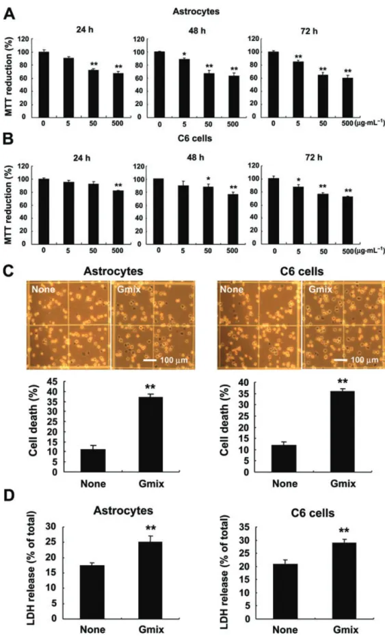
Experimental approach: We determined the effect of gangliosides on astrocyte death or survival using primary astrocyte cultures and astrocytoma/glioma cell lines as a model. Signalling pathways of ganglioside-induced autophagic cell death of astrocytes were examined using pharmacological inhibitors and biochemical and genetic assays.
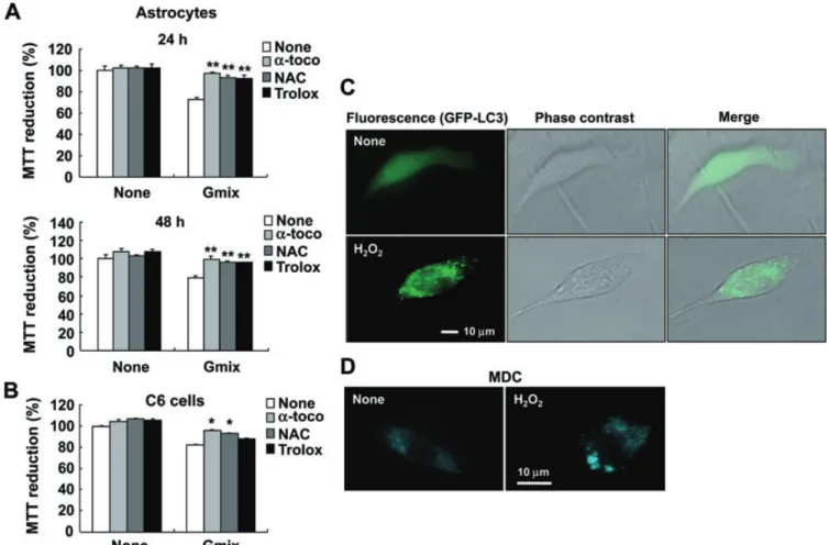
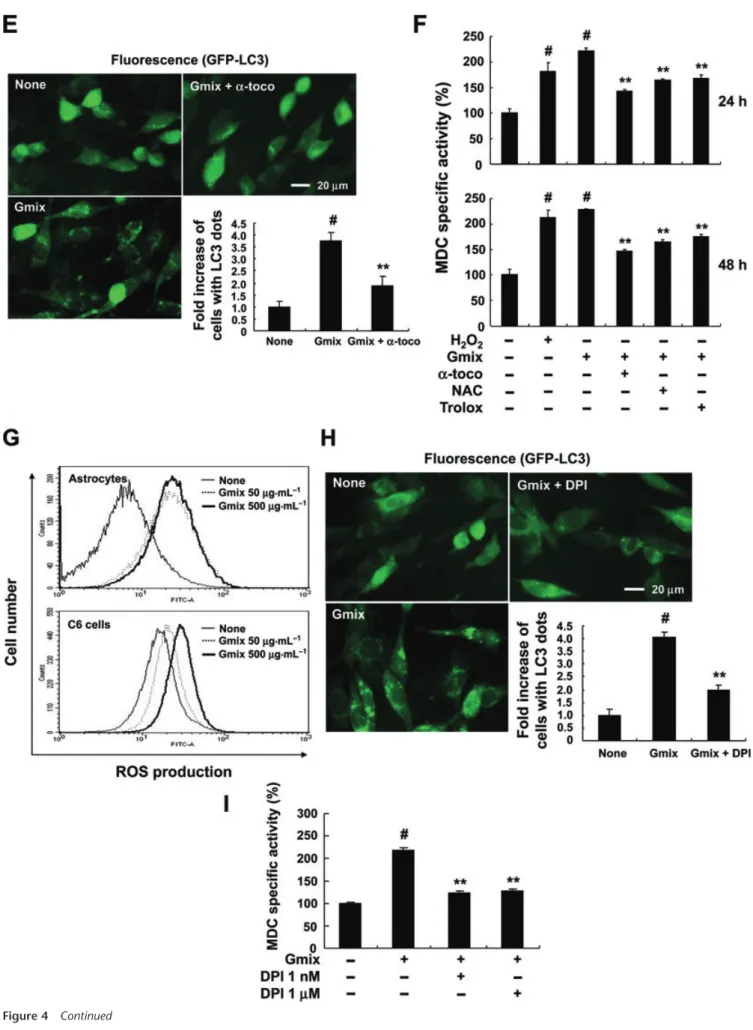
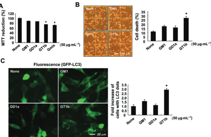
Key results: Gangliosides induced autophagic cell death in based on the following observations. Incubation of the cells with a mixture of gangliosides increased a punctate distribution of fluorescently labelled microtubule-associated protein 1 light chain 3 (GFP-LC3), the ratio of LC3-II/LC3-I and LC3 flux. Gangliosides also increased the formation of autophagic vacuoles as revealed by monodansylcadaverine staining. Ganglioside-induced cell death was inhibited by either a knockdown of beclin- 1/Atg-6 or Atg-7 gene expression or by 3-methyladenine, an inhibitor of autophagy. Reactive oxygen species (ROS) were involved in ganglioside-induced autophagic cell death of astrocytes, because gangliosides induced ROS production and ROS scavengers decreased autophagic cell death. In addition, lipid rafts played an important role in ganglioside-induced astrocyte death.
Conclusions and implications: Gangliosides released under pathological conditions may induce autophagic cell death of astrocytes, identifying a neuropathological role for gangliosides.
British Journal of Pharmacology (2010) 159, 586–603; doi:10.1111/j.1476-5381.2009.00563.x; published online 8 January 2010
Keywords: ganglioside; autophagy; astrocytes; reactive oxygen species; mTOR
Abbreviations: 3-MA, 3-methyladenine; EBSS, Earle’s balanced salt solution; GFP, green fluorescent protein; LC3, microtubule-associated protein 1 light chain 3; MAPKs, mitogen-activated protein kinases; MbCD, methyl b-cyclodextrin; MDC, monodansylcadaverine; mTOR, mammalian target of rapamycin; PARP, poly (ADP- ribose) polymerase; PI3K, phosphatidylinositol 3-kinase; ROS, reactive oxygen species
Introduction
Astrocytes, the major glial cell type in brain, provide meta- bolic and trophic support to neurons and also modulate syn- aptic activity (Barres and Barde, 2000). Astrocytes play an essential role in regulating neurotransmission and blood flow
as well as maintaining a normal brain physiology. In addition to these physiological roles, astrocytes have an important role in the processes of injury and disease in the CNS. Astrocytes are the main responder to CNS insults under various patho- logical conditions such as ischaemia, infection, autoimmu- nity and neurodegeneration (Giffard and Swanson, 2005;
Maragakis and Rothstein, 2006; Farina et al., 2007). Moreover, astrocytes play multiple roles ranging from passive support to the regulation of inflammation during brain injuries. Multiple signals have been shown to induce the cell death of astrocytes in vitro and in vivo including Ca
2+overload, oxidative stress,
Correspondence: Kyoungho Suk, Department of Pharmacology, KyungpookNational University School of Medicine, 101 Dong-In, Joong-gu, Daegu, 700- 422, Korea. E-mail: ksuk@knu.ac.kr
Received 8 July 2009; revised 3 September 2009; accepted 14 September 2009