Facial Exposure Dose Assessment During Intraoral Radiography by Radiological Technologists
- 구내 촬영시 방사선사의 안면부 피폭선량 측정 -
1)Department of International Radiological Science, Hallym University of Graduate Studies, Republic of Korea
2)Department of Radiology, Kyung Hee University Hospital at Gang-dong, Republic of Korea Hwan Yu1,2)․ Hanjoon Yang1)
― Abstract ―
The study examined the changes in the decreased facial exposure dose for radiological technologists depending on increased distance between the workers and the X-ray tube head during intraoral radiography. First, the facial phantom similar to the human tissues was manufactured. The shooting ex- amination was configured to the maxillary molars for adults (60kVp, 10mA, 50msec) and for children (60kVp, 10mA, 20msec), and the chamber was fixed where the facial part of the radiation worker would be placed using the intraoral radiography equipment. The distances between the X-ray tube head and the phantom were set to 10cm, 15cm, 20cm, 25cm, 30cm, 35cm, and 40cm. The phantom was radiated 20 times with each examination condition and the average scattered doses were examined. The rate at the distance of 40cm decreased by about 92.6% to 7.43% based on the scattered rays radiated at the distance of 10cm under the adult conditions. The rate at the distance of 40cm decreased by about 97.6% to 2.58% based on the scattered rays radiated at the distance of 10cm under the children conditions.
Protection from the radiation exposure was required during the dental radiographic examination.
Key words : Intraoral radiography, exposure dose, distance
교신저자: 양한준, (135-841) 서울시 강남구 역삼로 427 한림국제대학원 국제방사선학과
Tel : 010-2750-6181, FAX: 02-3453-6618 E-mail : [email protected]
* 접수일(2014년 8월 8일), 1차 심사일(2014년 8월 12일), 확정일(2014 년 9월 16일)
Ⅰ. INTRODUCTION
The ionizing radiation used for patient examinations can cause biological effects in the human body. Radiation absorbed by the body accumulates for life, necessitating keen attention to its use. In particular, radiological technologists, doctors, dentists, dental hygienists, medical image
experts, nurses, and nursing assistants may be over-exposed to ionizing radiation1,2). One study that analyzed individual radiation exposure and the management status of the 347 radiation involved personnel reported that all personnel were exposure to levels of radiation far below the legal dose limit (annual average exposure dose, 1.52±1.35 mSv), but that 125 of the 347 subjects (36.0%) were exposed to levels that greatly exceeded the average general level3). By departments, the exposure dose was highest for radiation and oncology, medical imaging, and nuclear medical science. The exposure was greatest in the isotope operation room and the injection room for the department of nuclear
medicine, with the highest exposure in examination rooms including the colon examination and the blood vessel examination rooms for the department of imaging medical science, which required direct radiation. Many studies have addressed radiation exposure for each department in the hospital.
However, few studies have focused on the radiation equipment. Radiographic examination generally performed in the dental clinics is the most frequently used and is used frequently with children4). Recently, interest in the exposure dose conferred by dental radiation has increased because of the increasing use of panoramic radiography and the cone beam-type computed tomography (CT) scan5-7). Patient exposure is extremely low for the intra-oral radiographic examination or the panorama radiographic examination. Nonetheless, the possibility of radiation-induced carcinogenesis cannot be ruled out. Exposure to dental radiation may increase the risk of tumors in the salivary gland and thyroid, and of meningioma8-10). The exposure risk is appreciably increased for patients during the intra-oral radiation examination, and as well for personnel involved in the examinations.
Intraoral radiography is required for the radiation examination patients, such as children, who may not maintain their postures or freely move their hands. The radiological technologists typically performs the intraoral radiography while wearing an apron in the examination room, which can expose the facial area to the radiation.
The purpose of the study was to investigate the radiation exposure to the face of radiation personnel and measure the scattered rays received by the radiation worker during the intraoral radiography.
Ⅱ. S UBJECTS AND METHOD
The experiment equipment was an IntraOral or Heliodent DS device (Sirona, Germany) and the dose measuring instrument was a Victoreen® NERO®
mAx X-ray test device, model 8000 (FLUKE
Biomedical, USA) equipped with a 400 cm3 external scatter ionchamber (FLUKE Biomedical) to measure the secondary scattered rays. The facial phantom was manufacture during material similar to human tissue using the Diagnostic X-Ray Phantom Model 76-2 Series (FLUKE Biomedical) The thickness of the human-like phantom is 4.6cm(Fig.1). The radiological technologists directly entered the examination room. The worker had a CCD sensor in the hand positioned inside the oral cavity and performed the examination while maintaining the X-ray tube head with the other hand, as would normally be done when examining children, elderly patients, or those who may not freely move their arms. The phantom was located near the facial area of the phantom to make the environment similar to the actual examination environment and the FOD was fixed at 5 cm after fixing the X-ray tube head toward the center of the phantom with a vertical and horizontal angle of 45 degrees and 0 degrees, respectively (Fig. 2). The shooting examination was configured to the maxillary molars for adults (60kVp, 10mA, 50msec) and children (60kVp, 10mA, 20msec), and the chamber was fixed where the facial part of the radiation worker would be placed.
The distances between the X-ray tube head and the phantom were set to 10cm, 15cm, 20cm, 25cm, 30cm, 35cm, and 40cm, and the phantom was radiated 20 times with each examination condition and the average scattered doses were examined.
Fiele size is 3×4 cm2. The average scattered dose was expressed as the irradiation dose. The equivalent doses were converted based on the measured irradiation dose. The conversion formula is given in Eq. (1):
……… Eq. 1
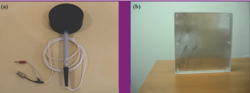
Fig. 1. The external scatter ion chamber was used to measure the secondary scattered rays (a) and the phantom used in the experiment was the facial phantom similar to the human tissues (b).
Fig. 2. The phantom was located to the facial part of the patientand the FOD was fixed at 5 cm after fixing the X-ray tube head toward the center of the phantom with a vertical and horizontal angle of 45 degrees and 0 degrees, respectively.
where 1Gy = 100 rad and the weighting factor on the radiation is multiplied (it because of the photon for the irradiation dose) to calculate the equivalent dose.
1 R = 0.877 rad = 0.00877 Gy = 0.00877 Sv = 0.00877 Sv
……… Eq. 2
Results were compared to the limit in the individual dose due to the planned exposure recommended by the International Commission on Radiological Protection(ICRP) 60 and 26 guidelines11,12). The individual dose limit was compared on the eyes and the thyroid on the facial part. The annual equivalent dose forecasts the annually received dose using the annual average cases for a year with the
daily received equivalent dose. The annual average number of patients under the intra-oral radiation examination for 3 years from 2011 to 2013 was 2,870 including 951 children. ANOVA test of SPSS Win 17.0 was used to investigate the difference in the average scattered rays for each distance and post-hoc analysis was performed with the Dunnett test to investigate more accurate differences. It was considered significant if the p value was < 0.05.
Ⅲ. RESULTS
Table 1 shows the averaged measured scattered X-rays after irradiation with the aforementioned exposure condition in the maxillary molars for adults for each distance. At 10cm, 20cm, 30cm, and 40cm, the scattered rays measured 16.67±0.18mR,
Table 1 The measured average scattered rays after the radiaton with the exposure condition in the maxillary molars for adults (60 kVp, 10 mA, 50 msec) for each distance.
Distance (cm)
Scatter dose (mR)
Reduction ratio (%)
Equivalent dose (mSv)
Annual equivalent dose
(mSv) P
10 16.67±0.18 100 0.15±0.00 419.58±4.53
0.025
15 9.32±0.12 55.9 0.08±0.00 234.52±3.02
20 5.51±0.13 33.05 0.05±0.00 138.68±3.27
25 3.52±0.08 21.11 0.03±0.00 88.59±2.01
30 2.21±0.04 13.25 0.019±0.00 55.62±1.02
35 1.57±0.08 9.41 0.013±0.00 39.51±2.01
40 1.24±0.04 7.43 0.01±0.00 31.21±1.02
Table 2 The measured average scattered rays after the radiation with the exposure condition in the maxillary molars for children (60 kVp, 10 mA, 20 msec) for each distance.
Distance (cm)
Scatter dose (mR)
Reduction ratio (%)
Equivalent dose (mSv)
Annual equivalent dose
(mSv) P
10 6.62±0.14 100 0.06±0.00 55.21±1.17
0.020
15 3.64±0.13 21.83 0.03±0.00 30.35±1.08
20 2.28±0.11 13.67 0.019±0.00 19.01±0.91
25 1.41±0.05 8.46 0.012±0.00 11.76±0.41
30 0.92±0.08 5.51 0.008±0.00 7.67±0.67
35 0.63±0.09 3.78 0.005±0.00 5.25±0.75
40 0.43±0.06 2.58 0.003±0.00 3.59±0.50
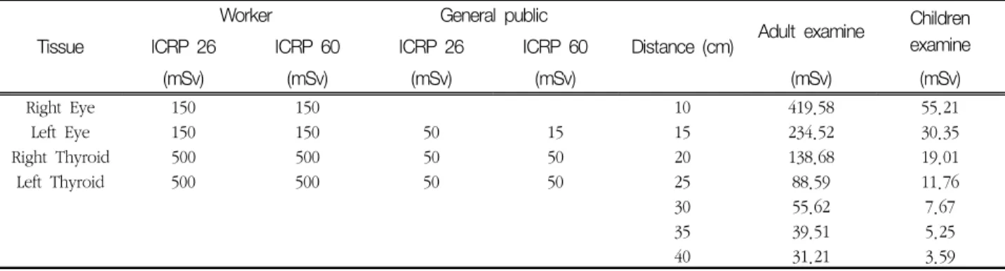
Table 3 The dose limit proposed by the ICRP 60 and 26, and comparison to the operator.
Tissue
Worker General public
Distance (cm) Adult examine Children examine
ICRP 26 ICRP 60 ICRP 26 ICRP 60
(mSv) (mSv) (mSv) (mSv) (mSv) (mSv)
Right Eye 150 150 10 419.58 55.21
Left Eye 150 150 50 15 15 234.52 30.35
Right Thyroid 500 500 50 50 20 138.68 19.01
Left Thyroid 500 500 50 50 25 88.59 11.76
30 55.62 7.67
35 39.51 5.25
40 31.21 3.59
5.51±0.13mR, 2.21±0.04mR, and 1.24±0.04mR, respectively, the equivalent dose was 0.15±0.00 mSv, 0.15±0.00mSv, and 0.01±0.00mSv, respectively, and the annual equivalent dose was 419.58±4.53mSv, 138.68±3.27mSv, 55.62±1.02mSv, and 31.21±1.02 mSv, respectively. The rate at 40cm decreased by about 92.6% to 7.43% based on the scattered rays radiated at the distance of 10cm (p<0.05).
Table 2 summarizes the measured average scattered rays after the radiation with the aforementioned
exposure conditions for children for each distance.
At 10cm, 20cm, 30cm, and 40cm, the scattered X-rays measured 6.62±0.14mR, 2.28±0.11 mR, 0.92±0.08mR, and 0.43±0.06mR, respectively, the equivalent dose was 0.06±0.00mSv, 0.019±0.00mSv, 0.008±0.00mSv, and 0.003±0.00mSv, respectively, and the annual equivalent dose was 55.21±1.17 mSv, 19.01±0.91mSv, 7.67±0.67mSv, and 3.59±0.50mSv.
The rate at the distance of 40cm decreased by about 97.6% to 2.58% based on the scattered rays
radiated at the distance of 10cm (p<0.05).
Table 3 shows the dose limit in the workers and the dose comparison of the examiners proposed by ICRP 60 and 26. The recommended dose limit of the eyes was 150mSv. The dose limits were exceeded at 10 and 15cm under the adult exposure conditions.
Radiation dose limit under the condition for the children was not exceeded for any distance. The recommended dose limit of the thyroid was 500mSv.
This limit was not exceeded for the adult and the children at any distance.
Ⅳ. DISCUSSION
Recently, the interest in the exposure dose in dental irradiation has increased due to increased use of panoramic radiography and the cone beam-type CT scan5-7). The dental radiographic examination confers less radiation exposure dose compared to other radiographic examinations, but involves a larger collective dose and difference in the radiation exposure dose due to differences in the features in the examination conditions and equipment for many dental clinics13,14). Underhill et al.15) measured the absorbed dose on the part 14 during the intra-oral radiographic examination and showed high values in the salivary gland with the highest value in the submaxillary gland. Also, the bone marrow in the 3rd maxillary molar was the highest in the lower jawbone. Avendanio et al.16). measured the absorbed dose during the intra-oral radiographic examination for 15 full mouths at part 27 and reported a high absorbed dose in the 3rd maxillary molars, premolar marrow, and skins in the lower jawbone and the skins of the jaw and the philtrum. The risk associated with exposure has emerged for patients during intra-oral radiation examination. However, the risk extends to radiological technologists. Against this backdrop, this study examined the changes in the decrease in the facial exposure dose for the radiological technologists depending on increasing the distance
between the radiological technologists and the X-ray tube head during the intraoral radiography.
The rate at 40cm decreased by about 92.6% to 7.43% based on the scattered rays radiated at 10 cm under the adult conditions, and by about 97.6%
to 2.58% based on the scattered rays radiated at the distance of 10 cm under the child conditions.
Similarly, Lim et al.17). measured the scattered ray dose at the distances of 50cm and 100cm for the intra-oral examination, and reported that the dose decreased by half at 100cm. However, difficulties existed in the comparison of figures in the study to existing studies, due to differences in dose because of all the conditions including the researchers, equipment, and differing examination conditions.
The study seeks to find out the optimal condition to protect from the risk in the exposure by comparing the measured values depending on the location and the distance after measuring the scattered rays. The experiment measured the scattered rays primarily hit and scattered from the object rather than the experiment measuring the risk in the exposure to the direct radiation18). Radiation intensity and dose decreased with increasing distance.
Ⅴ. CONCLUSION
Dental radiographic examination generates very low scattered rays due to low radiation exposure.
However, the biological impact on the human from the exposure due to the low radiation dose has not been clearly reported yet. The data indicate the wisdom of maintaining a distance from the radiation source as long as possible and wearing proper protection equipment. And the radiological technologists wear glasses and aprons when patient should not have posture maintenance or children.
REFERENCES
1. Lim HS: Health Disorders Caused by Physical Factors among Health Care Workers - Focusing
on Ionizing Radiation -, J Korean Med Assoc, 53, 483-491, 2010
2. Jeong GH, Lee HK, Cho WK et al.: Caregiver or Family Doses due to Discharged 131I Administrated Patient from the Hospital, Journal of radiological science and technology, 33, 149-154, 2010
3. Jeong TS, Shin BC, Moon CW et al.: The Analysis of radiation exposure of hospital ra- diologic technologists, Korean Soc Ther Radiol Oncol, 18, 157-166, 2000
4. Lee BD: Review of Radiographic Selection Criteria, Journal of Korean Dental Association, 51, 39-45, 2013
5. Price JB, Thaw KL, Tyndall DA et al.: ncidental findings from cone beam computed tomography of the maxillofacial region: a descriptive retro- spective study, Clin Oral Implants Res., 23, 1261-8, 2012
6. Fryback DG, Thornbury JR: The efficacy of di- agnostic imaging, Med Decis Making, 11, 88-94, 1991
7. An SY, An CH, Choi KS: Efficacy of panoramic radiography as a screening procedure in dental examination compared with clinical evaluation, Korean J Oral Maxillofac Radiol, 37, 83-6, 2007 8. Memon A, Godward S, Williams D et al.: Dental
x-rays and the risk of thyroid cancer: a case-control study, Acta Oncol., 49, 447-53, 2010 9. Claus EB, Calvocoressi L, Bondy ML et al.:
Dental x-rays and risk of meningioma, Cancer.
118, 4530-7, 2012
10. Ludlow JB: The Risks of Radiographic imaging, Dimensions of Dental Hygiene, 10, 59-61, 2012 11. ICRP 60: 1990 Recommendations of the International
Commission on Radiological Protection, Ann ICRP 60, 1991
12. ICRP 20: Alkaline Earth Metabolism in Adult Man, Ann ICRP 20, 1973
13. Cho JY, Han WJ, Kim EK et al.: Absorbed and effective dose from periapicalradiography by portable intraoral x-ray machine. Korean J Oral Maxillofac Radiol, 37, 149-156, 2007
14. Hatziioannou K, Psarouli E, Papanastassiou E et al.: Quality control and diagnostic reference levels in intraoral dental radiographic facilities, Dentomaxillofac Radiol., 34, 304-7, 2005
15. Underhill TE, Chilvarquer I, Kimura K et al.:
Radiobiologic risk estimation from dental radiology. Part I. absorbed doses to critical or- gans, Oral Surg Oral Med Oral Pathol, 66, 111-20, 1988
16. Avendanio B, Fredericksen NL, Benson BW et al.: Effective dose and risk assessment from detailed narrow beam radiography, Oral Surg Oral Med Oral Pathol Oral Radiol Endod., 82, 713-9, 1996
17. Lim CH, Kim SC, Jung HY et al.: The Study for Radio Protection According to a Possible Danger of Exposure During dental X-ray Examination J Kor Soc Radio., 5, 237-244, 2011
18. Park EC, Kim JD: A Study on the Scatter Radiation Affecting the Dental X-ray Film.
Korean J Oral Maxillofac Radiol, 22, 41-55, 1992
∙국문초록
구내 촬영시 방사선사의 안면부 피폭선량 측정
유 환1,2)‧ 양한준1)
1)한림국제대학원대학교 국제방사선학과 ‧2)강동 경희대 병원 영상의학과
본 연구에서는 구내 촬영시 방사선작업종사자와 X선관 사이 거리의 증가에 따라 방사선작업종사자의 안면 부 피폭선량 감소의 변화를 측정하고자 하였다. 우선 인체조직과 유사한 안면부 Phantom을 제작하였다. 구 내 촬영 장치를 이용하여 촬영조건을 성인 상악 대구치부 노출조건 (60kVp, 10mA. 50msec)과 소아 상악 대 구치부 노출조건 (60kVp, 10mA, 20msec)으로 설정 하고 방사선작업종사의 안면부의 위치에 Chamber를 고 정시켰다. X선 관구와 Phantom을 Chamber로 부터 10cm, 15cm, 20cm, 25cm, 30cm, 35cm, 40cm 위치시킨 후 각 검사조건으로 Phantom을 20회 조사한 후 평균 산란선량을 측정 하였다. 연구 결과 성인 조건으로 조 사한 후 감소율은 10cm에서 조사한 산란선량을 기준으로 하였을 때 40cm 에서는 감소율이 7.43%로 약 92.6%가 감소가 되었다. 또한 소아 조건으로 조사한 후 감소율은 10 cm에서 조사한 산란선량을 기준으로 하 였을 때 40 cm 에서는 감소율이 2.58 %로 약 97.6%가 감소가 되었다. 결론적으로 치과 방사선 검사 시 방 사선 피폭에 대한 방호가 필요할 것으로 사료된다.
중심단어 : 구내촬영, 피폭선량, 거리