www.earticle.net
반추동물에서의 보조생식기술 (총설)
Naresh Kumar Singh1· 이성진2*
인도 바나라스힌두대학교1, 강원대학교 동물생명과학대학2*
Assisted Reproductive Technology in Ruminants (Review)
Naresh Kumar Singh1 and Sung-Jin Lee2*
1 Dept. of Veterinary Surgery and Radiology, Faculty of Veterinary and Animal Sciences, Institute of Agricultural Sciences, Banaras Hindu University, Varanasi 221005, India,
2College of Animal Life Sciences, Kangwon National University, Chuncheon 24341, Korea
ABSTRACT1)
It has been now believed and documented that high producing animals can be exploited even best if the heat period expressed in them could be synchronized. Recently, the development in the field of animal science and the introduction of newer technology such as assisted reproductive technology (ART) has immensely rationalized the importance of certain techniques by improvising them in order to benefit the production part from animals. Assisted reproductive technologies have not only benefitted a lot by increasing the production but also by addressing several problems of reproduction failure in farm animals. These technologies have also assisted in improving them and in accelerating genetic improvement among animal population. Techniques such as improved follicular efficiencies, ovum pick up, in vitro embryo production, embryo and semen sexing, somatic cell nuclear transfer and production of transgenic and cloned embryos have been now taken up as cost effective shape in accelerating genetic improvement in animal population and all such technologies have been overviewed and highlighted in this review.
(Key words: Assisted reproductive technology, Ovum pick up, Transgenesis, Global heat, Ruminants)
INTRODUCTION
Reproductive inefficiencies invariably leads to major economic losses due to production loss in animals. To cope up with the future needs and to be able to sustain food production, agricultural research and its applications needs to rely on all the emerging modern reproductive technologies/ or biotechnologies. It has been documented and stated by several researchers at different
forums at different point of time that improvement in reproductive technologies will be crucial to meet out the challenges created by the anticipated increases in world population (nine billions) by 2050 (Mikawa et al., 2004).
Reproduction in animals is directly related to the production efficiency of an animal and it seems that reproductive technologies and production efficiencies of the animals could be improved further with assisted reproductive technology (ART). Moreover, ART’s would
* Corresponding author: Sung Jin Lee, College of Animal Life Sciences, Kangwon National University, Chuncheon 24341, Korea, Tel:
+82-33-250-8636, E-mail: [email protected]
This is an Open Access journal distributed under the teams of the Creative Commons Attribution Non-Commercial License (http://creativecommons.org/licenses(by-nc/3.0) which permits unrestricted non-commercial use, and reproduction in any medium, provided the original work is properly cited.
[Provider:earticle] Download by IP 118.70.52.165 at Monday, December 20, 2021 7:56 PM
www.earticle.net
lead us to develop strategies that could help to select animals genetically for traits that would improve production eventually (Mikawa et al., 2004).
ARTs assist and intervene in the reproductive process such as control of seasonal breeding, hormonal regulation of ovulation, oestrous cyclicity and pregnancy establishment, feeding to optimize reproduction, minimizing environmental stress, and selection of genes controlling reproduction and all of these steps pertaining towards the manipulation of male and female gametes, stem cells which are in fact progenitors of life. These technologies have influenced the commercial production of embryos by raising their number through super-ovulation /super-ovulated animals. The technology have gained momentum when freezing of embryo’s were established and this freezing of embryo’s have just been possible with the mere introduction of washing step/procedure that have made the intact zona pellucida specifically pathogen free.
The scenario in the field of veterinary research has changed drastically and dramatically particularly in the area of embryo technology and stem cell biology. The main concern on the embryo technology was invariably to optimize breeding for the improvement of production and health (Yovich and Grudzinskas, 1990). However, recently due to heat stress changes and global warming and its impact on animal resistance to health and diseases has also been influenced, therefore the focus of embryo technology and stem cell biology are also changing to actually develop animals through such technologies which could cope up harsher climatic situations without altering their production abilities.
Almost about 2-3 decades ago different schools of research have started to work on pluripotent embryonic stem cells with the focus to generate large number with the intension of genetically modified domestic animals.
However, such intension to produce large numbers of genetic modified animals could not succeed due to the failure and therefore many researchers abandoned such efforts. The discouraging results of the pluripotent stem cells was further hooked up with the ground breaking discovery that cultured embryonic or even somatic cells
could be reprogrammed into totipotency (Evans and Kaufman, 1981) by the egg cytoplasm, opened up the pandora box for the production of genetically modified animals by nuclear transfer for generations together and this approach would not only serve this purpose but also influence the improvement on the biomedical models for human diseases. This development has also given hope for in-depth research into fundamental aspects of developmental and stem cell biology in the larger domestic mammals, thus understanding of developmental biology and stem cell modulation got furthered in the arena of knowledge based research. The understanding of pre-implantation embryonic development is a key in optimizing the use of domestic animals as models for human diseases, e.g. via refinement by genetic modification and establishment of different stem cell tools, as well as for optimizing the use of embryo for different stem cells research investigations and grumbling, as well as for optimizing the use of embryo technologies for breeding and production. The present review is an attempt to highlight importance of assisted reproductive technologies in ruminants.
Follicular dynamics and reproductive efficiency
Functional oocytes in the mammalian species that give rise to the offsprings are sequestrated in the primordial follicles before birth and remain quiescent in the ovary until stimulated. Between the oocyte and the surrounding somatic cells of primordial follicles, complex bidirectional signalling occurs upon activation and that leads to the selection of follicles for further development and during all such activities specific cytokines and growth factors plays very pertinent role.
Functional unit of ovary are follicles and comprises an oocyte surrounded by one or more layer of somatic (theca and granulose) cells. A large pool of resting primordial follicle is laid down during the fetal development (12000-19000 in buffalo; 150000 in cattle) and remains in the resting stage until they are stimulated. Primordial follicle formation appears to be directed by the transcription factor FIGLA (a basic helix
[Provider:earticle] Download by IP 118.70.52.165 at Monday, December 20, 2021 7:56 PM
www.earticle.net
loop hole transcription factor that binds as a heterodimer to the Ebox elements found in the promoters of ZP genes to control their expression)’ which is expressed by the oocyte (Soyal et al., 2000). Figla is expressed in female mouse gonad at embryonic day 13 but its expression is found in the adult ovary. The total time required by an activated bovine primordial follicle to reach the pre-ovulatory size has been estimated to be 80-100 days. The oocyte progress into first meiotic prophase, onset of which occurs between days 75 and 80 post conception in cattle. On reaching the diplotene oocytes get surrounded by single layer of 4-8 squamous pre-granulosa cells and an intact basal lamina, effectively forming the first category of follicles, the resting primordial follicles. During LH surge just after standing estrus, a partial anoxia occurs in the stromal component of the ovary due to entry of blood plasma that stimulates a qualizecen flattened follicular cells around the oocyte e of primordial follicle to become round and start dividing. the oocytes increase in size and the surrounding squamous pre-granulosa cells become cuboidal and proliferate to form a layer of cuboidal cells
(11-12 cells in cattle) around the growing oocyte resulting follicle is known as primary follicle. Once follicles are recruited to grow, they are destined to undergo apoptosis unless protected by survival factors (Hsueh et al., 1984). The active life span of the dominant follicle during the estrous cycle with two follicular waves is approximately 5 to 7 days. Afterwards, the follicles either become atretic (first wave dominant follicles) or is ovulated (second wave dominant follicles).
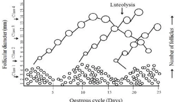
However, in case the animals (cows) are treated with a low level of progestogen to prevent ovulation after luteolysis, dominant follicles will remain active on the ovary for 15 to 20 days (Fig. 1, Lucy et al., 1991). The Fas antigen and Fas ligand system seem to play an important role in mediating apoptosis (Porter et al., 2011). Progression to the secondary follicle is characterized by the appearance of second layer of granulose cells and by the initial deposition of zona pellucid material around the oocyte. At the same time cortical granules are formed within the oocyte cytoplasm and at this stage first time oocyte RNA synthesis is detectable.
Fig. 1. Schematic representation of follicular development; During the class 1 stage there are pool in of different gonadotropin sensitive follicles which further upon growing becomes large and enters into the category of class 2 follicles. Class 2 follicles are the one that are selected further for maturation and fertilization. When class 2 follicles become even further and larger become dominant follicles which are categorized as class 3 follicles. From class 3 stage follicles starts to undergo atresia and hence class 4 follicles are considered atretic follicles.
[Provider:earticle] Download by IP 118.70.52.165 at Monday, December 20, 2021 7:56 PM
www.earticle.net
Table 4. Representation of Biochemical, physiological and functional characteristics of different size classes of bovine ovarian follicles Diameter, mm Function within follicular wave Physiological and biochemistry Class
3-5 Recruited pool of small follicles Below minimum size for ovulation after
luteolysis 1
6-9 Recruited follicles and selected follicle May be potential ovulatory follicle at luteolysis. Granulosa cells lack LH
receptors 2
10-15 Dominant follicle Granulosa cells have LH receptors 3
>15 Large dominant follicle Mature dominant or pre-ovulatory follicle 4
Follicular wave is the cohort of follicle that grows or gets recruited to the flow one after the other and more and more follicles continue to grow into dominant follicle. Here, the FSH has been observed to be the key hormone that induces the recruitment. However, the threshold to recruitment process gets bit easier with the LH surge. At the selection process, the dominant follicles are chosen while the remaining subordinate follicles enter into atresia. Dominant follicles are selected when the FSH is declining and LH has higher pulsatile secretion. Dominant follicles secrete large amount of oestradial and inhibin. Recruitment of a cohort of follicles are marked with the expressions of mRNA for P450scc and P450arom in granulose cells. Selections of dominant follicles are associated with expression of mRNA for LH receptors and 3Beta-HSD in granulose cells (Beg et al., 2003). Average numbers of follicles within four follicular sizes have been classified for ease to understand have been shown in Table. 4. Early during the estrous cycle (days 1 to 41, the average number of follicles detected by ultrasonography in Class 1 (3 to 5 d decreases, whereas the average numbers of follicles in Class2 (6 to 9 mm) increase. The shift apparently occurs because Class 1 follicles are growing into Class 2 and are not being replaced by smaller follicles (<3 mm in diameter). This movement represents the growth of follicles out of Class 1 into Class 2 during the recruitment phase. On approximately d 4 of the estrous cycle, the average number of Class 3 (10 to 15 mm) follicles increases as an average of one follicle grows into Class 3 as the selected follicle. At the same time, the average number of follicles in Class 2 decreases because follicles that did not become dominant decrease in size
and become atretic. This results in the detection of fewer Class 2 follicles and more Class 1 follicles after day 7 of the estrous cycle. Later during the first follicular wave (days 7 to 9 of the estrous cycle), the average number of Class 4 follicles increases because Class 3 follicles (dominant follicles) grow to a diameter >15 mm and are detected by ultrasonography as Class 4 follicles. The sustained suppression of Class 2 follicles (days 9 to 11 of the estrous cycle) is probably due to effects of dominance exerted by the first-wave dominant follicle.
Manipulation of follicular dynamics
Prostaglandin F-2 alpha hasbeen the most commonly used in the treatment for synchronisation of estrus in cattle. GnRH-PGF treatment protocol is Ovsynch protocol that helps in the synchronisation and dominant follicle selection. Super-ovulation is done by priming with progesterone for 12-18 days and then later administration of gonadotrophin (FSH or eCG) prior to or around the time of device removal, but this protocol does not involve current knowledge of follicular dynamics. Nutrition may also affect follicular growth directly through its effects on hypothalamic GnRH or pituitary goandotropin secretion or indirectly through effects on GH-IGH-insulin axis. Dietary restriction linearly decreases the IGF-1 concentration until the onset of anoestrus. Indirect effects of diet are mediated through altering GnRH pulse generator, which in-turn selectively reduces pulsatile LH secretionwithout any apparent adverse effect on FSH secretary pattern.
Endogenous opioid peptide (EOP), leptin, neuropeptide Y (NPY) and glucose all may play role in nutritional
[Provider:earticle] Download by IP 118.70.52.165 at Monday, December 20, 2021 7:56 PM
www.earticle.net
regulation of GnRH release.
MOET is a well established technology and is being used to obtain over 80% of the embryos produced for commercial purposes (Thibier, 2001). The initial use of undefined super-ovulation products like eCG has been replaced by pituitary extracts (porcine and ovine) and in some cases by human menopausal gonadotropins (HMG). These latter products have a shorter half life and require multiple injections but have the advantagesthat they with either no or little side effects such as over-stimulation, failure of ovulation and persistent follicular cyst. Most of the pituitary extracts available on the market have varying ratios of FSH and LH. They are administered in the mid-luteal phase of the estrus cycle of the donor over a 4 to 5 days period and are combined with induced luteolysis. At estrus the donor is inseminated, usually with at least two stress of semen 12 h apart, and 7 days later the uterus is flushed to recover the embryos and only oncetheysuper ovulates giving a large number of embryos (Boland et al., 1991). This is the main drawback of MOET because breeding companies usually require a few particular sire-dam combinations. Second limited attached with it is that it requires reproductively perfect donors and those which have actually passed their lactation peak and in many situation donors are dried off to gain better results.
Reproductive technologies are actually intended to shorten the generational intervals and to propagate genetic material among breeding animal populations and in particular it becomes more pertinent when it comes onto developing countries like India and surrounding countries the discussion over the application of reproductive biotechnologies in cattle and their impact for future achievements becomes more and more important. To achieve this goal, reproductive technologies such as artificial insemination (AI), embryo transfer (ET), manipulation of fertilization and embryo production in vitro and multiplication techniques (cloning) for the application of transgenesis. All these techniques along with the sperm separation techniques (Morrel and Rodriguez-Martinez, 2010), including that of selection of spermatozoa for chromosomal sex (sex
sorting) (Faber et al., 2003).
Though the preservation of semen for AI is well advanced but still a absolute understanding that how spermatozoa lose their capacity to remain fertile upon freezing and thawing are not known. Embryo transfer has revolutionized the area of research on super ovulation, embryo retrieval, transfer by low invasive methods as well as by the better and simpler cryopreservation methods but some of the techniques did not do well because of the nutrition and management constraint leading to reproductive failure.
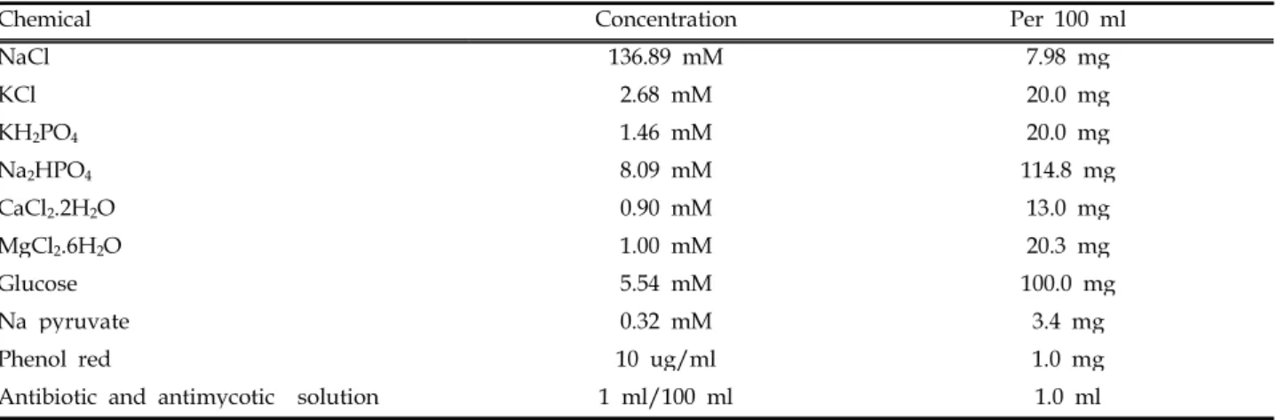
We still understand better that the use of AI and ET would help in preventing transmission of diseases provided intense research is carried out to address the issues related to determine risks of sperm and embryo-pathogen association. Gradual elimination of animal derived products during sperm and embryo handling, as well as during the production of in-vitro developed embryos is increasing. Inspite of the fact that in-vitro techniques for embryo production are well established for cattle, there is as yet a sub-optimal oocyte maturation that limits further developments. Production of offspring combining available reproductive technologies such as trans-vaginal ovum-pick up (OPU), in vitro embryo production and verification for direct ET in proper medium (Tables 1-3) appears as a promising combination for good applicability in breeding. Different compositions have been given for oocyte collection, embryo development and in vitro fertilization (IVF) in the Tables (1, 2, and 3). Since the time of oocyte collection till IVF the different mediums that have been used have many basic chemicals with little difference.
And these differences in their requirements actually speak about the importance of such chemicals at different stages such as during embryo development and fertilization. The addition of KH2PO4 (1.4 mM) and Na2HPO4 (8.09 mM) in the oocyte collection medium (Table 1) have been designated as essential. Similarly, the additional of NaHCO3 (24 mM) and NaH2.PO4.H2O (0.35 mM) have been documented as essential (Table 2).
During in vitro fertilization the changes takes place in the concentration of chemicals but the chemicals remain
[Provider:earticle] Download by IP 118.70.52.165 at Monday, December 20, 2021 7:56 PM
www.earticle.net
the same (Table 3). Cattle has been successfully used for reproductive cloning and for the production of transgenic clones, yet being affected by low effectiveness owing to epigenetic disarray. Beneficial outcome of the expanding gene targeting technology combined with
nuclear transfer and reproductive cloning have been seen as frontier research avenues along with the gene farming and genetic programmes that includes the genomic selection of future sires.
Table 1. Oocyte collection medium at pH 7.3-7.4 and osmolarity 280-290 mOsm.
Chemical Concentration Per 100 ml
NaCl 136.89 mM 7.98 mg
KCl 2.68 mM 20.0 mg
KH2PO4 1.46 mM 20.0 mg
Na2HPO4 8.09 mM 114.8 mg
CaCl2.2H2O 0.90 mM 13.0 mg
MgCl2.6H2O 1.00 mM 20.3 mg
Glucose 5.54 mM 100.0 mg
Na pyruvate 0.32 mM 3.4 mg
Phenol red 10 ug/ml 1.0 mg
Antibiotic and antimycotic solution 1 ml/100 ml 1.0 ml
Table 2. Embryo development medium at pH 7.3-7.4 and osmolarity 280-290 mOsm.
Chemical Concentration Per 100 ml
NaCl 89 mM 519 mg
KCl 3.2 mM 23.85 mg
CaCl2.2H2O 2.0 mM 29.4 mg
MgCl2.6H2O 0.5 mM 10.25 mg
NaHCO3 25 mM 210 mg
Na pyruvate 0.5 mM 5.5 mg
Na lactate - 170 ul
NaH2.PO4.H2O 0.35 mM 4.2 mg
Phenol red 10 ug/ml 1.0 mg
Antibiotic and antimycotic solution 1 ml/100 ml 1.0 ml
Table 3. Medium for IVF at pH 7.3-7.4 and osmolarity 280-290 mOsm.
Chemical Concentration Per 100 ml
NaCl 114 mM 666 mg
KCl 3.2 mM 23.85 mg
CaCl2.2H2O 2.0 mM 29.4 mg
MgCl2.6H2O 0.5 mM 10.25 mg
NaHCO3 25 mM 210 mg
Na lactate 1.86 ul/ml 186 ul
NaH2.PO4.H2O 0.34 mM 4.08 mg
Phenol red 10 ug/ml 1.0 mg
Antibiotic and antimycotic solution 1 ml/100 ml 1.0 ml
[Provider:earticle] Download by IP 118.70.52.165 at Monday, December 20, 2021 7:56 PM
www.earticle.net
Sex sorted semen
Pregnancy rate after transfer of in-vivo embryos produced with sex-sorted semen is lower than that after transfer of embryos produced with conventional semen (An et al., 2010). It has been observed and documented that sex sorted semen in super-ovulated donors consistently reveal a decrease in fertilization rate and impaired embryonic development during the 7 day period from insemination to embryo recovery (Hayakawa et al., 2009; Peippo et al., 2009) (Baruselli et al., 2008). Contrastingly, several reports have been published for in-vitro embryos and pregnancy rate associated with embryos fertilized with sex-sorted semen.
However, it has been reasonably documented that pregnancy rates can be achieved with sex sorted in vitro production (IVP) of embryo (Wheeler et al., 2006; Pontes et al., 2010 and Trigal et al., 2012). It was therefore documented that the effect of sex sorting is challenging to evaluate as the data of animals produced from such technologies (Trigal et al., 2012). Frequently, the bulls have been preselected for fertility under a laboratory conditions and the differences in the sex sorting process and in maturation, fertilization, and culture procedures complicate the interpretation of results. Wilson et al.
(2006) reported compromised conception rates after transfer of in-vitro produced embryos fertilized with sexed semen. On the other hand, acceptable pregnancy rates were achieved with vitrified sex sorted IVP embryos in a large scale transfer experiment (Xu et al., 2006).
Considering indicators of embryonic development other than pregnancy rate, sexed semen can compromise the cleavage rate of in-vitro embryos (Bermejo et al., 2008; Palma et al., 2008; Trigal et al., 2012). It has also been reported that blastocyts formation is lower with the sex semen (Wilson et al., 2005; Morton et al., 2007).
Other features that have been noted are delay in the embryonic development and ultra structural changes in the embryo, such as elevated proportion of immature mitochondria and deviations in the structure of rough endoplasmic reticulum and the nuclear envelope (Palma et al., 2008). Are impairments related sex sorting of
semen. Moreover, epigenetic changes can result in embryos produced with sex sorted semen. It has been reported that relative abundance of developmentally important genes in Day-7 embryos, Glut-3 and G6PD was higher in IVP embryos produced with unsorted semen (Morton et al., 2007). And such epigenetic variations could be involved in the compromised viability of embryos and the resulting offspring.
In addition to the impaired pregnancy with sorted semen, it was also seen that the males born out of the sorted semen were liable for more mortality than for male calves born from conventional semen (Tubman et al., 2004; Borchersen and Peacock, 2009). Insemination of super ovulated embryo donors with sex sorted semen has been show to be profitable, regardless of the reduction in embryo recovery rate. One of the important advantages of sexed semen in embryo production is that the recipient management can be optimised when transferring fewer embryo of the undesired gender (Kaimio et al., 2013; Soares et al., 2011). Despite the reduced embryo yield and pregnancy rate, the number of female’s calves is economically comparable with that for conventional semen. However, it was also seen that the higher mortality of male calves born from embryos produces with X sorted semen does not reflect the overall calf mortality of the sex sorted embryos because those embryos fertilized with Y sperm originating from the X fraction represent only a proportion, less than 10%, of the embryos (Kaimio et al., 2013). However, it can be concluded that pregnancy rates after transferring of embryos produced with sex sorted semen are compromised and the decline could range between 10-12 percent. Despite, this use of sex sorted semen decreases the number of transferable embryos recovered. Overall, the use of sex sorted semen is profitable, particularly for heifers. The rationale is that in heifers, more females calves can be produced with fewer embryo recipients (Larson et al., 2010).
In vitro production of embryos
IVP came to utilization first as a research tool and was
[Provider:earticle] Download by IP 118.70.52.165 at Monday, December 20, 2021 7:56 PM
www.earticle.net
applied to protect follicular oocyte of slaughtered donors. It is an alternative system to produce cattle embryos to use immature oocyte collected from the ovaries of donors of various age and physiological status (Galli and Lazzari, 1996). Subsequently, reliable procedures have allowed maturation and fertilization of bovine oocyte in-vitro and for the same several culture protocols that was developed by different laboratories can be used to grow them for about a week up to the stage suitable for transfer or freezing. In cattle the major activation of embryonic genome occurs at the 8 cell stage (Kues et al., 2008) accompanied by changes in chromatic structure such as acetylation of core histones (Memili and first, 1999). At 32 cell stage to 64 cell stage compaction occurs followed by blastulation at day 7-8, and hatching occurs at day 8-9 followed by elongation until implantation starts on day 20-21. Around hatching, the ICM cells differentiate into an inner layer facing the blastocyst cavity, the hypoblast, while the remaining cells from the epiblast (Vejlsted et al., 2006). The polar trophoblast (Rauber’s layer) soon gets degenerated and at the same time gene expression changes of key pluripotency transcription factors POU5F1, SOX2, and NANOG take place (Khan et al., 2012). However, in mouse, this core triad is not confined to the ICM in the early blastocytst, but it is also expressed by the trophoblast together with trophoblast-specific genes CDX2, HAND1, ETS2 and IFN-tau. After formation of the epiblast and hypoblast however, POU5F1, SOX2, and NANOG expression becomes restricted to the epiblast (Degrelle et al., 2005; Vejlsted et al., 2006).
First IVF calf derived from in vivo matured oocytes in 1982 (Brackett et al., 1982) and the discovery of heparin as capacitating agent for bull sperm came into existence in 1986 (Parrish et al., 1986). Such information and knowledge hastened the efficient production of IVP procedures including in vitro maturation (IVM), and subsequent in vitro culture (IVC) of embryos to the blastocyst stage. Initially the lack of knowledge on embryo requirements was overcome temporarily by in-vivo culture in the surrogate sheep oviduct (Galli et al., 2003a). Subsequently, co-culture technique was
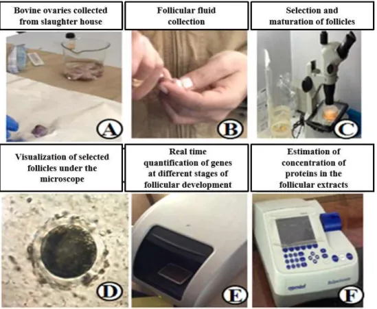
developed based on the synthetic oviductal fluid formulations (SOF, Gardner et al., 1994). At present the application of IVP combined with ovum pick up (OPU) from valuable donors is increasing due to developing breeding strategies based on genomic selection using SNP chips (Fig. 2), whereby thousands of genetic markers can be screened even on a small embryo biopsy before embryo transfer, with enough precision to anticipate that the need for progeny testing will hopefully gets dramatically reduced.
In vitro maturation and fertilization
Oocytes are very sensitive to temperature shock so it is important to monitor carefully the temperature during the collection procedures as fluctuations can easily occur.
During collection the oocyte are maintained in Dulbecco’s PBS or in TCM-199 (HEPES buffered). The maturation conditions used by the vast majority of the laboratories involve the use of TCM-199 supplemented with 10% FCS and gonadotropins (FSH, LH) in 5% CO2
in air at 38.5℃. After 20-24 hours of incubation the oocyte complete maturation with the extrusion of the first polar body and are ready to be fertilized. It is normally documented that if the donors are located far from the main laboratory, maturation can be completed during transport from the site of collection to the laboratory in test tubes in portable battery powered incubators. Under optimal conditions over 90% of the oocyte reaches metaphase II. Before fertilization the cumulus cells are partially removed to leave few corona cell layers surrounding the oocyte.
The methods delineated for cloning of animals using SCNT follows the protocol used by Wilmut et al. (1997) to produce Dolly, the sheep cloned from adult animals.
The main steps involved in the process of SCNT are: 1.
Oocyte enucleation to obtain the cytoplast, 2. Culture of somatic cell or nuclear donar cell called karyoplast, 3.
Transfer of karyoplast to the cytoplast, 4. Fusion and activation of the karyoplast-cytoplast couplets and 5.
Culture of cloned embryos up to blastocyst stage before transfer to the recipients. The technology has resulted in
[Provider:earticle] Download by IP 118.70.52.165 at Monday, December 20, 2021 7:56 PM
www.earticle.net
Fig. 2. Steps in from collection of ovaries, follicular fluid collection, selection, maturation and IVF and experimental analysis, investigation and interpretations. A, collection of ovary in PBS; B, collection of follicular fluid gently using proper gauge needle in standard medium; C, grading and selection of optimized follicle under the microscope; D, visualization of selected follicle; E, real time PCR machine for quantifying follicular genes; F, estimating the protein concentration in the follicular extracts.
Fig. 3. Cloning and embryo transfer technology; Objective of embryo transfer technology (ETT) focuses on to standardize ETT in Indian cows, buffaloes, sheep and goats and to create a seeding stock of high quality bulls and dams.
[Provider:earticle] Download by IP 118.70.52.165 at Monday, December 20, 2021 7:56 PM
www.earticle.net
many of cloned animals of the similar characteristics (Fig. 3). The intense efforts in the past have been carried out on embryo transfer and SCNT research is mostly due to the importance of transgenic research. Goats for example have been considered as having exceptional potential to serve as bioreactors to the produce milk containing recombinant proteins suitable for pharmacological usage. Goats produce from this technology would have short generation interval compared to cattle, two to three fold greater volume of milk production than sheep (Fig. 3). The concept has been also been seen in case of buffalos as well.
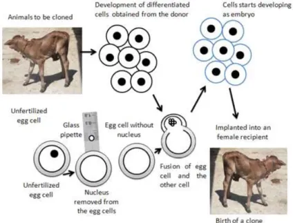
Furthermore, the SCNT technology involves the enucleation of a mature oocyte that creates a chromosome free cytoplast, which is then electro-fused with a diploid somatic cell (Fig. 4). This reconstructed embryo is then activated and further developed on the lines of embryonic development. Several steps which make this technology fruitful have been designed and refined with the contributions of many laboratories worldwide over several years.
Frozen semen is always used for IVF and a Percoll
based separation system is the most common method for isolating the motile sperm fraction after thawing (Galli et al., 2003b). Although other systems can be used (swim up, simple centrifugation), separation through a percoll gradient offers the consistency, flexibility and reliability that are required in a commercial setting, where new sires are required on a weekly basis by the clients. Two media are generally used for IVF: a TALP based medium or a SOF based medium both without glucose and with varying concentration of heparin. The concentration of spermatozoa that is needed for each bull, in order to achieve maximum fertilization with minimal polyspermy, is determined empirically by performing IVF tests with different sperm concentrations.
There fertilized oocytes are dixed 18-20 hours after co-incubation with the sperm and the chromatin configuration is analyzed following lacmoid staining. The results of this test indicate the optimal incubation of sperm and eggs. At this time the oocytes are completely denuded of the remaining cumulus cells and spermatozoa and are transferred to a culture system suitable for embryo development (Fig. 3).
Fig. 4. Cloning of cow calf by somatic cell nuclear transfer. Oocytes are collected from a donor, matured in vitro to metaphase II, and enucleated (with the aid of micromanipulation) to remove the first polar body and the portion of the oocyte containing the metaphase plate. The resulting cytoplast is combined with a donor cell (e.g. cultured fibroblast cell) originating from the animal to be cloned. The cytoplast and donor cell are submitted to electro fusion resulting in production of reconstructed embryos in which the oocyte cytoplasm is mixed with the donor cell cytoplasm and carries the donor cell nucleus. The reconstructed embryos are activated to further develop into blastocyst stage so that it can be transferred to the recipient for development into cloned offspring.
[Provider:earticle] Download by IP 118.70.52.165 at Monday, December 20, 2021 7:56 PM
www.earticle.net
Factors that affect the success of IVF are important to note as some of the bulls sometimes performs better than others and the other small proportion (<5%) performs really poor. A second factor that affects the IVF is the cumulus oocyte complex. Therefore, it is important to standardize the procedure for oocyte preparation before IVF, in order to obtain a homogenous population of oocytes with a cumulus cell mass (Do and Scholer, 2005). In fact the amount of cumulus is positively related to the sperm concentration required; oocytes with fewer cumulus cells are more susceptible to polyspermy while those with a large cumulus cell mass are less likely to be fertilized.
IVF for the non-infertile
Preimplantation genetic diagnosis IVF gives access to blastomere biopsy from a day 3 embryo for molecular analysis using the polymerase chain reaction.
Pre-implanation genetic diagnosis may be used to avoid the transmission of autosomal recessive genetic defects, such as cystic fibrosis and thalassaemia; autosomal conditions including Huntington’s disease and myotonic dystrophy and their molecular analysis using the polymerase chain reaction. Preimplantation genetic diagnosis may be used to avoid the transmission of autosomal recessive genetic defects, such as cystic fibrosis and thalassaemia; autosomal dominant conditions including Huntington’s disease and myotonic dystrophy; and X-linked disorders such as haemophilia and fragile-X syndrome. However, these diseases are of human concern and so far ruminant’s diseases have not been either studied or reported in the light of the current subject and therefore it needs to be investigated.
Pre-implantation aneuploidy screening
Aneuploidy is an abnormality in the number of chromosomes in an individual’s cells. Certain chromosomes (13, 15, 16, 18, 21, and 22) are particularly prone to aneuploidy. Aged ruminant animals undergoing IVF may have high number of neupolid embryos,
particularly with aneuploidies of chromosomes X, and Y, 13, 14, 15, 16, 18, 21, and 22. This phenomenon has been associated with recurrent implantation failures at IVF (Sartori et al., 2004). The pre-implantation testing of blastomeres using multiplex fluorescent in-situ hybridization before selecting normal blastocyst for transfer has not been shown to improve live birth rates in older ruminant animals so far and therefore the area needs to be researched further. There are possibilities that abnormalities may occur due to the small number of oocytes. It’s been known now that comparative genomic hybridizations through a newer PCR-based technology could screen all of the chromosomes in a cell. It is therefore mandated through studies which suggest that array-CGH has the potential to overcome several inherent limitations of FISH-based tests.
Future perspectives
The technology of embryo production in cattle is widely used both in research and for applied purposes.
As a research tool, the technology is of great value to address fundamental questions on the endocrine control, the molecular switches and the metabolic pathways that regulate early development. As an applied technology it provides the opportunity to increase the number of offspring from superior genotypes. However, in order to make the technology accessible to a wider base of breeders or even to commercial farmers, there are several aspects that need to be addressed at the clinical, cellular and molecular level to increase the efficiency and to reduce the costs involved.
The ovum pick up technique tends to be more consistent than MOET and it allows receptivity and safe embryo production without interfering with the reproductive cycle or milk production of dairy donors.
However, only a few of the collected oocyte develop into viable embryos. Several factors have a role to play in this context such as nutritional status of the donor (Boland et al., 2001) together with intra-ovarian factors such as the stage of the oocyte within the follicular wave, are likely to substantially affect embryo development.
[Provider:earticle] Download by IP 118.70.52.165 at Monday, December 20, 2021 7:56 PM
www.earticle.net
There is evidence that oocytes collected in the presence of a dominant follicle are of lesser quality than those collected during follicular growth. In addition, comparing in vitro with in vivo maturations recent studies (Rizos et al., 2002) have made it clear that the developmental potential of in vitro matured oocytes is generally lower than that of in vivo matured oocytes.
Such finding also reveals that herterogenous population of oocytes with different developmental potential would show diverse population of oocytes which would range from about to ovulate to the advance stage of atresia.
Researchers have shown the quality of the embryos developing following IVM-IVF is very much influenced by the culture conditions from zygote to blastocyts (Rizos et al., 2002). Therefore, improved oocyte maturation remains perhaps the most fundamental step to increase embryo production while embryo culture is the step that affects the quality/viabililty of the embryos (Kubelka et al., 2000).
A recent series of studies have attempted to improve oocyte quality by mimicking the last phase of in vivo oocyte growth with a period of pre-maturation in in vitro (24-48 hours) before final maturation. Several compounds have been used to maintain the oocyte in meiotic arrest during pre-maturation. Some of the tested compounds are namely; butyrolactone I and roscovitine, two protein kinase inhibitors, alone (Mermillod et al., 2000) or in combination (Ponderato et al., 2001) can maintain the oocytes in a fully reversible meiotic block for at least 24 hours without interfering with the following embryo and fetal development (Ponderato et al., 2002). However, all these studies have failed to show any improvement so far and are likely to limit any progress in the use of juvenile breeding as it was expected to get the most benefit from a pre-maturation step. It has been noted that with the flexibility in the embryo production gives the adjustment of the time of pre-maturation to maturation to the needs of the IVF laboratory or for cloning work (Laguita et al., 2002) and it is therefore important though.
Embryo quality and viability is mainly affected by the culture system following IVF (Rizos et al., 2002). This
means that the viability of an in-vitro produced embryo can vary considerably from one laboratory to the. It has been described earlier that embryo observation during culture especially at the compaction stage (day 5-6) is crucial for selecting good quality embryos that survive cryopreservation. Far better results can be obtained by the in-vivo culture of IVM-IVF embryos in the sheep oviduct. Besides the defined chemical composition of the medium, an important role is also played by the type of macromolecules present during IVC. Their presence of serum is usually associated with accumulation of lipids and a poor survival after freezing the supplementation of media with albumin, and PVA supplementation produces embryos with expression patterns similar to those grown in in-vivo (Wrenzycki et al., 1999). The culture system not only affects the viability of the embryos and their freezability, but also the characteristics of the newborn calf. It has been shown that IVC in the presence of high level of serum or BSA can induce a significant increase in birth-weight (Lazzari et al., 2002) and the occurrence of the complex condition known as large offspring syndrome.
Impact of heat stress on reproductive performance of the animals
Proper assessment of sexual behaviour and receptivity before breeding of animals may increase the possibility of fertile mating. High environmental temperature or the rapid and sudden fluctuations of temperature that often occur in arid and semi-arid regions have detrimental effect on sexual behaviour of animals (Maurya et al., 2005). The implication of thermal stress is such that even if an animal has a normal ovarian development leading to ovulation, there is a reduction in the full expression of oestrus behaviour that might lead to the failure of animal to mate and conceive. It has also been reported that heat stress suppresses the functions of the largest follicle, i.e. the dominant follicle. Under heat stress, the size of the dominant follicle is reduced during the first and second follicular waves the number of the follicles of the next largest size is increased. Thermal stress
[Provider:earticle] Download by IP 118.70.52.165 at Monday, December 20, 2021 7:56 PM
www.earticle.net
during folliculogenesis could lead to ovulation of low quality oocytes with lowered developmental competence.
The circulating concentrations of gonadotrophin, which plays an important role in regulating follicular dynamics, are also altered by heat stress. The heat stress induces alterations in the follicular LH receptors and adversely affects the rate of ovulation in cows (Alfujairi et al., 1993).
Most cells in the body produce heat shock proteins in response to heat stress that limit the damaging effects of elevated temperature on cell function. Unfortunately, in cattle, around the time of ovulation, the oocyte and /or the resulting early embryo are unable to produce heat shock proteins. Consequently, embryo viability is compromised resulting in lower conception rates.
Embryonic loss can occur when there is disruption in the physiological regulation of oviductal and uterine function. In cattle, exposure period leads to embryonic loss (Ealy et al., 1993) and fetal growth within the uterus is a complex biological event that is influenced by genetic, epigenetic, and environmental factors, as well as maternal maturity. These factors impact on the size and functional capacity of the placenta, utero-placental blood flow, transfer of nutrients and oxygen from mother to fetus, conceptus nutrient availability, the endocrine milieu, and metabolic pathways (Wu et al., 2006).
Heat stress drastically reduces the pregnancy rates in animals (Hahn et al., 2003). The reproductive efficiency of the animals in the environment in which they are kept should be fine-tuned to produce maximum number of offspring most efficiently. During the heat stress redistribution of blood flow from the viscera to the periphery increases for dissipation of heat, which leads to reduce blood supply to placentas and retards fetal growth (Collier et al., 1982). Fertility of animal is markedly prior to oestrus or immediately after mating.
The calf born from heat stressed cattle have lower birth weight that born from control cows maintained under comfortable environmental conditions. The animal exposed to thermal stress reduces the body condition score of animals, which affects the birth weight of new born animals. The experiments have also been conducted
in several institution in India where in digital device has been placed to monitor the wind velocity, environmental moisture, heat and so on that gets calculated and same could be applied over the animals to conduct experiments related to heat stress on animals (Fig. 5).
The same practice has benefitted a lot in terms of calibrating the thermo-neutral zone as it has been realised that it varies from one geographical point than others (Fig. 5a) (Sejian et al., 2012). Animal researchers have realized that global heat would have tremendous effect on animal’s health and production. Therefore, researchers are deciding to work on heat stress after creating the presumptive focus by creating pychrometric chamber (Fig. 5b) to draw immediate conclusions to improvise the animal health and production concern issues. Under such experiments one would not only perform proteomics or genomics but also can monitor hormonal changes assessed with different counters (Gamma and Beta counters) (Fig. 5c). Recently, nano-technologies have impacted many studies, the environmental biologists those concern heat stress have been striving to study the usage of nano-tech on stem cells. The application of nano-technology on stem cells could be fruitful as it would help us know the behaviour of such stem cells under high heat under laboratory conditions (Fig. 5d).
The male animals are most important for overall reproductive performance and productivity of the female herd and/or flocks. A highly fertile and adapted bull could produce greater number of cows early in the breeding season, and will be more likely to fertilize a higher proportion of eggs than less adapted and bull with poor fertility. It has also been shown that sephadex filtration of semen may increase the seminal quality of buffalo bulls (Hall et al., 2013). Bull that exhibit relatively rapid ejaculation rates are capable of inseminating a greater number of cows per unit of time than males of having poorer libido. Tests for ranking bull on sexual performance are repeatable and reliable predictors of sexual performance under field conditions, potentially allowing breeders to ejaculate the mating competence of individual males before they are
[Provider:earticle] Download by IP 118.70.52.165 at Monday, December 20, 2021 7:56 PM
www.earticle.net
Fig. 5. Impact of heat stress on follicular dynamics and vis-a-vis on fertility potential of animals. Experimental data on heat stress on cows, buffaloes, sheep and goat on their reproductive potential could be generated, calculated and interpreted. A, Monitoring of atmospheric temperature, humidity and wing velocity for heat stress experiments on cattle/or buffaloes; B, Psychrometeric chambers for study heat stress impact on the reproductive performance through follicular dynamics and functional proteomics; C, Estimation of different levels of hormones during heat stress in cattle/or buffaloes with Gamma counter; D, In vitro profiling of the inner cell mass stem cells to draw data on heat stress and reproductive efficiency of cattle/buffalo.
employed in a breeding programme. The stage of spermatogenesis that is most susceptible to elevated temperature is the primary spermatocytes but the damage to B spermatogonia can occur in bulls and prolonged exposure to heat can damage dividing spermatocytes and spermatids (Collier et al., 1982). Heat stress occurs when the scrotum is not able to reduce the temperature of the testicles below normal body temperature and resulted in loss of motility, increased proportion of abnormal sperm, decreased concentration of sperm and ultimately cessation of spermatogenesis. In addition to that, it would lead to higher percentage of abnormal spermatozoa and acrosomal damage. Researchers
have reported that heat stress on scrotum drastically affects the function of sertoli cells and release of androgen. The body condition score drastically affects the seminal attributes of animals (Collier et al., 1982).
Ruminants primarily adjust evaporative heat loss to maintain homeothermy during brief exposures to adverse weather, but will reduce feed intake to lower heat production during prolonged hot weather. In hot environment, energy exchanges by radiation are dominant. To alter the microclimate of an animal effectively through housing or environmental modification, one has to take into account the following factors: temperature of the surrounding; air temperature;
[Provider:earticle] Download by IP 118.70.52.165 at Monday, December 20, 2021 7:56 PM
www.earticle.net
air velocity; air vapour pressure; radiation or shade factors; and conductivity of surfaces that animals might contact. Grazing animals or animals giving birth will seek shelter from strong winds. Structures or trees can help protect wind exposure (especially newborn).
However, windbreaks have an importance much beyond these benefits, especially in the tropical and subtropical regions. A wind break acts as a barrier lowering the wind speed near the ground surface, deviating and splitting the air stream. The use of leguminous trees or shrubs can be a practical means to counteract the effects of the wind and the heat stress as well as to improve the animal diet.
ACKNOWLEDGEMENT
We are thankful to the Banaras Hindu University for facilitating and help in developing this review. The study was supported from research startup grant under XII plan (No. R/Dev/D/XII Plan/ Recurring/ Startup grant/95941, Dated: July 29, 2015), Banaras Hindu University, Varanasi-221005, Uttar Pradesh, India.
REFERENCES
1. Alfujairi, M. M., Albrahim, R. M. and Elnouty, E .D.
1993. Seasonal variations in superovulatory responses of Holstein cows treated with pregnant mare serum gonadotropin in Saudi Arabia. J. Reprod. Fertil. 11:75.
2. An, L., Wu, Z. H., Wu, Y. F., Zhang, X. L., Liu, X., Zhu, Y. B., Cheng, W. M., Gao, H. M., Guo, M. and Tian, J. H. 2010. Fertility in single-ovulating and superovulated dairy heifers after insemination with low dose sex-sorted sperm. Reprod. Domest. Anim.
45:e344-350.
3. Baruselli, P., Martins, C., Sales, J. and Ferreira, R.
2008. Recent advances in bovine superovulation. Acta Scientiae Veterinariae. 36:433-448.
4. Beg, M. A., Meira, C., Bergfelt, D. R. and Ginther, O.
J. 2003. Role of oestradiol in growth of follicles and
follicle deviation in heifers. Reproduction. 125:847-854.
5. Bermejo-Alvarez, P., Rizos, D., Rath, D., Lonergan, P.
and Gutierrez-Adan, A. 2008. Can bovine in vitro-matured oocytes selectively process X- or Ysorted sperm differentially? Biol. Reprod. 79:594-597.
6. Boland, M. P., Gouling, D. and Roche, J. F. 1991.
Alternative gonadotrophins for superovulation in cattle. Theriogenology. 35:5-17.
7. Boland, M. P., Lonergan, P. and O’Callaghan, D. 2001.
Effect of nutrition on endocrine parameters ovarian physiology and oocyte and embryo development.
Theriogenology. 55:1323-40.
8. Borchersen, S. and Peacock, M. 2009. Danish, A.I. field data with sexed semen. Theriogenology. 71:59-63.
9. Brackett, B. G., Bousquet, D., Boice, M. L., Donawick, W. J., Evans, J. F. and Dressel, M. A. 1982. Normal development following in vitro fertilization in the cow. Biol. Reprod. 27:147-158.
10. Collier, R. J., Doelger, S. G., Head, H. H., Thatcher, W. W. and Wilcox, C. J. 1982. Effects of heat stress during pregnancy on maternal hormone concentrations, calf birth weight and postpartum milk yield of Holstein cows. J. Anim. Sci. 54:309-319.
11. Degrelle, S. A., Campion, E., Cabau, C., Piumi, F., Reinaud, P., Richard, C., Renard, J. P. and Hue, I.
2005. Molecular evidence for a critical period in mural trophoblast development in bovine blastocysts.
Dev. Biol. 288:448-460.
12. Do, J. T. and Scholer, H. R. 2005. Comparison of neurosphere cells with cumulus cells after fusion with embryonic stem cells: reprogramming potential.
Reprod. Fertil. Dev. 17:143-149.
13. Ealy, A. D., Drost, M. and Hansen, P. J. 1993.
Developmental changes in embryonic resistance to adverse effects of maternal heat stress in cows. J.
Dairy Sci. 76:2899-2905.
14. Evans, M. J. and Kaufman, M. H. 1981.
Establishment in culture of pluripotential cells from mouse embryos. Nature. 292:154-156.
15. Faber, D. C., Molina, J. A., Ohlrichs, C. L., Van der Zwaag, D. F. and Ferre, L. B. 2003. Commercialization of animal biotechnology. Theriogenology. 59:125–138.
[Provider:earticle] Download by IP 118.70.52.165 at Monday, December 20, 2021 7:56 PM