Description and Application of a Marine Microalga Auxenochlorella protothecoides Isolated from Ulleung-do
Hyeong Seok Jang1, Nam Seon Kang2, Kyeong Mi Kim1, Byung Hee Jeon1, Joon Sang Park1 and Ji Won Hong1*
1Department of Taxonomy and Systematics, National Marine Biodiversity Institute of Korea, Seocheon 33662, Korea
2Marine Life-resources Management Department, National Marine Biodiversity Institute of Korea, Seocheon 33662, Korea Received July 26, 2017 /Revised September 8, 2017 /Accepted September 9, 2017
A unicellular green alga was axenically isolated from a tidal pool on Ulleung-do, Korea. Morphological, molecular, and biochemical analyses revealed that the isolate belonged to Auxenochlorella protothecoides.
The current study is the first record of this species in Korea. The microalgal strain was named as A.
protothecoides MM0011 and its growth, lipid and pigment compositions, and biomass properties were investigated. The strain is able to thrive in a wide range of temperatures (5~35°C) and to withstand up to 1.5 M NaCl. The results of GC/MS analysis showed that the isolate was rich in nutritionally important polyunsaturated fatty acids (PUFAs). Its major fatty acids were linoleic acid (27.6%) and α -linolenic acid (39.6%). Thus, this indigenous microalga has potential as an alternative source of ω3 and ω6 PUFAs, which currently come from fish and plant oils. Also, the HPLC analysis revealed that the value-added antioxidant, lutein, was biosynthesized as the accessory pigments by the microalga.
A proximate analysis showed that the volatile matter content was 85.6% and an ultimate analysis in- dicated that the gross calorific value was 20.3 MJ kg-1. Since 40.5% of total nitrogen and 27.9% of total phosphorus were removed from the medium, respectively, it also has potential as a feedstock for bio- fuel applications which could be coupled to wastewater treatment. In addition, the biomass may also serve as an excellent animal feed because of its high protein content (51.4%). Therefore, A. proto- thecoides MM0011 shows promise for application in production of microalgae-based biochemicals and as a biomass feedstock.
Key words : Auxenochlorella protothecoides, first record, lutein, PUFAs, Ulleung-do
*Corresponding author
*Tel : +82-41-950-0743, Fax : +82-41-950-0727
*E-mail : [email protected]
This is an Open-Access article distributed under the terms of the Creative Commons Attribution Non-Commercial License (http://creativecommons.org/licenses/by-nc/3.0) which permits unrestricted non-commercial use, distribution, and reproduction in any medium, provided the original work is properly cited.
Journal of Life Science 2017 Vol. 27. No. 10. 1152~1160 DOI : https://doi.org/10.5352/JLS.2017.27.10.1152
Introduction
Green microalgae (Chlorophyta) are photosynthetic mi- croorganisms that can be found in every conceivable aquatic habitat from seawater to freshwater. Marine microalgae are known to play indispensable roles in global carbon, nitro- gen, and phosphorus cycles as the major primary producers in the maritime ecosystem [1, 6, 11]. Since they are capable of taking up water (H2O) and carbon dioxide (CO2) and con- verting them into a variety of energy-rich organic com- pounds such as lipids, hydrocarbons, carbohydrates, and other valuable compounds via photosynthesis [7, 15, 20], mi- croalgae have recently received considerable interest and ac-
cordingly many studies have focused on the assessment of their biotechnological potential [4, 25, 32].
In this study, we have isolated and identified a unicellular microalga A. protothecoides MM0011 from a tidal pool on Ulleung-do, Ullenug-gun, Gyeonsangbuk-do, Korea. Auxeno- chlorella was ranked as a subgenus of Chlorella in 1965 [28], then it was upgraded to the genus rank based on physio- logical and biochemical characteristics by Kalina and Punčochářová in 1987 [12]. It was reported that A. proto- thecoides was isolated from the sap secreted from Populus and Ulmus trees in Germany [8, 30]. It was also found in munici- pal wastewater in Minnesota, USA [18]. Due to the fast cell growth and high lipid content of A. protothecoides, a number of heterotrophic cultivation researches have been carried out to produce biodiesel by sequestering CO2 [5, 10, 38, 42, 43].
In addition, many previous studies have also shown that A. protothecoides was able to produce relatively high concen- tration of lutein when grown under heterotrophic conditions [26, 27, 29, 34, 39].
The present report provides information on the first re-
cord of the species in Korea and its morphological, molec- ular, and chemotaxonomic features were investigated.
Materials and Methods
Sample collection and isolation
Seawater samples were collected in April 2016 from a ti- dal pool near Turtle Rock (37° 27’ 35.86’’N, 130° 51’ 23.49”E) on Ulleung-do, Seo-myeon, Ullenug-gun, Gyeonsangbuk-do, Korea. Samples were then transported to the laboratory and 1 ml of each sample was inoculated into 100 ml BG-11 me- dium [21]. The broad-spectrum antibiotics chloramphenicol (Sigma-Aldrich, St. Louis, MO, USA) and imipenem (JW Pharmaceutical, Seoul, Korea) were added to the medium at a concentration of 100 µg ml-1, respectively in order to prevent bacterial growth. The flasks were incubated in a stat- ic condition at 25℃ under cool fluorescent light (approxim- ately 40 μmole m-2 s-1) in a light:dark cycle (16:8 hr) until algal growth was apparent. Well-grown algal cultures (1.5 ml) were centrifuged at 3,000× g for 3 min to harvest algal biomass. The resulting pellets were streaked onto R2A agar (Becton, Dickinson and Company, Sparks, MD, USA) sup- plemented with imipenem (20 μg ml-1) and incubated under the aforementioned conditions. A single colony was then aseptically restreaked onto a fresh R2A agar plate to obtain an axenic culture.
Morphological identification
The isolate was grown in R2A medium for 8 days. Live cells were harvested by centrifugation at 3,000× g for 3 min, washed three times with sterile distilled H2O, and examined at 1,000× magnification under a Zeiss Axio Imager.A2 light microscope (Carl Zeiss, Göttingen, Germany). For scanning electron microscopy (SEM), 10 ml aliquots of cultures at ap- proximately 1,000 cells ml-1 were fixed for 10 min in osmium tetroxide (OsO4, Electron Microscopy Sciences, Hatfield, PA, USA) at a final concentration of 2%(v/v). The fixed cells were collected on a 3 µm pore size, polycarbonate membrane filter (Whatman, Kent, UK) and washed three times with distilled H2O to remove residual media components. The membranes were dehydrated in an ethanol series (Merck, Darmstadt, Germany) and immediately dried using an auto- mated critical point dryer (EM CPD300, Leica, Wetzlar, Germany). The dried filters were mounted on a stub and coated with gold-palladium in a low vacuum coater (EM ACE200, Leica, Wetzlar, Germany). Surface morphology was
observed with a field emission scanning electron microscopy (FE-SEM, SUPRA 55VP, Carl Zeiss, Jena, Germany).
For transmission electron microscopy (TEM), cells were transferred to a 10 ml tube and fixed in 2.5%(v/v) gluta- raldehyde (final concentrations) for 2 hr, the contents of the tube were placed in a 10 ml centrifuge tube and concentrated at 1,610 g for 10 min in a Vision Centrifuge VS-5500 (Vision, Bucheon, Korea). The resulting pellet was subsequently transferred to a 1.5 ml tube and rinsed in 0.2 M sodium cacodylate buffer (Electron Microscopy Sciences, Hatfield, PA, USA) at pH 7.4. After several rinses in 0.2 M sodium cacodylate buffer, cells were post-fixed for 90 min in 1%
(w/v) OsO4 in deionized H2O. The pellet was then em- bedded in agar. Dehydration was performed in a graded ethanol series (50, 60, 70, 80, 90, and 100% ethanol, followed by two changes in 100% ethanol). The material was em- bedded in Spurr’s resin (Electron Microscopy Sciences, Hatfield, PA, USA). Sections were prepared on an EM UC7 ultramicrotome (Leica, Wetzlar, Germany) and stained with 3%(w/v) aqueous uranyl acetate (Electron Microscopy Sci- ences, Hatfield, PA, USA) followed by lead citrate (Electron Microscopy Sciences, Hatfield, PA, USA). The sections were visualized on an H-7650 TEM (Hitachi, Tokyo, Japan) using a voltage of 100 kV.
Molecular identification
For molecular analysis, genomic DNA was extracted us- ing a DNeasy Plant Mini kit (Qiagen, Hilden, Germany). The universal primers NS1/NS8 and ITS1/ITS4 described by White et al. [35] were used to amplify the 18S rRNA se- quence and internal transcribed spacer (ITS) region, respectively. Phylogenetic analysis was performed with the 18S rRNA sequence of strain MM0011 using the software package MEGA ver. 6.0 [33]. Its closely related Auxenochlorella and Chlorella sequences were downloaded and aligned in the MEGA software, with the ClustalW tool. The best-fit nucleo- tide substitution model (Kimura 2-parameter) was selected using MEGA 6.0 based on the Bayesian information criterion.
This model was used to build a maximum likelihood (ML) phylogenetic tree with 1,000 bootstrap replicates. Due to the highly conserved nature of rRNA, the region RuBisCO rbcL was also amplified with primers, rbcL 7F and rbcL 1391R, described by Verbruggen et al. [37]. All analyses were car- ried out in triplicate, unless otherwise stated. DNA se- quences obtained in this study were deposited in the data- base of the National Center for Biotechnology Information
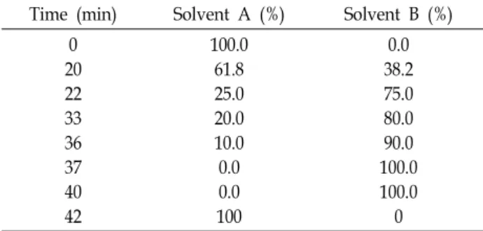
Table 1. Gradient profile and mobile phase composition Time (min) Solvent A (%) Solvent B (%)
0 20 22 33 36 37 40 42
100.0 61.8 25.0 20.0 10.0 0.0 0.0 100
0.0 38.2 75.0 80.0 90.0 100.0 100.0 0 under accession numbers MF040300, MF040301, and
MF043910 (Table 2).
Temperature and NaCl tolerance testing
Routine serial subculturing on R2A agar slant was per- formed to maintain the pure culture of A. protothecoides. A single colony of strain MM0011 was streaked onto R2A agar plates in triplicate and incubated for 14 days. Survival and growth of MM0011 cells maintained at temperatures ranging from 5℃ to 35℃ (at intervals of 5℃) were examined to de- termine the optimum culture temperature. NaCl tolerance test was conducted at 20℃ using R2A agar supplemented with 0.0 M, 0.5 M, 1.0 M, 1.5 M, and 2.0 M NaCl, respectively.
Gas chromatography/mass spectrometry (GC/MS) analysis
The isolate was heterotrophically grown in R2A medium for 8 days at 20℃ with shaking at 160 rpm on an orbital shaker (SH30, Fine PCR, Gunpo, Korea) and cells were har- vested by centrifugation at 2,063 g (1580R, Labogene, Daejeon, Korea) for lipid analysis. The samples were freeze-dried and pulverized to enhance the extraction efficiency. Lipid extraction was performed using the method developed by Breuer et al. [3]. The FAME composition was analyzed using a 7890A gas chromatograph equipped with a 5975C mass selective detector (Agilent, Santa Clara, CA, USA). GC runs were performed on a DB-FFAP column (30 m, 250 μm ID, 0.25 μm film thickness; Agilent, Santa Clara, CA, USA). The initial oven temperature of the gas chromato- graph was 50℃ and maintained for 1 min. The temperature was increased to 200℃ at a rate of 10℃ min-1 for 30 min and it was then increased to 240℃ at a rate of 10℃ min-1 and it was held for 20 min. The injection volume was 1 μl with a split ratio of 20:1. Helium was used as carrier gas at a constant flow rate of 1 ml min-1. The mass spectrometer parameters were as follows: injector and source temper- atures were 250℃ and 230℃, respectively, and the electron impact mode at an acceleration voltage of 70 eV was used for sample ionization, with an acquisition range from 50-550 m z-1. Compound identification was performed by matching the mass spectra with those in the Wiley/NBS libraries. The searches with a match value higher than 90% were consid- ered valid.
High pressure liquid chromatography (HPLC) analysis Pigment extraction was carried out using the method de-
veloped by Zapata et al. [41]. Briefly, 1 mg of freeze-dried biomass was extracted in 90% HPLC grade acetone (Dae- jung, Siheung, Korea) and filtered through a Whatman PTFE syringe filter with a pore size of 0.2 μm (Florham Park, NJ, USA) before injection. Samples were then analyzed on an Agilent 1260 Infinity HPLC system (Waldbronn, Germany) equipped with a Discovery C18 column (25 cm × 4.6 mm, 5 μm, Supelco, Bellefonte, PA, USA) at 33℃. The mobile phase gradient was programmed as described by Sanz et al. [24] at a constant flow rate of 1 ml min-1 consisted of a mixture methanol: 225 mM ammonium acetate (82:18, v:v) as solvent A and ethanol as solvent B (Table 1). HPLC grade methanol and ethanol were purchased from J. T. Baker (Avantor Performance Materials, Center Valley, PA, USA).
HPLC grade ammonium acetate was obtained from Fluka (Sigma-Aldrich, St. Louis, MO, USA). Samples were mixed with HPLC grade H2O (Fisher, Seoul, Korea) to avoid peak distortion [40] by adding 0.32 ml of HPLC grade H2O to 0.8 ml of each sample extract immediately before injection.
Pigment standards (Chlorophyll a, chlorophyll b, and lutein) were purchased from Sigma-Aldrich (St. Louis, MO, USA).
Lutein content was quantified by calculating the total peak areas of the lutein derived from a calibration curve.
Biomass characterization
The freeze-dried biomass samples were pulverized with a mortar and pestle and sieved through ASTM No. 230 mesh (opening = 63 μm). Ultimate analysis was conducted in order to determine the carbon (C), hydrogen (H), nitrogen (N), and sulfur (S) contents using a Flash 2000 elemental analyzer (Thermo Fisher Scientific, Milan, Italy) in duplicate. Oxygen (O) was calculated by subtracting the sum of percent of C, H, N, S, and ash from 100%. Gross calorific value (GCV) was estimated by the following equation developed by Friedl et al. [9]: [GCV = 3.55C2 – 232C – 2,230H + 51.2C
× H + 131N + 20,600 (MJ kg-1)].
A B
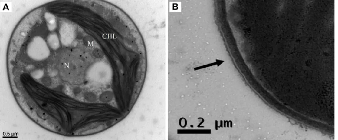
Fig. 3. (A) TEM of A. protothecoides MM0011. CHL: chloroplast, M: mitochondrion, N: nucleus. (B) Magnification of the cell wall structure. The arrow indicates the trilaminar outer layer.
Fig. 1. Light microscopy of A. protothecoides MM0011.
Fig. 2. FE-SEM of A. protothecoides MM0011.
Proximate analysis was carried out on a DTG-60A thermal analyzer (Shimadzu, Kyoto, Japan). Platinum pans were used to contain 30 mg of α-alumina (α-Al2O3) powder (Shimadzu, Kyoto, Japan) as a reference material and ap- proximately 10 mg of each sample, respectively. Nitrogen (> 99.999%, N2) was supplied as the carrier gas at a rate of 25 ml min-1 to protect the microalgae powder from oxidation. Samples were heated from 50℃ to 900℃ at a rate of 10°C min-1. Thermogravimetric analysis (TGA) data were analyzed by ta60 Ver. 2.21 software (Shimadzu, Kyoto, Japan). Protein content was calculated from the N content in the ultimate analysis by using the conversion factor (×
6.25).
Total nitrogen (TN) and total phosphorus (TP) con- sumption
TN and TP concentrations of the day 0 and day 8 R2A media were estimated using HS-TN(CA)-L and HS-TP-L wa- ter test kits (Humas, Daejeon, Korea) according to the manu- facturer’s instruction.
Results
Identification of the strain MM0011
The cells were solitary, non-motile, and round in shape with a diameter of approximately 3-4 μm (Fig. 1, Fig. 2).
The cell walls were composed of a trilaminar layer (Fig. 3B) and a prominent cup-shaped chloroplast was present (Fig.
1, Fig. 3). It was also shown that the isolate lacked a pyrenoid in the chloroplasts (Fig. 3A). Overall, the strain MM0011 showed typical morphology of the species A. protothecoides.
Molecular characterization inferred from sequence analyses of the 18S rRNA, ITS region, and rbcL gene also showed
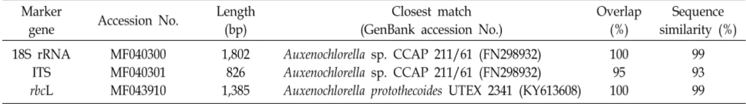
Table 2. Results from BLAST searches using the sequences of the 18S rRNA, ITS, and rbcL genes of strain MM0011 Marker
gene Accession No. Length
(bp)
Closest match (GenBank accession No.)
Overlap (%)
Sequence similarity (%) 18S rRNA
ITS rbcL
MF040300 MF040301 MF043910
1,802 826 1,385
Auxenochlorella sp. CCAP 211/61 (FN298932) Auxenochlorella sp. CCAP 211/61 (FN298932) Auxenochlorella protothecoides UTEX 2341 (KY613608)
100 95 100
99 93 99
Fig. 4. The phylogenetic relationship of strain MM0011 and its closely related species inferred from the 18S rRNA sequence data.
The tree was generated by the ML method with 1,000 bootstrap replicates. The scale bar represents a 1% difference in nucleo- tides sequences.
Table 3. Growth of strain MM0011 at various temperatures and NaCl concentrations
Temperature (°C) 5 10 15 20 25 30 35
Growth + ++ +++ +++ +++ +++ +++
NaCl 0.0 M 0.5 M 1.0 M 1.5 M 2.0 M
Growth +++ ++ + + -
+++ : good growth, ++ : moderate growth, + : poor growth, - : no growth that the isolate belonged to the A. protothecoides group; fur-
thermore, all results were in agreement (Table 2, Fig. 4).
Therefore, this marine microalga was identified as A. proto- thecoides MM0011. The isolate was deposited at the Korean Collection for Type Cultures under the accession number KCTC13291BP.
Optimal growth temperatures and NaCl tolerance of strain MM0011
A. protothecoides MM0011 could grow at temperatures ranging from 5℃ to 35℃; its maximum growth was ob- served at ambient temperatures (Table 3). Growth was sup- pressed when incubated at lower temperatures. Also, the isolate was able to withstand up to 1.5 M NaCl even though poor growth was noticed in the presence of NaCl. It did
not survive at 2.0 M NaCl.
GC/MS analysis of strain MM0011
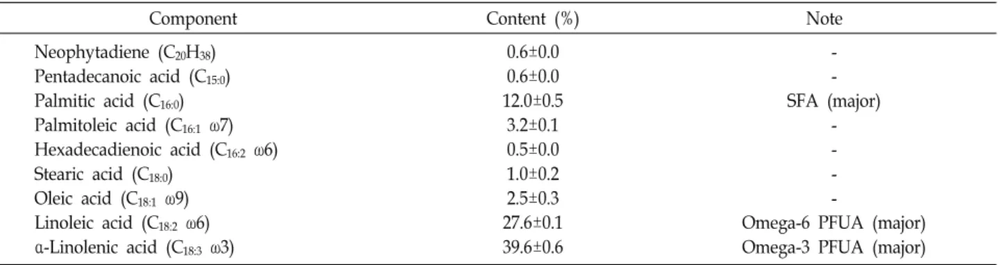
The FAME profile of A. protothecoides MM0011, on the ba- sis of the average ± standard deviation of three determi- nations, is summarized in Table 4. The major cellular fatty acids of the isolate were C16:0 (12.0%±0.5%), C18:2 ω6 (27.6%±0.1%), and C18:3 ω3 (39.6%±0.6%). In addition, trace amounts of unsaturated fatty acids such as C16:1 ω7 (3.2%±0.1%), C18:1 ω9 (2.5%±0.3%), and C16:2 ω6 (0.5%±0.0%) were detected in this photosynthetic microorganism.
HPLC analysis of strain MM0011
The pigment profile of A. protothecoides MM0011 is re- ported in Table 5. The major pigments of the isolate were
Table 4. Lipid profile of strain MM0011
Component Content (%) Note
Neophytadiene (C20H38) Pentadecanoic acid (C15:0) Palmitic acid (C16:0) Palmitoleic acid (C16:1 ω7) Hexadecadienoic acid (C16:2 ω6) Stearic acid (C18:0)
Oleic acid (C18:1 ω9) Linoleic acid (C18:2 ω6) α-Linolenic acid (C18:3 ω3)
0.6±0.0 0.6±0.0 12.0±0.5
3.2±0.1 0.5±0.0 1.0±0.2 2.5±0.3 27.6±0.1 39.6±0.6
- - SFA (major)
- - - -
Omega-6 PFUA (major) Omega-3 PFUA (major)
Table 5. Pigment profile of strain MM0011
Peak number Retention time Compound Content (%) 1
2 3 4 5 6 7
16.0 16.4 24.4 26.3 27.4 29.4 37.9
- - Lutein
- Chl b Chl a
-
7.0±0.1 4.2±0.0 31.3±0.1 1.9±0.1 14.4±0.1 38.4±0.1 2.7±0.1
Fig. 5. TGA profiles of A. protothecoides MM0011.
chlorophyll a (Chl a, 38.4%±0.1%), chlorophyll b (Chl b, 14.4%±0.1%), and lutein (31.3%±0.1%). The other minor peaks were not identified. Lutein content of strain MM0011 was 3.5±0.1 mg g-1 DW.
Biomass properties
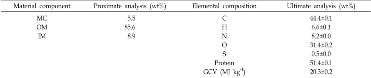
In proximate analysis by TGA, the moisture content (MC) is determined by the mass loss before 110℃ under N2 atmos- phere, the organic matter (OM) refers to the mass loss be- tween 110-900℃ under N2 as a result of thermal decom- position, and the remaining mass represents the inorganic matter (IM). The TGA profile is shown in Fig. 5 and the
material composition of strain MM0011 is presented in Table 6. The GCV and protein content based on the ultimate analy- sis were 20.3 MJ kg-1 and 51.4%, respectively (Table 6).
Total nitrogen (TN) and total phosphorus (TP) removal It was found that 40.5% of TN and 27.9% of TP from R2A were utilized by the microalga, respectively (Table 7).
Discussion
Because of its simple morphology, A. protothecoides was previously regarded as Chlorella protothecoides for a long peri- od even though it is one from the oldest algal strains de- posited in the culture collections [8]. Auxenochlorella was ranked as a subgenus of Chlorella in recognition of its heavy dependence on organic compounds such as sugars [17] and vitamins [28] and absence of a pyrenoid [14]. Later, it was ranked as a genus [12] and there are currently three tax- onomically accepted species, A. protothecoides (Krüger) Kalina & Punčochářová, A. pyrenoidosa (H.Chick) Molinari
& Calvo-Pérez and A. symbiontica Darienko & Pröschold.
Light and electron microscopic analyses suggested that the isolate shared very similar morphological characteristics with A. protothecoides, including the presence of trilamianr outer layer in the cell wall and absence of a pyrenoid in the chloroplast (Fig. 1, Fig. 3). Molecular identification re- sults also confirmed that MM0011 belonged to the species.
A. protothecoides which is known as a terrestrial species [8], but it was also isolated from wastewater [18] although its origin is uncertain. This manuscript describes the first record of the species isolated from seawater in Korea.
Analysis of the cellular fatty acid composition of the strain MM0011 revealed that it was rich in C16:0 (12.0%) saturated fatty acid (SFA) and C18:2 (27.6%) and C18:3 (39.6%) un- saturated fatty acids. Numerous studies have demonstrated
Table 6. Proximate and ultimate analysis results of A. protothecoides MM0011
Material component Proximate analysis (wt%) Elemental composition Ultimate analysis (wt%) MC
OM IM
5.5 85.6 8.9
C H N O S Protein GCV (MJ kg-1)
44.4±0.1 6.6±0.1 8.2±0.0 31.4±0.2
0.5±0.0 51.4±0.1 20.3±0.2
Table 7. TN and TP consumption by A. protothecoides MM0011 TN (mg L-1) TN (mg L-1) Day 0
Day 8 Consumption
(removal %)
135.0±3.7 80.3±2.9 54.7±6.6 (40.5%±3.8%)
72.8±0.8 52.5±0.5 20.3±0.3 (27.9%±0.2%)
that these essential PUFAs have many beneficial health ef- fects [16] and a variety of commercial products are available worldwide [19]. Omega-3 PUFAs are usually derived from fish oils and omega-6 PUFAs are primarily obtained from plant sources such as sunflower, corn, and soybean oils.
Therefore, this isolate may have the potential to be used as an alternative to fish- and/or plant-based sources. The 16- carbon saturated palmitic acid suitable for biodiesel pro- duction was also biosynthesized by strain MM0011 as one of the major fatty acids.
The OM is defined as the part of solid fuel that is driv- en-off as a gas by heating and typical biomass generally has a OM content of up to 80%(crop residue: 63-80%; wood:
72-78%). The OM content of the microalga (85.6%) used in this study was higher than the range of wood-based biomass feedstocks. The GCV was also calculated to understand the potential of algal biomass as a biofuel feedstock (Table 6).
The results showed that the GCV was also higher than the range of the terrestrial energy crops (17.0-20.0 MJ kg-1) [22].
In addition, the biomass itself may serve as an excellent ani- mal feed because of its high protein content (51.4%).
Pigment analysis revealed that the isolate was able to pro- duce a high concentration of lutein. Lutein is a naturally occurring carotenoid and it has been extensively used in the food, pharmaceutical, and cosmetics industries. It has re- cently received a significant attention due to its protective roles in the human eyes [13]. These antioxidant properties make lutein one of the fastest growing carotenoids on the market estimated to grow to $309 million by 2018 with an
annual growth rate of 3.6% [31]. Commercial sources are cur- rently extracted from marigold petals (Tagetes erecta), but the lutein content is very low (0.3 mg g-1 DW) and it is also difficult to separate from other esters [2, 23, 36]. It should be stated that the lutein content of A. protothecoides MM0011 (3.5 mg g-1 DW) is lower than those of the previously re- ported microalgal strains (4.6-5.4 mg g-1 DW) [31]. However, improved productivity could be achieved through the evalu- ation of the effects of various culture conditions including media components on stain MM0011.
In this study, we report the first record of A. protothecoides in Korea and it was added to the public culture collections.
In conclusion, this indigenous microalga could serve as po- tential biological resource to produce compounds of bio- chemical interest. The real potential of this maritime micro- alga should be evaluated through further cultivation studies at molecular, laboratory, and field scales.
Acknowledgement
This work was supported by the Securement, Analysis, and Evaluation of Marine Plant Bioresources (2017M00700), funded by the National Marine Biodiversity Institute of Korea (MABIK).
References
1. Arrigo, K. R. 2005. Marine microorganisms and global nu- trient cycles. Nature 437, 349-355.
2. Ausich, R. L. 1997. Commercial opportunities for carotenoid production by biotechnology. Pure Appl. Chem. 69, 2169- 2173.
3. Breuer, G., Evers, W. A. C., de Vree, J. H., Kleinegris, D.
M. M., Martens, D. E., Wijffels, R. H. and Lamers, P. P. 2013 Analysis of fatty acid content and composition in microalgae.
J. Vis. Exp. 80, e50628.
4. Chew, K. W., Yap, J. Y., Show, P. L., Suan, N. H., Juan, J. C., Ling, T. C., Lee, D. J. and Chang, J. S. 2017. Microalgae biorefinery: High value products perspectives. Bioresour.
Technol. 229, 53-62.
5. Ceron-Garcia, M. C., Macias-Sanchez M. D., Sanchez-Miron A., Garcia-Camacho F., Molina-Grima E. 2013. A process for biodiesel production involving the heterotrophic fermenta- tion of Chlorella protothecoides with glycerol as the carbon source. Appl. Energy 103, 341-349.
6. Cloern, J. E., Foster, S. Q. and Kleckner, A. E. 2014. Phyto- plankton primary production in the world's estuarine-coast- al ecosystems. Biogeosciences 11, 2477-2501.
7. Cuellar-Bermudez, S. P., Aguilar-Hernandez, I., Cardenas- Chavez, D. L., Ornelas-Soto, N., Romero-Ogawa, M. A. and Parra-Saldivar, R. 2015. Extraction and purification of high-value metabolites from microalgae: essential lipids, as- taxanthin and phycobiliproteins. Microb. Biotechnol. 8, 190- 209.
8. Darienko, T. and Pröschold, T. 2015. Genetic variability and taxonomic revision of the genus Auxenochlorella (Shihira et Krauss) Kalina et Puncocharova (Trebouxiophyceae, Chlor- ophyta). J. Phycol. 51, 394-400.
9. Friedl, A., Padouvas, E., Rotter, H. and Varmuza, K. 2005.
Prediction of heating values of biomass fuel from elemental composition. Anal. Chim. Acta 544, 191-198.
10. Hu, B., Min, M., Zhou, W., Li, Y., Mohr, M., Cheng, Y., Lei, H., Liu, Y., Lin, X., Chen, P. and Ruan, R. 2012.
Influence of exogenous CO2 on biomass and lipid accumu- lation of microalgae Auxenochlorella protothecoides cultivated in concentrated municipal wastewater. Appl. Biochem.
Biotechnol. 166, 1661-1673.
11. Hutchins, D. A., Mulholland, M. R. and Fu, F. 2009. Nutrient cycles and marine microbes in a CO2-enriched ocean.
Oceanography 22, 128-145.
12. Kalina, T. and Punčochářová, M. 1987. Taxonomy of the subfamily Scotiellocystoideae Fott 1976 (Chlorellaceae, Chlorophyceae). Algol. Stud. 45, 473-521.
13. Krinsky, N. I., Landrum, J. T. and Bone, R. A. 2003. Biologic mechanisms of the protective role of lutein and zeaxanthin in the eye. Annu. Rev. Nutr. 23, 171-201.
14. Krüger, W. 1894. Kurze Charakteristik einiger niederer Organismen im Saftflusse der Laubbäume. Hedwigia 33, 241- 66.
15. Leu, S. and Boussiba, S. 2014. Advances in the production of high-value products by microalgae. Ind. Biotechnol. 10, 169-183.
16. Mehta, L. R., Dworkin, R. H. and Schwid, S. R. 2009.
Polyunsaturated fatty acids and their potential therapeutic role in multiple sclerosis. Nat. Clin. Pract. Neurol. 5, 82-92.
17. Miao, X. and Wu, Q. 2006. Biodiesel production from hetero- trophic microalgal oil. Bioresour. Technol. 97, 841-846.
18. Min, M., Hu, B., Zhou, W., Li, Y., Chen, P. and Ruan, R.
2012. Mutual influence of light and CO2 on carbon seques- tration via cultivating mixotrophic alga Auxenochlorella pro- tothecoides UMN280 in an organic carbon-rich wastewater.
J. Appl. Phycol. 24, 1099-1105.
19. Packaged Facts. 2012. The Global Market for EPA/DHA Omega-3 Products. Published online at: http://www.
packagedfacts.com/Global-EPA-DHA-7145087/ (accessed on
26 July 2017).
20. Pulz, O. and Gross, W. 2004. Valuable products from bio- technology of microalgae. Appl. Microbiol. Biotechnol. 65, 635- 648.
21. Rippka, R., Deruelles, J., Waterbury, J. B., Herdman, M. and Stanier, R. 1979. Genetic assignments, strain histories and properties of pure cultures of cyanobacteria. J. Gen.
Microbiol. 111, 1-61.
22. Ross, A. B., Jones, J. M., Kubacki, M. L. and Bridgeman, T. 2008. Classification of macroalgae as fuel and its thermo- chemical behaviour. Bioresour. Technol. 99, 6494-6504.
23. Sánchez, J. F., Fernández, J. M., Acién, F. G., Rueda, A., Pérez-Parra, J. and Molina, E. 2008. Influence of culture con- ditions on the productivity and lutein content of the new strain Scenedesmus almeriensis. Process Biochem. 43, 398-405.
24. Sanz, N., García-Blanco, A., Gavalás-Olea, A., Loures, P. and Garrido, J. L. 2015. Phytoplankton pigment biomarkers:
HPLC separation using a pentafluorophenyloctadecyl silica column. Methods Ecol. Evol. 6, 1199-1209.
25. Schirmer, A., Rude, M. A., Li, X., Popova, E. and Del Cardayre, S. B. 2010. Microbial biosynthesis of alkanes.
Science 329, 559-562.
26. Shi, X. M., Liu, H. J., Zhang, X. W. and Chen, F. 1999.
Production of biomass and lutein by Chlorella protothecoides at various glucose concentrations in heterotrophic cultures.
Process Biochem. 34, 341-347.
27. Shi, X. M. and Chen, F. 2002. High-yield production of lu- tein by the green microalga Chlorella protothecoides in hetero- trophic fed-batch culture. Biotechnol. Prog. 18, 723-727.
28. Shihira, I. and Krauss, R. W. 1965. Chlorella. Physiology and taxonomy of forty-one isolates, pp. 1-97. Maryland University of Maryland, College Park.
29. Shinde, S. and Lele, S. S. 2010. Statistical media optimization for lutein production from microalgae Auxenochlorella proto- thecoides SAG 211-7A. Int. J. Adv. Biotechnol. Res. 1, 104-114.
30. Silva, P. C. (1996-to date). Index Nominum Algarum, University Herbarium, University of California, Berkeley http://ucjeps.berkeley.edu/INA.html.
31. Sun, Z., Li, T., Zhou, Z. G. and Jiang, Y. 2015. Microalgae as a source of lutein: Chemistry, biosynthesis, and caroteno- genesis. pp. 37-58. In: Posten, C. and Chen S. F. (eds.), Microalgae Biotechnology. Springer International Publishing AG: Cham, Switzerland.
32. Spolaore, P., Joannis-Cassan, C., Duran, E. and Isambert, A.
2006. Commercial applications of microalgae. J. Biosci.
Bioeng. 101, 87-96.
33. Tamura, K., Stecher, G., Peterson, D., Filipski, A. and Kumar, S. 2013. MEGA6: molecular evolutionary genetics analysis version 6.0. Mol. Biol. Evol. 30, 2725-2729.
34. Wei, D., Chen, F., Chen, G., Zhang, X., Liu, L. and Zhang, H. 2008. Enhanced production of lutein in heterotrophic Chlorella protothecoides by oxidative stress. Sci. China C. Life Sci. 51, 1088-1093.
35. White, T. J., Bruns, T., Lee, S. and Taylor, J. 1990. Amplifica- tion and direct sequencing of fungal ribosomal RNA genes for phylogenetics, pp. 315-322. In: Innis, M. A., Gelfand, D.
초록:울릉도 거북바위 조수웅덩이에서 분리된 해양 미세조류 옥세노클로렐라 프로토테코이드 균주의 기술 및 응용
장형석1․강남선2․김경미1․전병희1․박준상1․홍지원1*
(1국립해양생물자원관 분류연구실, 2국립해양생물자원관 해양생명자원관리부)
단세포 녹조류 균주를 경상북도 울릉군 울릉도 거북바위 주변 조수웅덩이로부터 순수분리하여 형태적, 분자적, 및 생화학적 특성을 분석한 결과 옥세노클로렐라 프로토테코이드에 속하는 것으로 밝혀졌다. 본 종은 현재까지 한국에서 공식 기록이 없는 미기록종으로 옥세노클로렐라 프로토테코이드 MM0011 균주라고 명명하였으며, 생 장, 지질/광합성 색소 조성 및 바이오매스 특성에 대해 조사를 실시하였다. 분리균주는 광범위한 온도(5-35°C)에 서 생장할 수 있었으며 1.5 M 염화나트륨 농도까지 생존할 수 있었다. 가스크로마토그래프/질량분석기를 이용한 분석 결과, 본 균주에는 영양적으로 중요한 불포화지방산이 풍부한 것으로 나타났으며, 특히 리놀네산(27.6%) 및 알파 리놀렌산(37.2%)이 주요 지방산 성분으로 확인되었다. 따라서 본 토착 미세조류 균주는 어유 또는 식물성유 를 대체할 수 있는 잠재적인 오메가-3 및 오메가-6 불포화지방산 원료가 될 수 있을 것으로 사료된다. 또한, 고부 가가치 항산화 물질인 루테인이 보조색소로서 본 균주에 의해 생합성 되는 것으로 밝혀졌다. 일반성분분석 결과
MM0011 균주의 휘발성물질 함량은 85.6%였으며, 원소분석 결과 총 발열량은 20.3 MJ kg-1으로 나타났다. 또한
배지로부터 40.5%의 전질소와 27.9%의 전인을 각각 제거할 수 있어 향후 바이오연료 원료물질 생산과 오·폐수 처리를 연계할 수 있는 가능성 역시 제시하였다. 추가적으로 MM0011 바이오매스는 높은 단백질 함량(51.4%)을 갖고 있어 우수한 동물사료의 원료가 될 수 있는 가능성도 보여주고 있다. 따라서, 본 균주는 미세조류 기반 생화 학 물질 생산 및 바이오매스 원료로서 상업적인 이용 가능성이 높음을 시사한다.
H., Sninsky, J. J. and White, T. J. (eds.), PCR Protocols: A Guide to Methods and Applications. Academic Press: San Diego, CA, USA.
36. Vachali, P., Bhosale, P. and Bernstein, P. S. 2012. Microbial carotenoids. pp. 41-59. In: Barredo, J. (ed.), Microbial Carotenoids from Fungi: Methods and Protocols. Springer International Publishing AG: Cham, Switzerland.
37. Verbruggen, H., Ashworth, M., LoDuca, S. T., Vlaeminck, C., Cocquyt, E., Sauvage, T., Zechman, F. W., Littler, D. S., Littler, M. M., Leliaert, F. and DeClecrk, O. 2009. A multi-lo- cus time-calibrated phylogeny of the siphonous green algae.
Mol. Phylogenet. Evol. 50, 642-653.
38. Xiao, Y., Lu, Y., Dai, J. and Wu, Q. 2015. Industrial fermenta- tion of Auxenochlorella protothecoides for production of bio- diesel and its application in vehicle diesel engines. Front.
Bioeng. Biotechnol. 3, 164
39. Xiong, W., Li, X., Xiang, J. and Wu, Q. 2008. High-density fermentation of microalga Chlorella protothecoides in bio- reactor for microbiodiesel production. Appl. Microbiol.
Biotechnol. 78, 29-36.
40. Zapata, M. and Garrido, J. L. 1991. Influence of injection conditions in reversed-phase high-performance liquid of
chromatography of chlorophylls and carotenoids. Chroma- tographia 31, 589-594.
41. Zapata, M., Rodríguez, F. and Garrido, J .L. 2000. Separation of chlorophylls and carotenoids from marine phyto- plankton: a new HPLC method using a reversed phase C8 column and pyridine-containing mobile phases. Mar. Ecol.
Prog. Ser. 195, 29-45.
42. Zhou, W., Li, Y., Min, M., Hu, B., Chen, P. and Ruan, R.
2011. Local bioprospecting for high-lipid producing micro- algal strains to be grown on concentrated municipal waste- water for biofuel production. Bioresour. Technol. 102, 6909- 6919.
43. Zhou, W., Min, M., Li, Y., Hu, B., Ma, X., Cheng, Y., Liu, Y., Chen, P. and Ruan, R. 2012. A hetero-photoautotrophic two-stage cultivation process to improve wastewater nu- trient removal and enhance algal lipid accumulation.
Bioresour. Technol. 110, 448-455.