New Four-herb Formula Ameliorates Memory Impairments via Neuroprotective Effects on Hippocampal Cells
Sung Min Ahn
1, Young Whan Choi
2, Hwa Kyoung Shin
1,3and Byung Tae Choi
1,3*
1Department of Korean Medical Science, School of Korean Medicine, Pusan National University, Yangsan 50612, Korea
2Department of Horticultural Bioscience, College of Natural Resource and Life Science, Pusan National University, Miryang 50463, Korea
3Division of Meridian and Structural Medicine, School of Korean Medicine, Pusan National University, Yangsan 50612, Korea Received November 16, 2015 /Revised January 21, 2016 /Accepted March 15, 2016
The current study was conducted to evaluate beneficial effects of a new formula (CWC-9) using four traditional Oriental medicinal herbs, Cynanchum wilfordii, Rehmannia glutinosa, Polygala tenuifolia, and Acorus gramineus, on hippocampal cells and memory function. To examine the neuroprotective effects of a new four-herb extract, cell viability, cytotoxicity, and reactive oxygen species (ROS) assays were performed in HT22 cells and behavioral tests (Morris water maze and passive avoidance retention), Western blot, and immunohistochemistry were performed in a mouse model of focal cerebral ischemia.
In HT22 hippocampal cells, pretreatment with CWC-9 resulted in significantly reduced glutamate-in- duced cell death with suppression of ROS accumulation caused by glutamate. In a mouse model of focal cerebral ischemia, we observed significant improvement of spatial and short-term memory func- tion by treatment with CWC-9. Phosphorylated p38 mitogen-activated protein kinases (MAPK) in hip- pocampus of ischemic mice were decreased by treatment with CWC-9, but phosphorylated phosphati- dylinositol-3 kinase (PI3K) and cAMP response element binding protein (CREB) were significantly enhanced. By immunohistochemical analysis, we confirmed higher expression of phosphorylation of CREB in the hippocampal neurons of CWC-9 treated mice. These results suggest that new multi-herb formula CWC-9 mainly exerted beneficial effects on cognitive function through regulation of neuro- protective signaling pathways associated with CREB.
Key words :
Focal cerebral ischemia, hippocampal neurons, memory, neuroprotection
*Corresponding author
*Tel : +82-51-510-8475, Fax : +82-51-510-8437
*E-mail : [email protected]
This is an Open-Access article distributed under the terms of the Creative Commons Attribution Non-Commercial License (http://creativecommons.org/licenses/by-nc/3.0) which permits unrestricted non-commercial use, distribution, and reproduction in any medium, provided the original work is properly cited.
Journal of Life Science 2016 Vol. 26. No. 4. 475~483 DOI : http://dx.doi.org/10.5352/JLS.2016.26.4.475
Introduction
As an aging population is occurring on a global scale, the incidence of memory and cognitive impairments includ- ing stroke is likely to show a significant increase in the near future [12]. Damage of hippocampal neurons can explain the progress of learning and memory impairment in a wide number of brain disorders [6, 16]. Effective new therapeutic strategies are needed to prevent hippocampal neuronal deaths, which can ameliorate cognitive dysfunction of many neurological disorders.
The neuroprotective function has been verified in HT22 hippocampal cells and middle cerebral artery occlusion (MCAO)-induced brain injury [10, 13]. Neuronal death is the
result of excessive oxidative stress in a variety of neuro- degenerative disorders of the central nervous system [5, 26].
Prolonged exposure to high concentrations of glutamate shows reactive oxygen species (ROS)-mediated oxidative toxicity in HT22 and cerebral ischemic models [8, 24].
Neurons contain low levels of endogenous antioxidant [5], consequently, the brain is unusually vulnerable to ROS mediated neuronal cell death and cerebral ischemic injury [18, 25]. Functional deficits associated with ischemic neuro- nal damage in MCAO models are regulated by an anti- oxidative mechanism [13]. Activation of p38 mitogen-acti- vated protein kinase (MAPK) is generally implicated in neu- ronal death, while the phosphatidylinositol-3 kinase (PI3K) pathways lead to cell survival against oxidative stress [22].
Expression of cyclic AMP response element binding protein (CREB) in different brain areas plays a role in neuronal plas- ticity, memory, and learning as well as promoting neuronal survival in the brain [14].
After systematic literature investigation of Donguibogam,
published by Joon Hur in the early 17th century in Korean,
we prepared a multi-herb extract composed of Cynanchum
wilfordii Hemsley, Rehmannia glutinosa (Gaertn) Libosch., Polygala tenuifolia Willd., and Acorus gramineus Soland. to demonstrate the synergistic effects. Approach to prevention and treatment using a multi-herb formula may be more ef- fective in age-related dementia and memory impairment [20]. A new multi-herb extract may be a potential candidate for protection of hippocampal neuronal cells with sub- sequent improvement of learning and memory impairment.
Therefore, we investigated the beneficial effects of herbal mixture extract on hippocampal neurons in HT22 cells and hippocampus of focal cerebral ischemia mice.
Material and Methods
Chemicals and antibodies
L-glutamate, 3-(4,5-dimethylthiazol-2-yl)-2,5-diphenylte- trazolium bromide (MTT), and β-actin antibody were pur- chased from Sigma-Aldrich (St. Louis, MO, USA). Dulbecco's modified Eagle's medium (DMEM), fetal bovine serum (FBS), and other cell culture reagents were purchased from Gibco-Invitrogen (Carlsbad, CA, USA). Antibodies recogniz- ing p38 and phospho-p38 (pp38, Thr180/Tyr182) were sup- plied by Santa Cruz Biotechnology (Santa Cruz, CA, USA), and PI3K, phospho-PI3K (pPI3K, Tyr458), CREB, and phos- pho-CREB (pCREB, Ser133) were supplied by Cell Signaling (Danvers, MA, USA). Antibody recognizing neuronal nuclei (NeuN) was supplied by Millipore Corporation (Billerica, MA, USA). Secondary antibodies were supplied by ENZO Life Sciences (Farmingdale, NY, USA). A lactate dehydroge- nase (LDH) cytotoxicity assay kit was purchased from Promega (Madison, WI, USA), and ROS detection reagent, 5-(and-6)-carboxy-2′,7′-dichlorodihydrofluorescein diac- etate (carboxy-H
2DCFDA), was purchased from Invitrogen (Carlsbad, CA, USA).
Preparation of four herbal mixture extract
The dried roots of Cynanchum wilfordii Hemsley, Rehman- nia glutinosa (Gaertn) Libosch., Polygala tenuifolia Willd., and Acorus gramineus Soland. were purchased from Dongnam Co. (Busan, Korea) and were authenticated by Professor Y.W. Choi, Department of Horticultural Bioscience, College of Natural Resources and Life Science, Pusan National University. A voucher specimen (accession number CWCWSD2; CWC-9) was deposited at the Plant Drug Research Laboratory of Pusan National University (Miryang, Korea). Dried powdered Cynanchum wilfordii (25.5 kg),
Rehmannia glutinosa (9.5 kg), Polygala tenuifolia (7.5 kg), and Acorus gramineus roots (7.5 kg) were immersed in 450 L of distilled water and boiled at 120±5℃ for 150 min. The re- sultant extract was centrifuged (2,000× g for 20 min at 4℃) and filtered through a 0.2-μm filter. The filtrate was then concentrated in vacuo at 70±5℃ under reduced pressure and then converted into a fine spray-dried powder at a yielding rate of 6.0% (3.0 kg) in a vacuum drying apparatus. Finally, the solid form of the spray-dried powder was dissolved with dimethyl sulfoxide (DMSO) for use as CWC-9 in experiments.
Cell culture
HT22 cells were cultured in DMEM supplemented with 10% FBS and 1% penicillin/streptomycin in a 5% CO
2hu- midified incubator at 37℃. The cells were incubated for 24 hr prior to experimental treatments. After incubation, cells were treated with various concentrations of CWC-9 for 24 hr, followed by exposure to 5 mM glutamate for 24 hr.
CWC-9 was removed from the medium during exposure to glutamate.
Determination of cell viability and cytotoxicity For MTT assay, cells were incubated with 0.5 mg/ml MTT in culture medium for 4 hr at 37℃ and absorbance was read at 595 nm using a SpectraMax 190 spectrophotometer (Molecular Devices, Sunnyvale, CA, USA). Results were ex- pressed as a percentage of controls. Release of LDH from damaged cells was performed according to the manu- facturer’s instructions for the CytoTox96 Non-Radioactive Cytotoxicity Assay Kit (Promega). Absorbance was meas- ured using a SpectraMax 190 spectrophotometer (Molecular Devices) at 490 nm. Data represent the percentage of LDH released relative to the assay positive controls.
ROS measurement
HT22 cells were cultured in 96-well white plates at a den- sity of 5×10
3cells per well. After adherence, cells were pre- treated with CWC-9 for 24 hr and then exposed to 5 mM glutamate for 24 hr. Treated cells were washed with PBS.
Carboxy-H
2DCFDA (20 μM) (Invitrogen) was applied to the cells, followed by incubation for 1 hr in a 37℃ incubator.
Fluorescence was measured using a Mutilabel counter
(Perkin Elmer 1420, MA, USA).
Focal cerebral ischemia and CWC-9 administration To confirm beneficial effects of CWC-9 on hippocampal cells, we used middle cerebral artery occlusion (MCAO) model. Male C57BL/6 mice (20-25 g) were obtained from Dooyeol Biotech (Seoul, Korea). The mice were housed at 22℃ under alternating 12 hr cycles of dark and light, and were fed a commercial diet and allowed tap water ad libitum.
All experiments were approved by the Pusan National University Animal Care and Use Committee (Approval number, PNU 2014-0517). Each group consisted of six mice and all treatments were administered under isoflurane (Choongwae, Seoul, Korea) anesthesia, which was provided using a calibrated vaporizer (Midmark VIP3000, Orchad Park, OH, USA).
Focal cerebral ischemia was induced by occluding the middle cerebral artery using the intraluminal filament technique. A fiber-optic probe was affixed to the skull over the middle cerebral artery for measurement of regional cere- bral blood flow using a PeriFlux Laser Doppler System 5000 (Perimed, Stockholm, Sweden). Middle cerebral artery occlu- sion model was induced by a silicon-coated 4-0 monofila- ment in the internal carotid artery and the monofilament was advanced to occlude the middle cerebral artery. The fila- ment was withdrawn 30 min after occlusion and reperfusion was confirmed using laser Doppler. Mice in the CWC-9 groups received oral administration daily at the doses of 100 and 500 mg/kg for three weeks after MCAO, while mice in the control and vehicle groups were only given distilled water at the same intervals.
Behavioral assessment
In experiment 1, acquisition training for the Morris water maze was performed on four consecutive days from 10 days to 7 days before MCAO (5 trials per day) and basal time was measured at 6 days before MCAO. The tank had a diam- eter of 100 cm and an altitude of 50 cm. The platform was placed 0.5 cm beneath the surface of the water. Each trial was performed for 90 s or until the mouse arrived on the platform. After final administration of CWC-9, results of the experiment were recorded using SMART 2.5.18 (Panlab S.L.U.).
In experiment 2, a passive avoidance test was performed in an apparatus containing an illuminated (light) and a dark compartment separated by an automatic guillotine door (Med-Associates, Inc., Albans, VT). During acquisition trials, once the mouse crossed into the dark compartment, it re-
ceived a 0.5 mA electric foot shock for 3 s. Approximately 24 hr later, a retention trial was administered by placing the mouse in the light compartment and recording the step- through latency, during which time no shocks were ad- ministered. Maximal testing time was 600 s. Animals who failed to enter the dark compartment within 600 s were as- signed a maximum test latency score of 600 s. Results of the experiment were recorded using MED-PC software inter- faced to chambers.
Western blot analysis
Tissue punches from each hippocampus were sonicated in ice cold lysis buffer [200 mM Tris (pH 8.0), 150 mM NaCl, 2 mM EDTA, 1 mM NaF, 1% NP40, 1 mM PMSF, 1 mM Na
3VO
4, protease inhibitor cocktail]. Equal amounts of pro- teins were separated by 10 or 12% sodium dodecyl sul- fate-polyacrylamide gel electrophoresis (SDS-PAGE) and then transferred to a nitrocellulose membrane (Whatman GmbH, Dassel, Germany). The membranes were blocked with 5% skim milk in PBST for 1 h, followed by overnight exposure to appropriate antibodies. Membranes were then incubated with appropriate horseradish peroxidase-con- jugated antibodies for 1 hr. All specific bands were vi- sualized using an enhanced chemiluminescence system (Pierce Biotech, Rockford, IL, USA) and imaged using an Image Quant LAS-4000 imaging system (GE Healthcare Life Science, Uppsala, Sweden). Results of the Western blot assay reported here are representative of at least three experiments.
Immunofluorescence staining
Mice anesthetized with isoflurane received intracardial perfusion with saline and then 4% paraformaldehyde in PBS.
Brains were removed, post-fixed in the same fixative for 4 hr at 4℃, and immersed in 30% sucrose for 48 hr at 4℃
for cryoprotection. Frozen 20 μm-thick sections were in- cubated for blocking with a blocking buffer (1X PBS/5% nor- mal serum/0.3% Triton X-100) for 1 hr at room temperature.
The sections were incubated with the following primary an-
tibodies to NeuN (Millipore Corporation), and pCREB (Cell
Signaling) for 20 hr at 4℃. After washes with PBS, the sec-
tions were incubated with the fluorescent secondary anti-
body (Vector Laboratories, Inc., Burlingame, CA, USA) for
2 hr at room temperature in the dark, respectively, and then
washed three times with PBS. Subsequently, slides were
mounted in the mounting medium (Vector Laboratories,
Inc.) and captured using a fluorescence microscope.
A B
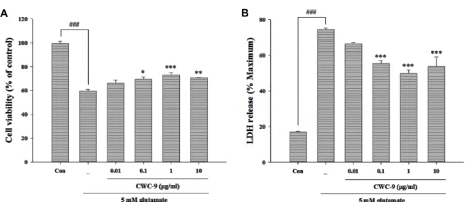
Fig. 1. Effect of CWC-9 on glutamate-induced cell death in HT22 cells. Cell viability and toxicity were determined by MTT (A) and LDH assay (B). Cells were pretreated with 0.01, 0.1, 1, and 10 μg/ml of CWC-9 for 24 hr, followed by exposure to 5 mM glutamate for 24 hr. ###p<0.001 vs. control; *p<0.05, **p<0.01 and ***p<0.001 vs. glutamate-treated cells. All data are represented as the mean±SEM of three independent experiments.
Fig. 2. Effect of CWC-9 on ROS generation in glutamate-treated HT22 cells. Cells were pretreated with 0.01, 0.1, 1, or 10 μg/ml of CWC-9 for 24 hr, followed by exposure to 5 mM glutamate for 24 hr. The oxidation sensitive fluo- rescence dye, carboxy-H2DCFDA (20 μM), was used in measurement of ROS levels. Production of ROS was an- alyzed using a fluorescence plate reader. ###p<0.001 vs.
control; **p<0.01 and ***p<0.001 vs. glutamate-treated cells. All data are represented as the mean±SEM of three independent experiments.
Data analysis
All data were expressed as mean±SEM and analyzed us- ing the SigmaStat statistical program version 11.2 (Systat Software, San Jose, CA, USA). Statistical comparisons were performed using one-way analysis of variance (ANOVA) for repeated measures followed by Tukey’s test of least sig- nificant difference. A P-value<0.05 was considered to in- dicate a statistically significant result.
Results
Pretreatment with CWC-9 protects hippocampal cells from oxidative glutamate toxicity
To assess the neuroprotective effect of CWC-9 under con- ditions of glutamate toxicity, HT22 was challenged with 5 mM glutamate with and without pretreatment of CWC-9.
Cell viability in the glutamate treated group declined by ap- proximately 40.4% compared with the control. Pretreatment of cells with CWC-9 at a concentration range of 0.1 to 10 μg/ml resulted in reduced glutamate-induced cytotoxicity (Fig. 1A). The levels of LDH release showed a significant increase to 74.5% after exposure to glutamate, while pretreat- ment with CWC-9 resulted in a marked decrease of gluta- mate-induced release of LDH (Fig. 1B).
Because oxidative stress mediates glutamate-induced cell death in HT22 cells via ROS accumulation, we examined the effect of CWC-9 on oxidative stress promoted by glutamate toxicity. Pretreatment with CWC-9 resulted in a significant
decrease in ROS production, which prevented elevation of
ROS level caused by exposure to glutamate (Fig. 2). These
results suggest that pretreatment with CWC-9 exerts a po-
tent neuroprotective effect against oxidative toxicity caused
by exposure to glutamate in HT22 hippocampal cells.
A B
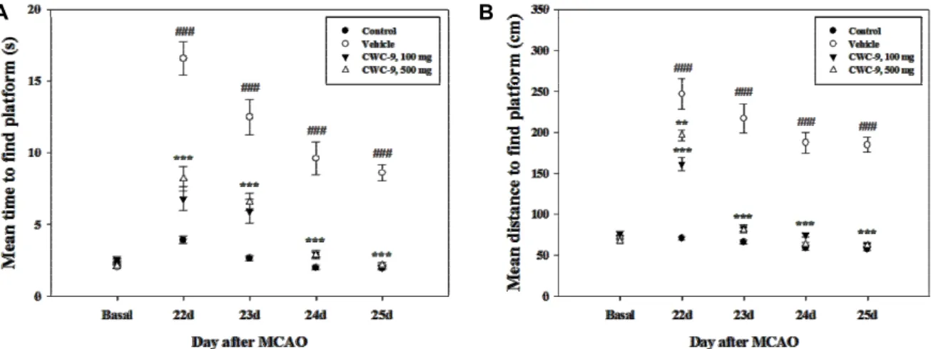
Fig. 3. Beneficial effects of CWC-9 on the spatial memory function in MCAO mice. Morris water maze test was performed from 22 d to 25 d after MCAO. Administration of CWC-9 resulted in significantly improved memory function of MCAO mice during the late phase of the experiment. Mean±SEM. ### p<0.001 vs. control; ** p<0.01 and *** p<0.001 vs. MCAO mice (vehicle).
Fig. 4. Beneficial effects of CWC-9 on the short-term memory function in MCAO mice. Retention of the passive avoid- ance task was assessed 24 days after MCAO. The re- tention test was preceded by a training session on days 22(habituation) and 23(acquisition). Data are presented as the latency to cross into a compartment in which the mice have previously received an electric foot-shock. No shock was applied during the testing phase, and the maximal testing time was 600 s. Administration of CWC-9 resulted in significantly improved short-term memory function of MCAO mice. Mean±SEM. ## p<0.01 vs. control (acquisition); ** p<0.01 vs. control (retention);
$$p<0.01 vs. MCAO mice (retention).
Treatment with CWC-9 ameliorates memory im- pairment in MCAO mice
Spatial memory was assessed using the water maze test.
MCAO mice took a longer time and distance on average to find the platform than the basal group. However, CWC-9- administered mice attained a significantly lower time and total distance at both concentrations from 22 to 25 days after MCAO compared to the vehicle group (Fig. 3). We assessed whether CWC-9 improves short-term memory in the passive avoidance task. A significant group effect was observed in the step-through latency in the retention trial. The MCAO group exhibited a decreased step-through latency time com- pared with the control group. However, treatment with CWC-9 at a dose of 100 or 500 mg/kg resulted in signifi- cantly prolonged step-through latency compared with the MCAO group (Fig. 4). During the acquisition trials, no sig- nificant inter-group differences in step-through latency were observed. These results suggest that treatment with CWC-9 may induce beneficial effects for improvement of memory function in a focal cerebral ischemia model.
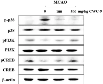
Treatment with CWC-9 enhances CREB phos- phorylation via signaling pathway of p38 MAPK and PI3K in the hippocampus of MCAO mice
Western blot analysis was performed to determine wheth- er CWC-9 could modulate glutamate-mediated cell death or survival signaling. Protein levels of p38 MAPK phosphor- ylation were significantly decreased by treatment with CWC-9 compared to the MCAO group, while the levels of phosphorylated PI3K and CREB phosphorylation were in-
creased by treatment with CWC-9 in a dose-dependent man-
ner (Fig. 5). Positive neuronal cells of pCREB/NeuN in the
hippocampal CA1 and dentate gyrus (DG) regions were
counted after double-label immunofluorescence staining at
26 days after MCAO. Treatment with CWC-9 followed by
MCAO surgery resulted in a significant increase in the num-
Fig. 5. Effect of CWC-9 on expression of intracellular kinases in the hippocampus at 26 days after MCAO. Equal amounts of proteins and each sample were subjected to Western blot analysis using the indicated antibodies.
Equal protein loading was confirmed by β-actin ex- pression.
A B
Fig. 6. Effect of CWC-9 on pCREB protein expression in the hippocampus at 26 days after MCAO. Photomicrograph (A) and its histogram for pCREB/NeuN (B) double-positive cells in the DG and CA1 regions of the hippocampus. Total number of pCREB+/NeuN+ cells was significantly increased by administration of CWC-9 in DG and CA1 compared to the MCAO group.
### p<0.001 vs. control; * p<0.05 and *** p<0.001 vs. MCAO mice. Scale bar = 100 μm.
ber of pCREB/NeuN double-positive cells in the CA1 and DG region of the ipsilateral hippocampus compared to the MCAO group (Fig. 6). These results suggest that CWC-9 may prevent hippocampal neuronal death via both p38 MAPK and PI3K signaling with enhanced phosphorylation of CREB in MCAO mice.
Discussion
Here we investigated the neuroprotective effects of a new herbal mixture extract on hippocampal neuron and memory function in HT22 hippocampal cells and MCAO mice.
Screening for selection of functional herbs on memory im- pairment was performed using HT22 cells. Among many herbal candidates, we found significant neuroprotective ef- fects of Cynanchum wilfordii, which is known to play a key role in preventing vascular disorders, ischemia-induced dis- ease, and aging progress by Joon Hur in Donguibogam [17].
Therefore, we used CWC-9 including Cynanchum wilfordii and the above mentioned multi-herb formulae to enhance medical efficiency.
Exposure of HT22 cells to glutamate evokes neuronal cell
death via non-receptor-mediated oxidative stress [19]. High
levels of ROS production caused by exposure to glutamate
lead to both apoptotic and necrotic processes [23] and the
subsequent oxidative stress can cause cellular damage and
death in cerebral ischemia and reperfusion [9]. In particular,
hippocampus, one of the brain regions most sensitive to is-
chemic injury, plays a key role in learning and memory [3,
4]. Our results indicate that cell death promoted by gluta-
mate toxicity could be ameliorated by the suppression of
ROS formation after treatment with CWC-9 and support a
neuroprotective role and therapeutic potential for a new
herbal mixture extract. In addition, the Morris water maze
and passive avoidance task were performed to assess
MCAO-induced deficits of memory. Treatment of MCAO mice with CWC-9 resulted in a significant reduction in time and distance from 22 to 25 days compared with the MCAO group by the water maze test. In the passive avoidance test, the step-through latency time was prolonged by treatment with CWC-9 compared with the MCAO group. These results suggest that CWC-9 may be a good candidate for recovery of damaged memory and cognitive function in a focal cere- bral ischemia model.
MAPK and PI3K signaling pathways in neuronal cell death or neurodegenerative diseases play an important role in neuroprotective effects against oxidative stress [1]. In par- ticular, roles of cell death (p38 MAPK) [15] and survival pathways (PI3K) [8, 21] are essential for cellular defense un- der oxidative stress conditions. Our results showed that neu- roprotective effects of CWC-9 are regulated by signaling pathways of p38 MAPK and PI3K after cerebral ischemic injury.
Critical transcription factor CREB modulates adaptive neuronal responses [2] and impaired CREB phosphorylation in hippocampus may be a pathological component in neuro- degenerative disorders [7, 27]. In our examination of the hip- pocampus using Western blot analysis, protein levels of CREB phosphorylation were recovered in CWC-9-treated MCAO mice compared with vehicle. Immunohistochemical detection of the activation of CREB resulted in significant expression in the hippocampal CA1 and DG regions after CWC-9 treatment, suggesting that enhanced CREB phos- phorylation by CWC-9 treatment plays a central role in sur- vival of hippocampal neuronal cells.
At present, the main functional components of multi-herb formula CWC-9 are not known. However, a methanolic ex- tract from dried roots of Cynanchum wilfordii significantly mitigates the neurotoxicity induced by exposure to gluta- mate that participates in the cellular defense against oxida- tive stress [17]. Neuroprotective compound, cynandione A, from Cynanchum species (C. wilfordii and C. auriculatum) can attenuate ischemic brain injury and promote functional re- covery [11, 28]. Moreover, the multi-herb formulae, includ- ing the roots of Rehmannia glutinosa, Polygala tenuifolia, and Acorus gramineus can improve the mental or physical symp- toms in the elderly [20]. CWC-9 may have synergistic memo- ry function to target primary neuroprotective effects by di- verse roles within multi-herb formulae. Our results demon- strate that CWC-9 exerts significant beneficial effects on glu- tamate-induced HT22 cell death and ischemia-reperfusion
injury model.
In conclusion, this study demonstrates that CWC-9, a new herbal extract mixture, protects hippocampal neuronal cells via suppression of ROS generation with regulation of the signaling pathways of p38 MAPK and PI3K associated with CREB phosphorylation. These effects can also improve mem- ory and cognitive function against cerebral ischemic stroke.
These results have shown that multi-herb formula CWC-9 may be a useful therapeutic agent in treatment of various brain disorders associated with changes in memory and cognition.
Acknowledgement
This work was supported by the R&D program of MOTIE/KIAT (N0000697, Establishment of Infra Structure for Anti-aging Industry Support).
References
1. Anderson, C. N. and Tolkovsky, A. M. 1999. A role for MAPK/ERK in sympathetic neuron survival: protection against a p53-dependent, JNK-independent induction of apoptosis by cytosine arabinoside. J. Neurosci. 19, 664-673.
2. Bonni, A., Brunet, A., West, A. E., Datta, S. R., Takasu, M.
A. and Greenberg, M. E. 1999. Cell survival promoted by the Ras-MAPK signaling pathway by transcription-depend- ent and -independent mechanisms. Science 286, 1358-1362.
3. Cechetti, F., Worm, P. V., Elsner, V. R., Bertoldi, K., Sanches, E., Ben, J., Siqueira, I. R. and Netto, C. A. 2012. Forced tread- mill exercise prevents oxidative stress and memory deficits following chronic cerebral hypoperfusion in the rat. Neuro- biol. Learn. Mem. 97, 90-96.
4. Collino, M., Aragno, M., Mastrocola, R., Gallicchio, M., Rosa, A. C., Dianzani, C., Danni, O., Thiemermann, C. and Fantozzi, R. 2006. Modulation of the oxidative stress and inflammatory response by PPAR-gamma agonists in the hippocampus of rats exposed to cerebral ischemia/
reperfusion. Eur. J. Pharmacol. 530, 70-80.
5. Coyle, J. T. and Puttfarcken, P. 1993. Oxidative stress, gluta- mate, and neurodegenerative disorders. Science 262, 689-695.
6. Debette, S. 2013. Vascular risk factors and cognitive dis- orders. Rev. Neurol. (Paris) 169, 757-764.
7. Dineley, K. T., Westerman, M., Bui, D., Bell, K., Ashe, K.
H. and Sweatt, J. D. 2001. Beta-amyloid activates the mi- togen-activated protein kinase cascade via hippocampal al- pha7 nicotinic acetylcholine receptors: In vitro and in vivo mechanisms related to Alzheimer's disease. J. Neurosci. 21, 4125-4133.
8. Fukui, M., Choi, H. J. and Zhu, B. T. 2010. Mechanism for the protective effect of resveratrol against oxidative stress- induced neuronal death. Free Radic. Biol. Med. 49, 800-813.
9. Ghosh, A., Sarkar, S., Mandal, A. K. and Das, N. 2013.
Neuroprotective role of nanoencapsulated quercetin in com- bating ischemia-reperfusion induced neuronal damage in young and aged rats. PLoS One 8, e57735.
10. Gim, S. A. and Koh, P. O. 2014. Ferulic acid prevents the injury-induced decrease of gamma-enolase expression in brain tissue and HT22 cells. Lab. Animal Res. 30, 8-13.
11. Hwang, B. Y., Kim, Y. H., Ro, J. S., Lee, K. S. and Lee, J.
J. 1999. Acetophenones from the roots of Cynanchum wilfor- dii H(EMSLEY). Arch. Pharm. Res. 22, 72-74.
12. Itua, I. and Naderali, E. K. 2010. Review: omega-3 and mem- ory function: to eat or not to eat. Am. J. Alzheimers Dis. Other.
Demen. 25, 479-482.
13. Jamarkattel-Pandit, N., Pandit, N. R., Kim, M. Y., Park, S.
H., Kim, K. S., Choi, H., Kim, H. and Bu, Y. 2010.
Neuroprotective effect of defatted sesame seeds extract against in vitro and in vivo ischemic neuronal damage.
Planta Med. 76, 20-26.
14. Kitagawa, K. 2007. CREB and cAMP response element- mediated gene expression in the ischemic brain. FEBS J. 274, 3210-3217.
15. Lasa, M., Abraham, S. M., Boucheron, C., Saklatvala, J. and Clark, A. R. 2002. Dexamethasone causes sustained ex- pression of mitogen-activated protein kinase (MAPK) phos- phatase 1 and phosphatase-mediated inhibition of MAPK p38. Mol. Cell. Biol. 22, 7802-7811.
16. Lee, K. Y., Jeong, E. J., Huh, J., Cho, N., Kim, T. B., Jeon, B. J., Kim, S. H., Kim, H. P. and Sung, S. H. 2012.
Cognition-enhancing and neuroprotective activities of the standardized extract of Betula platyphylla bark and its ma- jor diarylheptanoids. Phytomedicine 19, 1315-1320.
17. Lee, M. K., Yeo, H., Kim, J., Markelonis, G. J., Oh, T. H.
and Kim, Y. C. 2000. Cynandione A from Cynanchum wil- fordii protects cultured cortical neurons from toxicity in- duced by H2O2, L-glutamate, and kainate. J. Neurosci. Res.
59, 259-264.
18. Lo, E. H., Dalkara, T. and Moskowitz, M. A. 2003. Mechan- isms, challenges and opportunities in stroke. Nat. Rev.
Neurosci. 4, 399-415.
19. Maher, P. and Davis, J. B. 1996. The role of monoamine
metabolism in oxidative glutamate toxicity. J. Neurosci. 16, 6394-6401.
20. May, B. H., Lu, C., Bennett, L., Hugel, H. M. and Xue, C.
C. 2012. Evaluating the traditional Chinese literature for herbal formulae and individual herbs used for age-related dementia and memory impairment. Biogerontology 13, 299- 312.
21. Qin, R., Li, X., Li, G., Tao, L., Li, Y., Sun, J., Kang, X. and Chen, J. 2011. Protection by tetrahydroxystilbene glucoside against neurotoxicity induced by MPP+: the involvement of PI3K/Akt pathway activation. Toxicol Lett. 202, 1-7.
22. Stanciu, M., Wang, Y., Kentor, R., Burke, N., Watkins, S., Kress, G., Reynolds, I., Klann, E., Angiolieri, M. R., Johnson, J. W. and DeFranco, D. B. 2000. Persistent activation of ERK contributes to glutamate-induced oxidative toxicity in a neu- ronal cell line and primary cortical neuron cultures. J. Biol.
Chem. 275, 12200-12206.
23. Tan, S., Sagara, Y., Liu, Y., Maher, P. and Schubert, D. 1998.
The regulation of reactive oxygen species production during programmed cell death. J. Cell Biol. 141, 1423-1432.
24. Tan, S., Schubert, D. and Maher, P. 2001. Oxytosis: A novel form of programmed cell death. Curr. Top. Med. Chem. 1, 497-506.
25. Taylor, C. P., Weber, M. L., Gaughan, C. L., Lehning, E.
J. and LoPachin, R. M. 1999. Oxygen/glucose deprivation in hippocampal slices: altered intraneuronal elemental com- position predicts structural and functional damage. J.
Neurosci. 19, 619-629.
26. Xu, J., Xilouri, M., Bruban, J., Shioi, J., Shao, Z., Papazoglou, I., Vekrellis, K. and Robakis, N. K. 2011. Extracellular pro- granulin protects cortical neurons from toxic insults by acti- vating survival signaling. Neurobiol. Aging 32, 2326.
27. Yamamoto-Sasaki, M., Ozawa, H., Saito, T., Rosler, M. and Riederer, P. 1999. Impaired phosphorylation of cyclic AMP response element binding protein in the hippocampus of dementia of the Alzheimer type. Brain Res. 824, 300-303.
28. Yue, R., Yuan, X., Liu, X., Zhang, J., Jiang, P., He, C., Shan, L., Yu, Y. and Zhang, W. 2012. Cynandione A mitigates is- chemic injuries in rats with cerebral ischemia. J. Neurochem.
121, 451-464.
초록:한약재 4종 복합추출물의 해마신경세포 보호를 통한 기억력 개선
안성민
1․최영완
2․신화경
1․최병태
1*
(1부산대학교 한의과학과, 2부산대학교 원예생명과학과)