A LuxR-type Transcriptional Regulator, PsyR, Coordinates Regulation of Pathogenesis-related Genes in Pseudomonas syringae pv. tabaci
Yeon Hee Choi, Jun Seung Lee, Sora Yun and Hyung Suk Baik*
Department of Microbiology, College of Natural Science, Pusan National University, Busan 609-735, Korea Received January 20, 2015 /Revised February 4, 2015 /Accepted February 6, 2015
Pseudomonas syringae pathovar tabaci is a plant pathogenic bacterium that causes wildfire disease in tobacco plants. In P. syringae pv. tabaci, PsyI, a LuxI-type protein, acts as an AHL synthase, while pri- mary and secondary sequence analysis of PsyR has revealed that it is a homolog of the LuxR-type transcriptional regulator that responds to AHL molecules. In this study, using phenotypic and genetic analyses in P. syringae pv. tabaci, we show the effect of PsyR protein as a quorum-sensing (QS) tran- scriptional regulator. Regulatory effects of PsyR on swarming motility and production of siderophores, tabtoxin, and N-acyl homoserine lactones were examined via phenotypic assays, and confirmed by quantitative real-time reverse transcription-polymerase chain reaction (qRT-PCR). Further qRT-PCR showed that PsyR regulates expression of these virulence genes in response to environmental signals.
However, an upstream region of the gene was not bound with purified MBP-PsyR protein; rather, PsyR was only able to shift the upstream region of psyI. These results suggested that PsyR may be indirectly controlled via intermediate-regulatory systems and that auto-regulation by PsyR does not occur.
Key words :
Electrophoretic mobility shift assay (EMSA), MBP-psyR expression, Pseudomonas syringae, Quorum sensing, Real-time reverse transcription-polymerase chain reaction (qRT-PCR)
*Corresponding author
*Tel : +82-51-510-2271, Fax : +82-51-514-1778
*E-mail : [email protected]
This is an Open-Access article distributed under the terms of the Creative Commons Attribution Non-Commercial License (http://creativecommons.org/licenses/by-nc/3.0) which permits unrestricted non-commercial use, distribution, and reproduction in any medium, provided the original work is properly cited.
Journal of Life Science 2015 Vol. 25. No. 2. 136~150 DOI : http://dx.doi.org/10.5352/JLS.2015.25.2.136
Introduction
Bacteria undergo a variety of physiological and morpho- logical adaptations in reaction to physical and chemical changes that arise from the environment. Importantly, some of the most active signaling molecules are not only metabolic wastes but also diffusible molecules released from a bacterial population for cell-to-cell communication [5]. This strategy, called quorum sensing (QS), regulates the specific expression of a group of genes according to the bacterial population density. The QS system coordinates physiological reactions such as virulence, production of secondary metabolites, and antibiotic resistance, based on the local density of the bacte- rial population [12]. When QS signaling molecules reach a threshold, QS bacteria can detect a QS signaling molecule called an autoinducer, resulting in coordinated expression of specific target genes.
In gram-negative bacteria, various QS systems have been found, such as the Agrobacterium tumefaciens TraIR system, the Erwinia carotovora CarIR system, and the Pseudomonas aer- uginosa LasIR and RhlIR systems [14, 23, 43, 46, 49]. The QS systems in gram-negative bacteria are similar to the conven- tional QS circuit of Vibrio fischeri that was described in the early 1970s [29, 30]. The QS system consists of two homo- logues of V. fischeri regulatory proteins, LuxI and LuxR. The LuxI family proteins play a role as AHL synthases and pro- duce a specific AHL known as the QS signaling molecule.
The LuxR-type proteins regulate QS-related genes. Most of
the LuxR-type regulators act as transcriptional activators,
but some act as repressors or play a dual role. The amino-ter-
minal domain of LuxR acts as an AHL-binding domain,
while its carboxyl-terminal domain acts as a DNA-binding
domain. The DNA-binding domain is assumed to form a
helix-turn-helix (HTH) motif with a cluster of four helical
structures. The HTH motif binds to the major groove of
DNA in the presence of AHL [11, 35, 52]. In the absence
of AHL, the LuxR-type protein is in the monomeric, inactive
conformation. AHL can interact with the amino-terminal do-
main of LuxR-type transcriptional regulators; this complex
then dimerizes or multimerizes [9, 13]. Consequently, the
carboxyl-terminal of AHL-bound LuxR binds to a palin-
dromic promoter region, the lux box, which affects ex-
pression of QS-controlled target genes.
Pseudomonas is a noteworthy genus, because it includes a human pathogen (P. aeruginosa), a phytopathogen (P. sy- ringae), and a nonpathogenic bioremediation agent (P. puti- da). Among these strains, P. syringae is an important plant pathogen used for studying plant–bacterial interactions. P.
syringae is a gram-negative bacterium that causes disease in a wide range of plant species. P. syringae consists of more than 50 pathovars, based on host-specificities. P. syringae pathovar tabaci ATCC 11528 causes wildfire disease in tobac- co plants and a hypersensitive response (HR) in non-host plants. The AHL molecules produced in P. syringae pv. tabaci are N-(3-oxohexanoyl)-l-homoserine lactone and N-(3-oxooc- tanoyl)-l-homoserine lactone. In P. syringae species, the gene psyI has a putative lux box sequence with dyad symmetry.
In P. syringae pv. tabaci, PsyI, a LuxI-type protein, acts as an AHL synthase, while primary and secondary sequence analysis of PsyR has revealed that it is a homolog of the LuxR-type transcriptional regulator that responds to AHL molecules [28, 40].
The QS system has been studied intensively in recent years. According to a recent study, the QS system both pos- itively and negatively regulates diverse putative virulence factors. The QS system in P. syringae pv. syringae was re- ported to positively regulate production of extracellular pol- ysaccharides (EPSs), as well as tolerance to oxidative stress and maceration of plant tissue, and also to negatively regu- late swarming motility [36, 45]. Other phenotypes are de- termined by QS upon sensing of environmental cues such as nutrient deficiency or iron limitation [42, 50]. Therefore, the QS system is closely associated with other regulons.
The regulation of virulence genes by several regulators has been studied [6, 7, 24, 51]. The iron-uptake regulator Fur controls the expression of sigma factors and operon genes associated with iron uptake, acid tolerance, oxidative stress response, chemotaxis, and metabolic pathways [4, 27, 34, 44, 47, 48]. GacA, a response regulator of the two-compo- nent system, controls a variety of phenotypes, including mo- tility, biofilm formation, and production of secondary metab- olites such as antibiotics, siderophores, QS autoinducers, tox- ins, and extracellular polysaccharides, although the signals that activate the GacS/GacA two-component system have not been identified [8, 28, 37].
Another significant virulence factors are the type III effec- tor proteins that are essential for disease progress in host plants and for the HR in non-host plants. These effector pro-
teins translocate into plant cells via the type III secretion system (TTSS) [10, 16]. According to a recent study, the TTSS in plant pathogens is required for the suppression of plant defense during the first step of interaction with their hosts [19]. Several transcriptional factors that mediate the environ- mental regulation of P. syringae hrp genes have been identi- fied; however, transcription of the hrp genes is mediated by different mechanisms in different species [17, 18]. The TTSS activator HrpR is required for the expression of hrp regu- latory genes. The activation of hrp regulatory genes, includ- ing hrpA, which encodes the key hrp pili protein, is not understood. A TTSS-linked regulatory system has been elu- cidated in Ralstonia solanacearum. Specifically, it was found that PrhI (an extracytoplasmic function sigma factor), PrhR (a periplasmic domain of the inner membrane protein), and PrhA (an outer membrane receptor for unknown plant sig- nals [26]) comprise this hrp gene regulatory cascade. PrhA activates transcription of the hrp genes after recognition of tobacco plant cell-contact in P. syringae pv. tabaci ATCC 11528 [24]. PrhA transduces the signal to PrhR, which con- trols hrp genes encoding the TTSS, along with PrhI. When PrhR receives the signals, PrhI is released from PrhR into the cytoplasm. The regulatory cascade driving the ex- pression of the type III secretion determinants would there- fore start with PrhI [3, 15].
The purpose of this study was to determine the effect of PsyR protein as a transcriptional regulator of various viru- lence factors in P. syringae pv. tabaci ATCC 11528. In this study, phenotypes of the psyR deletion mutant were com- pared with those of the wild-type. We showed that PsyR has effects on pathogenesis, such as production of side- rophores, AHLs, tabtoxin, swarming motility, and pathoge- nicity to tobacco leaf.
Materials and Methods
Bacterial strains, plasmids, and culture conditions
The bacterial strains and plasmids used in this study are
shown in Tables 1 and 2, respectively. P. syringae pv. tabaci
ATCC 11528 was obtained from the American Type Culture
Collection (Manassas, VA, USA). P. syringae pv. tabaci was
cultured in King’s B (KB) medium at 28°C with aeration via
shaking [22]. Escherichia coli Top10 and DH5α were used for
cloning experiments, E. coli S17-1 λ
pirwas used as a donor
for conjugation, while E. coli BL21 (DE3) was used as host
for overexpression of psyR; these strains were grown in
Table 1. Bacterial strains used in this study
Strains Relevant characteristicsa Reference or source
Escherichia coli Top10 DH5α S17-1 λpir
BL21(DE3) BL759
BL792
Agrobacterium tumefaciens NT1
Pseudomonas syringae pv. tabaci ATCC 11528
BL37 BL779
Transformation host for cloning vector Transformation host for cloning vector
S17-1 derivative, RK2 tra regulon, host for pir-dependent plasmids Overexpression host for expression vector
E. coli BL21(DE3)/pBL318 [P. syringae pv. tabaci psyR overexpression (pMAL-c5×)]
BL759/pBL337 [P. syringae pv. tabaci psyR overexpression (pMAL-c5x) with psyI expression vector]
A. tumefaciens carrying plasmid pDCI41E33 containing a traG::lacZ fusion and traR cloned into pDSK519, spontaneous Gmr
Plant pathogen, wild-type strain ATCC 11528
Δ
psyR (678-bp deletion) BL37 containing pBL336Invitrogen, USA Takara, Japan
[6]
Novagen, USA This study
This study
[40]
ATCC, USA Lab collection
This study
aGmr, gentamycin resistance.
Table 2. Plasmids used in this study
Plasmids Relevant propertiesa Reference or source
pGEM-T easy T-blunt pDMS197 pRK415 pMAL-c5x
pBL108
pBL111 pBL122
pBL186 pBL278 pBL318 pBL336 pBL337
Cloning vector, Apr Cloning vector, Apr, Kmr Suicide vector, Tcr
Broad host range cloning vector, Tcr
Expression vector carrying an N-terminal maltose binding protein sequence and lacI, Apr
1.7-kb DNA containing 5′-flanking and 3′-flanking regions of psyR in pGEM-T easy vector, Apr
Derivative of pDMS197 containing the insert DNA of pBL108, DAP required, Kmr Derivative of pGEM-T Easy containing the 1.34-kb wild type psyR with promoter
region, Apr
Derivative of pGEM-T Easy containing the 946-bp wild type psyI, Apr 749-bp psyR-coding region in T-blunt vector, Apr, Kmr
Derivative of pMAL-c5x containing the 749-bp psyR, Apr Derivative of pRK415 containing the 1.34-kb wild-type psyR, Tcr Derivative of pRK415 containing the 946-bp wild-type psyI, Tcr
Promega, USA Solgent, Korea
[6]
[20]
NEB, UK
This study
This study This study
This study This study This study This study This study
aApr, ampicillin resistance; Kmr, kanamycin resistance; Tcr, tetracycline resistance; DAP; diaminopimelic acid.
Luria-Bertani (LB) medium at 37°C [2, 25]. The Agrobacterium tumefaciens NT1 (pDCI41E33) indicator strain was main- tained in AB minimal medium at 28°C [33].
Media were solidified using 1.5% (w/v) agar. When re- quired, antibiotics were added to media at the following con- centrations: 100 μg/ml ampicillin (Ap), 20 μg/ml kanamycin (Km), 20 μg/ml tetracycline (Tc), and 15 μg/ml gentamicin (Gm).
DNA manipulation
Plasmid DNA was prepared with a Plasmid Mini Prep kit (Solgent; Daejeon, Korea) and DNA fragments were ex-
tracted from agarose gels using a QIAquick Gel Extraction kit (Qiagen; Hilden, Germany). Transformation of E. coli or P. syringae strains was done by either RbCl-CaCl
2heat shock or electroporation (Bio-Rad; Hercules, CA, USA) at 2,500 volts, 200 ohms, and 25 μF. Transfer of the recombinant sui- cide plasmid to Pseudomonas was accomplished by con- jugation with E. coli S17-1 λ
piras the plasmid donor.
Oligonucleotide primers used in this study are described
in Table 3. Polymerase chain reaction (PCR) amplification
was performed with Taq or Pfu polymerase from Solgent,
under the following reaction conditions: denaturation at
95°C for 20 s, primer annealing according to melting temper-
Table 3. Primers used in this study
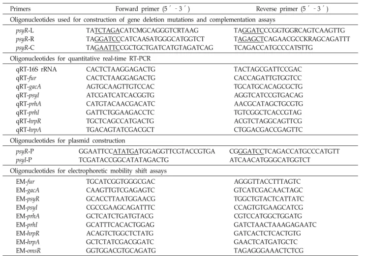
Primers Forward primer (5′–3′) Reverse primer (5′–3′)
Oligonucleotides used for construction of gene deletion mutations and complementation assays psyR-L
psyR-R psyR-C
TATCTAGACATCMGCAGGGTCRTAAG TAGGATCCCATCAASATGGGCATGGTCT TAGAATTCCGCTGCTGATCATGTAGATCAG
TAGGATCCCGGTGGRCAGTCAAGTTG TAGAGCTCAGAACGCCKRAGCAGATTT TCAGACCATGCCCATSTTG
Oligonucleotides for quantitative real-time RT-PCR qRT-16S rRNA
qRT-fur qRT-gacA qRT-psyI qRT-prhA qRT-prhI qRT-hrpR qRT-hrpA
CACTCTAAGGAGACTG CACTCTAAGGAGACTG AGTGCAAGTTGTCCAC ATCGATCATCACGGTG CATGTACAACGACATC GATTCTGGAAGACCTC TGCTCAGCCATGACTG TGACAGTATCGACGCT
TACTAGCGATTCCGAC CACCAGATTGTGGTCC TGCATGCACAGCGCTG AGGTCATCCGTGACAG AACGCATAGCTGCGTG TGTCGGCTCACCGTAG ACGTCTAGGCAGTTCG CTGGACGACCGAGTTC Oligonucleotides for plasmid construction
psyR-P psyI-P
GGAATTCCATATGATGGAGGTTCGTACCGTGA TCGATACCGGCATATAGACTG
CGGGATCCTCAGACCATGCCCATGTT ATCAACATGGGCATGGTCT
Oligonucleotides for electrophoretic mobility shift assays EM-fur
EM-gacA EM-psyR EM-psyI EM-prhA EM-prhI EM-hrpR EM-hrpA EM-omsR
TGCATCGGTGGGCGAC CAAGTTGTCGAGAGTC GCACCTTAATGGAACG CGCCGAAGCAGATTTC GCTCATCTGATGTACG GCATTTCACACTGGAG ACAGTCTGGCTCTATG GCTCTATCGACGGATC GGTGGACGTGCAGATG
AGGGTTACCTTTAGTC GTCATCGACAACTAGC TGGCTGTACTCATTATC CCAGTGTGAAGCATCG CGTCCATGGCTGGATG GATCTAACTAAAGAGAATC GATCACTCTCACTGTG GAACTCATGATGCTC TAGAGGGAAACTCTCG
ature for 40 s, polymerization at 72°C for 1-3 min depending on the length of DNA fragment; these steps were repeated for 25 cycles, and were followed by a final extension at 72°C for 5 min.
Construction of the psyR mutant strain and com- plementation strain
The psyR deletion mutant of P. syringae pv. tabaci ATCC 11528 was constructed by the allelic-exchange method [38].
The 5′-flanking region of psyR was amplified by PCR using psyR-L forward and reverse primers designed from the P.
syringae pv. tabaci sequence (GenBank Accession No.
AF110468). In addition, the 3′-flanking region of psyR was amplified using psyR-R forward and reverse primers.
Amplified DNA fragments were cloned into the pGEM-T easy vector, yielding pBL108. A 1.7-kb DNA fragment from pBL108 was digested with XbaI and SacI and cloned into the suicide vector pDMS197, creating pBL111. Then, the re- combinant suicide plasmid was introduced into E. coli S17-1 λ
pircarrying the plasmid as the plasmid donor, via
electroporation. Cloning was verified by analyses of the DNA fragment profile after restriction enzyme digestion.
pBL111 was introduced into P. syringae pv. tabaci strain by mating. Colonies carrying the chromosomal insertion of the recombinant suicide plasmid were selected on plates supple- mented with kanamycin. Loss of the suicide plasmid after the second recombination between two homologous regions was selected using the sacB-based sucrose sensitivity counter selection system. Finally, the psyR deletion mutant strain was confirmed via PCR amplification using psyR-L forward and psyR-R reverse primers.
To generate a construct for complementation of the psyR deletion, the DNA fragments containing the intact psyR gene were amplified from the chromosomal DNA using the psyR-C primer set (Table 3). The purified PCR product was cloned into the pGEM-T easy vector, resulting in pBL122.
Then, the 1.34-kb DNA fragment from pBL122 was digested
with EcoRI and cloned into the broad-host-range cloning
vector pRK415, creating pBL336. pBL336 was transferred in-
to P. syringae pv. tabaci strain from E. coli S17-1 λ
pirby means
of conjugation [20]. Finally, the complement strain, BL779, was constructed using the above procedures.
Phenotypic tests and plant pathogenicity assays We performed chrome azurol S (CAS) universal side- rophore detection assays, as described by Schwyn and Neiland [39]. Swarming motility was determined using KB agar (0.4% agar) plates [7]. Tabtoxins were detected using standard bioassays with A. tumefaciens NT1 as the indicator bacteria. To detect AHLs production in a liquid medium, AHLs extracts were prepared as described by Shaw et al.
[41]. The concentrated extracts were characterized by thin-layer chromatography (TLC) using A. tumefacience NT1 as the AHL-sensor strain. Plant pathogenicity assays were performed using tobacco plants (Nicotiana tabacum L. cv.
Samsun) as described previously [24]. Each treatment was repeated three times. All assays were repeated in three sepa- rate experiments.
RNA isolation and quantitative real-time reverse transcription (qRT)-PCR analysis
Cells were grown to the early (OD
6000.5) and late (OD
6003.5) exponential phases. Total RNA isolation and qRT-PCR were conducted as described previously [6]. Briefly, total RNA was extracted from bacteria using an RNAspin Mini RNA Isolation kit (GE Healthcare; Pittsburgh, PA, USA) ac- cording to the manufacturer’s instructions. The total RNA concentration was quantified using a Nanodrop 2000 spec- trometer (Thermo Fisher Scientific; Waltham, MA, USA).
cDNAs were synthesized using a Reverse Transcription Premix kit (ElpisBiotech; Daejeon, Korea) according to the manufacturer’s protocols, using target gene-specific primers, and were stored at -20°C until required for use.
Real-time RT-PCR was carried out using SYBR Primix Ex Taq
TM(TaKaRa; Osaka, Japan) with a StepOne Real-Time PCR System (Applied Biosystems; Foster City, CA, USA), as previously described [6]. Specific primer sets used to am- plify the genes of interest were designed based on sequences in the GenBank database. The PCR conditions were as fol- lows: 94°C for 5 min, followed by 40 cycles each consisting of 10 s at 94°C, 10 s at 55°C, and 15 s at 72°C, followed by a 7-min incubation at 72°C. To normalize the fold-in- duction of mRNA, the expression level of 16S rRNA (endogenous control) was quantified and then normalized to the threshold value acquired for the wild-type strain [1].
The relative expression ratios were calculated using a mathe-
matical model, which contained an efficiency correction for the real-time RT-PCR efficiency of each transcript [32]. The data are shown as the average of triplicate samples.
Overproduction and purification of the PsyR protein The psyR-coding region was amplified from the genomic DNA of P. syringae pv. tabaci by PCR, using the psyR-P for- ward and reverse primers. The 749-bp DNA fragment was cloned into the T-blunt vector, creating pBL278; this was then digested with NdeI and BamHI and the digested frag- ment was cloned into the pMAL-c5x vector, resulting in pBL318. To support the production of soluble PsyR protein, a co-expression vector carrying the AHL-encoding psyI was constructed. The psyI fragment, including its promoter re- gion, was amplified by PCR using psyI-P forward and re- verse primers. Amplified DNA fragments were cloned into the pGEM-T Easy vector, resulting in pBL186. A 946-bp DNA fragment from pBL186 was digested with EcoRI and cloned into the broad host range cloning vector pRK415, cre- ating pBL337. The integrity of the psyR and psyI sequences was confirmed by DNA sequencing.
For PsyR overexpression, pBL318 expressing the C-termi- nally maltose-binding protein (MBP)-tagged PsyR proteins, was introduced into E. coli BL21 (DE3) by electroporation, yielding strain BL759. To enhance the production of soluble PsyR protein, pBL337 expressing AHL synthase from psyI was transformed into the BL759 strain. In this way, the PsyR overproduction strain, harboring the pBL318 and pBL337 plasmids, was constructed. The PsyR overproduction strain, BL792, was cultured overnight in 5 ml LB broth containing tetracycline (20 μg/ml), ampicillin (50 μg/ml), and 0.2%
glucose. The overnight culture was diluted 100-fold with 50 ml LB medium containing ampicillin (50 μg/ml) and in- cubated at 37°C. At the exponential phase (OD
6000.4-0.6), the cell cultures were induced with 0.1 mM IPTG and in- cubated at 15°C for 12 hr. After incubation, cells were har- vested by centrifugation and the cell pellet was suspended in 5 ml column buffer (20 mM Tris-HCl, 200 mM NaCl, 1 mM EDTA, pH 7.4). The suspended cells were lysed with 1 mg/ml lysozyme and using an Ultrasonic generator US-300 (Nissei; Osaka, Japan). Cell debris was removed by centrifugation at 20,000×g for 20 min at 4°C. The 1.5×10-cm column, filled with amylose resin (New England Biolabs;
Ipswich, MA, USA), was washed with five column volumes
of column buffer and the cleared cell lysate was loaded. The
column was washed with 12 column volumes of column buf-
fer and subsequently eluted with five column volumes of elution buffer (20 mM Tris-HCl (pH 7.4), 200 mM NaCl, 1 mM EDTA, 10 mM maltose). The protein-containing frac- tions were concentrated to about 1 mg/ml in an Amicon Ultra-15 (Millipore; Billerica, MA, USA).
Electrophoretic mobility shift assay
To investigate the interaction of PsyR protein with the virulence gene upstream region, an electrophoretic mobility shift assay (EMSA) was performed using an EMSA kit (Invitrogen; Waltham, MA, USA) [19]. DNA fragments were generated by PCR amplification of the regulatory regions of omsf, psyR, psyI, gacA, fur, prhA, prhI, hrpR, and hrpA using the primer pairs listed in Table 3. As a positive control for the DNA fragment, we used the promoter region of psyI, encoding N-acyl homoserine lactone synthase, whose ex- pression is known to be regulated by PsyR. The EMSA re- action mixture (final volume of 10 μl) contained 0.16 pM DNA, 0~2.4 nM protein, and 5× binding buffer (50 mM Tris-HCl [pH 7.4], 750 mM KCl, 0.5 mM dithiothreitol, 0.5 mM EDTA), and was incubated at room temperature for 20 min. After the addition of 2 μl of 6× EMSA gel-loading sol- ution, the reaction mixtures were analyzed by electro- phoresis in 0.5× TBE buffer (1.1 M Tris-HCl [pH 8.3], 9 M boric acid, 25 mM EDTA). All reactions were loaded onto native 5% polyacrylamide gels (1:30 bis-acrylamide to acryl- amide ratio) at 110 V (15 V/cm) for 55 min at 4°C. The gel was stained in SYBR Green EMSA staining solution at room temperature for 20 min, and then washed twice with sterile distilled water to remove excess stain. The stained DNA gel was visualized using a Lumi-imager F1 (Roche; Basel, Switzerland).
Results
Phenotypes of the psyR deletion mutant
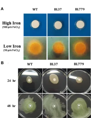
CAS agar plates were used to detect production of side- rophores in P. syringae pv. tabaci ATCC 11528. As shown in Fig. 1A, both wild-type (WT) and ΔpsyR mutant (BL37) strains did not produce siderophores under high-iron con- dition, but produced siderophores under low-iron condition.
In the ΔpsyR mutant strain, the orange halo was slightly smaller than in the WT strain. These results suggest that the production of siderophores may be co-regulated by PsyR in P. syringae pv. tabaci ATCC 11528.
Swarming motility is the rapid and coordinated move-
ment of a bacterial population across a semi-solid medium, which is driven by flagella. In comparison with the WT strain, the ΔpsyR mutant strain demonstrated enhanced swarming motility (Fig. 1B). These results indicated that PsyR represses the swarming motility of P. syringae pv. tabaci ATCC 11528, and functions as a negative regulator of this activity.
To determine whether the ΔpsyR mutation affects tabtoxin synthesis, tabtoxin production was measured using the A.
tumefaciens NT1 growth inhibition assay. The produced tab- toxin formed zones of inhibition in the case of the WT strain of P. syringae pv. tabaci ATCC 11528 (Fig. 1C). The zones of inhibition of the ΔpsyR mutant strain were slightly greater than that of its parent strain. Therefore, PsyR may be a neg- ative regulator of tabtoxin synthesis in P. syringae pv. tabaci ATCC 11528.
The A. tumefaciens NT1 indicator strain carrying a lacZ- traG fused plasmid was used for the detection of AHL. Upon TLC analysis, two spots were observed on the TLC plates with a culture of the A. tumefaciens NT1 strain (Fig. 1D). The top spot represented N-(3-oxohexanoyl)-l-homoserine lac- tone and the bottom spot N-(3-oxooxtanoyl)-l-homoserine lactone. In the ΔpsyR mutant strain, production of AHL was considerably reduced as compared to the wild-type strain.
These results indicated that synthesis of AHL in P. syringae pv. tabaci ATCC 11528 is regulated by PsyR.
The WT and ΔpsyR mutant strains were observed at 1, 3, 5, and 7 days after inoculation onto tobacco leaves (Fig.
2A). Disease symptoms, such as brown necrotic lesions sur- rounded by chlorosis, were on the tobacco leaves inoculated with the WT strain. However, inoculation with the ΔpsyR mutant strain resulted in a slight delay in disease symptom development. Consequently, symptom development of wild-fire disease appears to be regulated by PsyR.
The wild-type and ΔpsyR mutant strains were grown in LB medium. As shown in Fig. 2B, the ΔpsyR mutant strain grew slightly faster than did the WT strain.
Regulation of gene expression by PsyR
To choose regulatory factors related with the regulation
of virulence-associated gene expression, qRT-PCR was per-
formed using total RNA from the ΔpsyR mutant and WT
strains, which were incubated until the early and late ex-
ponential phases. The relative levels of mRNA were de-
termined from the threshold values that were normalized
to 16S rRNA gene expression. The genes chosen for this anal-
C
D
Fig. 1. (A) Siderophore assay with wild-type (WT),
Δ
psyR mutant (BL37), andΔ
psyR mutant harboring pBL336 (BL779) strain of Pseudomonas syringae pv. tabaci ATCC 11528. P. syringae pv. tabaci strains were grown overnight and diluted to 2×10⁸ CFU/ml in sterile water. Aliquots of 20 μl were spotted on chrome azure S (CAS) agar plates containing high or low iron concentrations.The inoculated plates were incubated for 48 hr at 30°C. (B) Comparison of swarming motility in a wild-type (WT) strain of P. syringae pv. tabaci,
Δ
psyR mutant (BL37), and aΔ
psyR mutant-carrying pBL336 (BL779) strain. Overnight cultures of bacteria were inoculated onto 6-mm sterile filter discs in KB medium containing 0.4% agar. The plates were incubated at 30°C and observed at 24 hr and 48 hr post-inoculation. (C) Detection of tabtoxin-productive capacity with WT,Δ
psyR mutant (BL37), andΔ
psyR mutant-harboring pBL336 (BL779) strain of P. syringae pv. tabaci ATCC 11528. Overnight cultures of bacteria were inoculated onto 6-mm sterile filter discs in KB agar plates containing Agrobacterium tumefaciens NT1 indicator bacteria.The plates were incubated at 30°C and the zones of inhibition were observed at 48 hr. (D) Thin layer chromatography analysis of N-acetyl homoserine lactone (AHL) production. A. tumefaciens NT1 was used for the detection of AHL produced in P. syringae pv. tabaci ATCC 11528 (WT);
Δ
psyR mutant (BL37) and theΔ
psyR mutant-harboring pBL336 (BL779) strain.A, N-(3-oxohexanoyl)-l-homoserine lactone; B, N-(3-oxooxtanoyl)-l-homoserine lactone.
A
B
Time (hr)
Fig. 2. (A) Test for disease symptoms on tobacco leaves. Disease symptoms generated by the wild-type (WT),
Δ
psyR mutant- (BL37), andΔ
psyR mutant-harboring pBL336 (BL779) strain of Pseudomonas syringae pv. tabaci ATCC 11528. The tobacco leaves were photographed 1, 3, 5, and 7 days post-inoculation. 1, WT; 2, BL37. (B) Growth characteristics of the WT, BL37, and BL779 P. syringae pv. tabaci ATCC 11528 strains.A
B
24 hr
48 hr
A B
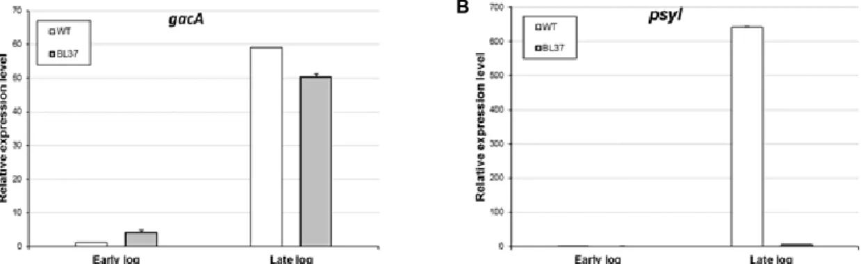
Fig. 3. The relative mRNA expression levels of gacA and psyI in wild-type (WT) and the
Δ
psyR mutant Pseudomonas syringae pv.tabaci ATCC 11528 strains. The relative mRNA levels of each gene were measured by quantitative real-time RT-PCR and normalized to 16S rRNA gene expression as the endogenous control. Bacterial cells were incubated until the early (OD600
of 0.5) (A) and late (OD600 of 3.5) exponential phases (B). The data are expressed as the average of three replicates ± the standard deviation.
A B
C
Fig. 4. Relative mRNA expression levels of fur in wild-type (WT) and ΔpsyR mutant Pseudomonas syringae pv. tabaci ATCC 11528 strains (A). Relative fur mRNA levels were measured by quantitative real-time RT-PCR and normalized to 16S rRNA gene expression as an endogenous control. Bacterial cells were cultured until early (OD600 of 0.5) and late (OD600
of 3.5) exponential phases under low- (B) or high-iron con- ditions (C). The data are expressed as the average of three replicates ± the standard deviation.
ysis were those encoding a global regulator (gacA), a QS sig- nal synthase (psyI), a ferric uptake regulator (fur), a TTSS regulator, structural components (hrpR, hrpA), and a TTSS- linked regulatory system (prhI, prhA).
The WT and ΔpsyR mutant strains showed a higher tran- script level of gacA at the late-exponential phase in compar- ison to levels at the early exponential phase (Fig. 3A). At the early exponential phase, the mRNA levels of gacA in the ΔpsyR mutant strain were increased approximately 3.9-fold. At the late exponential phase, the expression levels of gacA in the ΔpsyR mutant strain was decreased approx- imately 0.8-fold compared to that in the WT strain. These
results showed that expression of gacA was not significantly different between WT and ΔpsyR mutant strains.
The expression level of psyI in ΔpsyR mutant strain was decreased approximately 160-fold at the late-exponential phase (Fig. 3B). Therefore, expression of psyI at a high cell-density is positively regulated by PsyR.
As shown in Fig. 4A, in the wild-type strain, the fur
mRNA levels at the late exponential phase was higher than
that at the early exponential phase, indicating that bacterial
cell density affects the mRNA expression of fur. At the early
exponential phase, the transcript levels of fur were slightly
higher in the ΔpsyR mutant strain than in the WT strain.
A B
C D
Fig. 5. Relative mRNA expression levels of prhA, prhI, hrpR, and hrpA in wild-type (WT), and ΔpsyR mutant in Pseudomonas syringae pv. tabaci ATCC 11528 strains. (A) prhA, (B) prhI, (C) hrpR, (D) hrpA. Other details are as described in the legend to Fig. 4.
Furthermore, the fur mRNA levels of the ΔpsyR mutant strain at the late exponential phase was slightly higher than that of the WT strain.
In addition, the fur mRNA levels were also assayed in P. syringae pv. tabaci strains grown under high- or low-iron conditions (Fig. 4B and 4C). Under low-iron conditions, the mRNA levels of fur in the ΔpsyR mutant strain at the early exponential phase were higher than that in the WT strain.
However, these mRNA expression levels were reversed at the late exponential phase. A lower amount of fur mRNA was observed in the ΔpsyR mutant strain grown under high-iron conditions in comparison with the WT strain, but this was again reversed at the late exponential phase.
In order to investigate the correlation between TTSS-asso- ciated regulatory factors and the QS regulator PsyR, qRT- PCR was conducted as described previously [6]. P. syringae pv. tabaci PrhI, corresponding to R. solanacearum PrhI, ex- hibited sequence similarity to other sigma factors.
The expression levels of prhA in the WT strain at the early exponential phase were greater than that at the late ex- ponential phase (Fig. 5A). In comparison with the WT strain, the prhA mRNA levels in ΔpsyR mutant strain were in- creased approximately 0.8-fold at the early exponential phase; slightly greater levels of prhA mRNA were also ob- served at the late exponential phase. These results showed
that the mRNA expression levels of prhA are not sig- nificantly different between WT and ΔpsyR mutant strains.
For prhI, the mRNA levels in the WT strain were increased at the late exponential phase (Fig. 5B). As compared with the WT strain, the ΔpsyR mutant strain exhibited higher lev- els of this transcript at the early exponential phase. The mRNA levels of prhI in the ΔpsyR mutant strain were de- creased at the late exponential phase. Therefore, expression of prhI is regulated optimally by PsyR in a cell den- sity-dependent manner.
Along with PrhA and PrhI, TTSS regulatory factors influ- encing expression of HrpR (an activator of TTSS) and HrpA (a major protein of TTSS pili) were investigated in the WT and ΔpsyR mutant strains (Fig. 5C). In the WT strain, the mRNA levels of hrpR decreased at the late exponential phase. In general, lower hrpR mRNA levels were observed in the ΔpsyR mutant strain as compared with the WT strain.
However, the mRNA expression levels of hrpR were not sig- nificantly different between WT and ΔpsyR mutant strains.
The WT strain showed a higher hrpA mRNA levels at the
late exponential phase (Fig. 5D). In the ΔpsyR mutant strain,
the mRNA levels of hrpA at the late exponential phase was
greater than that of the WT strain. These observations sug-
gested that PsyR may repress hrpA expression at a high cell
density. Therefore, hrpA expression at the late exponential
Fig. 6. Alignment of the upstream regions of virulence-related genes in Pseudomonas syringae pv. tabaci ATCC 11528 with lux box sequences; alignments were created using CLUSTAL X. Arrows above the sequences indicate dyad areas.
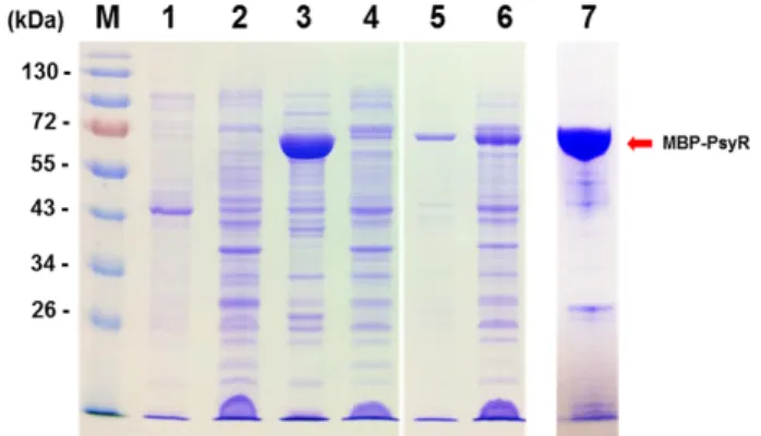
Fig. 7. SDS-PAGE of the PsyR-tagged maltose-binding protein (MBP-PsyR) of Pseudomonas syringae pv. tabaci ATCC 11528. Lane M, prestained protein ladder (Fermentas);
lane 1, insoluble proteins in cell lysates (BL792) without IPTG; lane 2, soluble proteins in cell lysates (BL792) without IPTG; lane 3, insoluble proteins in cell lysates (BL759) with 0.1 mM IPTG; lane 4, soluble proteins in cell lysates (BL759) with 0.1 mM IPTG; lane 5, insoluble proteins in cell lysates (BL792) with 0.1 mM IPTG; lane 6, soluble proteins in cell lysates (BL792) with 0.1 mM IPTG; lane 7, MBP-PsyR purified from BL792.
phase was negatively regulated by PsyR.
Putative promoter region related to virulence genes of P. syringae pv. tabaci ATCC 11528
A specific palindromic sequence, called a lux box, has been reported in the promoter regions of many QS-regulated genes [31]. In this study, we searched for a putative lux box sequence in the promoter regions of eight genes of interest (Fig. 6). Interestingly, this regulatory box lies between the -35 and -10 position, and overlaps the putative -10 sequence.
Thus, all these genes have a putative lux box with homolo- gous dyad symmetry.
DNA-binding activity of PsyR
PsyR was purified from BL792 containing the plasmids pBL318 and pBL337, as described in the Materials and Methods. Protein samples from each phase of purification, as well as the eluted protein fractions, were analyzed by SDS-PAGE (Fig. 7). The molecular weight of the purified MBP-PsyR was determined as being about 70 kDa, which matched the predicted molecular weights of this protein.
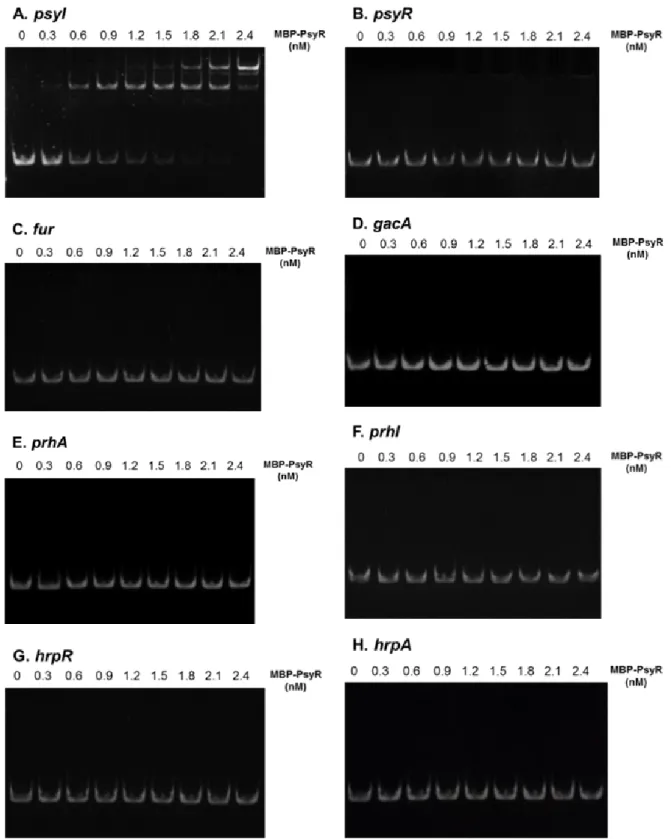
The ability of P. syringae pv. tabaci 11528 PsyR to bind the lux box in vitro was investigated using EMSA (Fig. 8).
The purified MBP-PsyR caused a band mobility shift of the PCR fragment on a 5% polyacrylamide gel. The DNA frag- ment was amplified from the upstream region of viru- lence-related genes in assays using as little as 160 fM of the purified DNA fragment. PsyR derived from the BL792 har- boring pBL318 and pBL337 was not able to shift DNA frag-
ments containing the lux box of any of the genes, except for the upstream region of psyI. These results confirmed that PsyR binds specifically to the lux box region in psyI, but does not directly regulate virulence-related genes.
Discussion
Pseudomonas syringae pv. tabaci is a plant pathogenic bacte- rium that causes wildfire disease in tobacco plants. Studies on the genetic basis of the pathogenesis of P. syringae patho- vars has been reported, and have revealed the role of TTSS-related genes, global regulatory genes, and motility and virulence-associated factors such as various phytotoxins, EPSs, and siderophores [3]. P. syringae pv. tabaci exhibits QS that is based on the production of diffusible signal mole- cules; however, the role of QS in this organism has not yet been clearly defined. In this study, the effect of PsyR protein as a QS transcriptional regulator was demonstrated using phenotypic and genetic analyses in P. syringae pv. tabaci ATCC 11528. We investigated the regulation of virulence-re- lated genes by PsyR and observed differences in the patterns of regulation of psyI, fur, prhI, and hrpA in the ΔpsyR mutant strain as compared to that in the WT strain.
We also investigated the regulatory effects of PsyR on
swarming motility and production of siderophores, tabtoxin,
and AHLs, as well as symptoms of wildfire disease in tobac-
Fig. 8. Electrophoretic mobility shift assays (EMSA) of binding of MBP-PsyR to the putative promoter-containing regions of target genes: (A) psyI, (B) psyR, (C) fur, (D) gacA, (E) prhA, (F) prhI, (G) hrpR, (H) hrpA. All lanes contained 160 fM of DNA and the indicated concentration of MBP-PsyR. The DNA–protein reaction mixtures were loaded onto 5% polyacrylamide gels.
co leaves by comparing the mutant and WT strains. Previous studies of tabtoxin reported that tabtoxin causes chlorosis around necrotic lesions and assists the development of
lesions. Our results demonstrated that PsyR coordinated the
production of tabtoxin. In addition, swarming motility was
increased in the ΔpsyR mutant strain, indicating that PsyR
was involved in the regulation of swarming motility. The ΔpsyR mutant strain caused a delay in the development of disease symptoms compared to the WT strain. However, PsyR did not affect the growth rate of P. syringae pv. tabaci 11528. Thus, the phenotypic characteristics of the ΔpsyR mu- tant may not be due to differences in growth rates.
Furthermore, the ΔpsyR mutant strain produced less side- rophores than the WT strain under low-iron conditions and did not produce siderophores under high-iron conditions.
This was confirmed by the results from real-time RT-PCR.
Under low-iron conditions, the fur mRNA level in the ΔpsyR mutant strain at the early exponential phase was higher than that in the WT strain, suggesting that the expression of fur repressed production of siderophores and may be positively regulated by PsyR. Moreover, the fur mRNA level in the ΔpsyR mutant strain was lower under high-iron conditions than in the WT strain. This may suggest that expression of fur in the presence of high iron concentrations is controlled by PsyR, using another mechanism.
Production of AHL was regulated by PsyR in response to cell density; this finding was supported by the real-time RT-PCR results. The psyI mRNA levels in the ΔpsyR mutant strain were significantly reduced at the late exponential phase. These results suggested that expression of psyI at a high density of bacteria is positively controlled by PsyR.
Thus, psyI expression is tightly controlled by PsyR in re- sponse to bacterial cell density. There was no marked differ- ence in the gacA mRNA levels between WT and ΔpsyR mu- tant strains. Previous studies have reported that GacA pos- itively controls QS signaling and PsyR [6, 8, 31]. More specif- ically, GacA regulates expression of psyR, according to a re- cent study, but PsyR does not regulate expression of gacA.
Expression levels of prhI in the ΔpsyR mutant strain were higher at the late exponential phase. In addition, the mRNA levels of hrpA at the late exponential phase were greater than those in the WT strain. These results showed that prhI and hrpA are expressed via regulation by PsyR in response to bacterial cell density; thus, QS influences expression of TTSS genes. These results suggest that PsyR has a dual role as an activator or repressor.
To determine whether PsyR binds upstream of patho- genesis-related genes in order to regulate their expression, EMSA was performed. A PsyR-overproduction strain was constructed specifically for use in EMSA in this study. A recent study reported that cognate AHLs are necessary for protein stability and solubility during the purification of sev-
eral LuxR-type regulators [21]. psyI, encoding an AHL syn- thase, was co-expressed in the PsyR-overproduction strain to enhance the production of soluble protein. Soluble PsyR proteins were successfully overproduced when the over- production strain harbored both psyI and psyR. The identi- fication of cis-regulatory sequences and the binding of PsyR to the upstream region of each virulence gene provided evi- dence of their involvement in the regulation of these genes.
No binding of a purified MBP-PsyR protein to the upstream region of these genes, except psyI, was detected. Surprisingly, PsyR did not bind to the upstream region of psyR. These results suggested that PsyR may be indirectly controlled via intermediate-regulatory systems and that auto-regulation by PsyR does not occur.
In conclusion, this study shows that PsyR controls pro- duction of the effectors by modulating transcript levels.
These findings indicate that PsyR-mediated regulation plays important roles in infection of plants by P. syringae pv. tabaci.
Integration of the regulatory network including PsyR is re- quired in order to obtain a better understanding of the path- ogenetic mechanism common to all P. syringae pathovars and to identify the major components of each regulatory system. Elucidating pathogenetic mechanisms in phytopath- ogens will offer valuable insight into diseases caused by plant pathogens and will lay the basis for studies into anti- microbial therapies, as well as in the medical, food, and agri- culture industries.
Acknowledgement
This work was supported by a 2-year Research Grant of Pusan National University.
References
1. Allen, S. S. and McMurray, D. N. 2003. Coordinate cytokine gene expression in vivo following induction of tuberculous pleurisy in guinea pigs. Infect. Immun. 71, 4271-4277.
2. Bertani, G. 1951. A method for detection of mutations, using streptomycin dependence in Escherichia coli. Genetics 36, 598- 611.
3. Brito, B., Aldon, D., Barberis, P., Boucher, C. and Genin, S. 2002. A signal transfer system through three compart- ments transduces the plant cell contact-dependent signal controlling Ralstonia solanacearum hrp genes. Mol. Plant Microbe Interact. 15, 109-119.
4. Calderwood, S. B. and Mekalanos, J. J. 1987. Iron regulation of Shiga-like toxin expression in Escherichia coli is mediated by the fur locus. J. Bacteriol. 169, 4759-4764.
5. Camilli, A. and Bassler, B. L. 2006. Bacterial small-molecule signaling pathways. Science 311, 1113-1116.
6. Cha, J. Y., Lee, D. G., Lee, J. S., Oh, J. I. and Baik, H. S.
2012. GacA directly regulates expression of several virulence genes in Pseudomonas syringae pv. tabaci 11528. Biochem.
Biophys. Res. Commun. 417, 665-672.
7. Cha, J. Y., Lee, J. S., Oh, J. I., Choi, J. W. and Baik, H. S.
2008. Functional analysis of the role of Fur in the virulence of Pseudomonas syringae pv. tabaci 11528: Fur controls ex- pression of genes involved in quorum-sensing. Biochem.
Biophys. Res. Commun. 366, 281-287.
8. Chatterjee, A., Cui, Y., Yang, H., Collmer, A., Alfano, J. R.
and Chatterjee, A. K. 2003. GacA, the response regulator of a two-component system, acts as a master regulator in Pseudomonas syringae pv. tomato DC3000 by controlling regu- latory RNA, transcriptional activators, and alternate sigma factors. Mol. Plant Microbe Interact. 16, 1106-1117.
9. Choi, S. H. and Greenberg, E. P. 1992. Genetic dissection of DNA binding and luminescence gene activation by the Vibrio fischeri LuxR protein. J. Bacteriol. 174, 4064-4069.
10. Deng, W. L., Rehm, A. H., Charkowski, A. O., Rojas, C. M.
and Collmer, A. 2003. Pseudomonas syringae exchangeable ef- fector loci: sequence diversity in representative pathovars and virulence function in P. syringae pv. syringae B728a. J.
Bacteriol. 185, 2592-2602.
11. Ducros, V. M., Lewis, R. J., Verma, C. S., Dodson, E. J., Leonard, G., Turkenburg, J. P., Murshudov, G. N., Wilkin- son, A. J. and Brannigan, J. A. 2001. Crystal structure of GerE, the ultimate transcriptional regulator of spore for- mation in Bacillus subtilis. J. Mol. Biol. 306, 759-771.
12. Dulla, G., Marco, M., Quinones, B. and Lindow, S. 2005.
A Closer Look at Pseudomonas syringae as a Leaf Colonist - The pathogen P. syringae thrives on healthy plants by em- ploying quorum sensing, virulence factors, and other traits.
Asm News 71, 469-475.
13. Fuqua, C. and Greenberg, E. P. 2002. Listening in on bac- teria: Acyl-homoserine lactone signalling. Nat. Rev. Mol. Cell.
Biol. 3, 685-695.
14. Fuqua, C., Parsek, M. R. and Greenberg, E. P. 2001. Regula- tion of gene expression by cell-to-cell communication: ac- yl-homoserine lactone quorum sensing. Annu. Rev. Genet.
35, 439-468.
15. Genin, S., Brito, B., Denny, T. P. and Boucher, C. 2005.
Control of the Ralstonia solanacearum Type III secretion sys- tem (Hrp) genes by the global virulence regulator PhcA.
FEBS Lett. 579, 2077-2081.
16. Hauck, P., Thilmony, R. and He, S. Y. 2003. A Pseudomonas syringae type III effector suppresses cell wall-based ex- tracellular defense in susceptible Arabidopsis plants. Proc.
Natl. Acad. Sci. USA 100, 8577-8582.
17. Hendrickson, E. L., Guevera, P., Penaloza-Vazquez, A., Shao, J., Bender, C. and Ausubel, F. M. 2000. Virulence of the phytopathogen Pseudomonas syringae pv. maculicola is rpoN dependent. J. Bacteriol. 182, 3498-3507.
18. Jin, Q., Thilmony, R., Zwiesler-Vollick, J. and He, S. Y. 2003.
Type III protein secretion in Pseudomonas syringae. Microbes
Infect. 5, 301-310.
19. Jing, D., Agnew, J., Patton, W. F., Hendrickson, J. and Beechem, J. M. 2003. A sensitive two-color electrophoretic mobility shift assay for detecting both nucleic acids and pro- tein in gels. Proteomics 3, 1172-1180.
20. Keen, N. T., Tamaki, S., Kobayashi, D. and Trollinger, D.
1988. Improved Broad-Host-Range Plasmids for DNA Cloning in Gram-Negative Bacteria. Gene 70, 191-197.
21. Kim, J. and Park, W. 2013. Identification and character- ization of genes regulated by AqsR, a LuxR-type regulator in Acinetobacter oleivorans DR1. Appl. Microbiol. Biotechnol. 97, 6967-6978.
22. King, E. O., Ward, M. K. and Raney, D. E. 1954. Two simple media for the demonstration of pyocyanin and fluorescin.
J. Lab. Clin. Med. 44, 301-307.
23. Lazdunski, A. M., Ventre, I. and Sturgis, J. N. 2004.
Regulatory circuits and communication in Gram-negative bacteria. Nat. Rev. Microbiol. 2, 581-592.
24. Lee, J. S., Cha, J. Y. and Baik, H. S. 2011. Plant cell contact- dependent virulence regulation of hrp genes in Pseudomonas syringae pv. tabaci 11528. J. Life Sci. 21, 227-234.
25. Lennox, E. S. 1955. Transduction of linked genetic characters of the host by bacteriophage P1. Virology 1, 190-206.
26. Marenda, M., Brito, B., Callard, D., Genin, S., Barberis, P., Boucher, C. and Arlat, M. 1998. PrhA controls a novel regu- latory pathway required for the specific induction of Ralstonia solanacearum hrp genes in the presence of plant cells. Mol. Microbiol. 27, 437-453.
27. Mey, A. R., Wyckoff, E. E., Kanukurthy, V., Fisher, C. R.
and Payne, S. M. 2005. Iron and fur regulation in Vibrio chol- erae and the role of fur in virulence. Infect. Immun. 73, 8167-8178.
28. Miller, M. B. and Bassler, B. L. 2001. Quorum sensing in bacteria. Annu. Rev. Microbiol. 55, 165-199.
29. Nealson, K. H. and Hastings, J. W. 1979. Bacterial bio- luminescence: its control and ecological significance. Micro- biol. Rev. 43, 496-518.
30. Nealson, K. H., Platt, T. and Hastings, J. W. 1970. Cellular control of the synthesis and activity of the bacterial lumines- cent system. J. Bacteriol. 104, 313-322.
31. Parsek, M. R. and Greenberg, E. P. 2000. Acyl-homoserine lactone quorum sensing in gram-negative bacteria: a signal- ing mechanism involved in associations with higher organisms. Proc. Natl. Acad. Sci. USA 97, 8789-8793.
32. Pfaffl, M. W. 2001. A new mathematical model for relative quantification in real-time RT-PCR. Nucleic Acids Res. 29, e45.
33. Piper, K. R., Beck von Bodman, S. and Farrand, S. K. 1993.
Conjugation factor of Agrobacterium tumefaciens regulates Ti plasmid transfer by autoinduction. Nature 362, 448-450.
34. Prince, R. W., Storey, D. G., Vasil, A. I. and Vasil, M. L.
1991. Regulation of toxA and regA by the Escherichia coli fur gene and identification of a Fur homologue in Pseudomonas aeruginosa PA103 and PA01. Mol. Microbiol. 5, 2823-2831.
35. Pristovsek, P., Sengupta, K., Lohr, F., Schafer, B., von Trebra, M. W., Ruterjans, H. and Bernhard, F. 2003. Structural analy-
sis of the DNA-binding domain of the Erwinia amylovora RcsB protein and its interaction with the RcsAB box. J. Biol.
Chem. 278, 17752-17759.
36. Quinones, B., Dulla, G. and Lindow, S. E. 2005. Quorum sensing regulates exopolysaccharide production, motility, and virulence in Pseudomonas syringae. Mol. Plant Microbe Interact. 18, 682-693.
37. Schwartz, T., Walter, S., Marten, S. M., Kirschhofer, F., Nusser, M. and Obst, U. 2007. Use of quantitative real-time RT-PCR to analyse the expression of some quorum-sensing regulated genes in Pseudomonas aeruginosa. Anal. Bioanal.
Chem. 387, 513-521.
38. Schweizer, H. P. 1992. Allelic exchange in Pseudomonas aeru- ginosa using novel ColE1-type vectors and a family of cas- settes containing a portable oriT and the counter-selectable Bacillus subtilis sacB marker. Mol. Microbiol. 6, 1195-1204.
39. Schwyn, B. and Neilands, J. B. 1987. Universal chemical- assay for the detection and determination of siderophores.
Anal. Biochem. 160, 47-56.
40. Shaw, P. D., Ping, G., Daly, S. L., Cha, C., Cronan, J. E., Jr., Rinehart, K. L. and Farrand, S. K. 1997. Detecting and characterizing N-acyl-homoserine lactone signal molecules by thin-layer chromatography. Proc. Natl. Acad. Sci. USA 94, 6036-6041.
41. Shaw, P. D., Ping, G., Daly, S. L., Cha, C., Cronan, J. E., Rinehart, K. L. and Farrand, S. K. 1997. Detecting and char- acterizing N-acyl-homoserine lactone signal molecules by thin-layer chromatography. Proc. Natl. Acad. Sci. USA 94, 6036-6041.
42. Sircili, M. P., Walters, M., Trabulsi, L. R. and Sperandio, V. 2004. Modulation of enteropathogenic Escherichia coli vir- ulence by quorum sensing. Infect. Immun. 72, 2329-2337.
43. Smith, R. S. and Iglewski, B. H. 2003. Pseudomonas aeruginosa quorum sensing as a potential antimicrobial target. J. Clin.
Invest. 112, 1460-1465.
44. Thompson, D. K., Beliaev, A. S., Giometti, C. S., Tollaksen, S. L., Khare, T., Lies, D. P., Nealson, K. H., Lim, H., Yates,
J., 3rd, Brandt, C. C., Tiedje, J. M. and Zhou, J. 2002.
Transcriptional and proteomic analysis of a ferric uptake regulator (fur) mutant of Shewanella oneidensis: possible in- volvement of fur in energy metabolism, transcriptional regu- lation, and oxidative stress. Appl. Environ. Microbiol. 68, 881- 892.
45. Ulrich, R. L., Deshazer, D., Brueggemann, E. E., Hines, H.
B., Oyston, P. C. and Jeddeloh, J. A. 2004. Role of quorum sensing in the pathogenicity of Burkholderia pseudomallei. J.
Med. Microbiol. 53, 1053-1064.
46. Von Bodman, S. B., Bauer, W. D. and Coplin, D. L. 2003.
Quorum sensing in plant-pathogenic bacteria. Annu. Rev.
Phytopathol. 41, 455-482.
47. Watnick, P. I., Eto, T., Takahashi, H. and Calderwood, S.
B. 1997. Purification of Vibrio cholerae fur and estimation of its intracellular abundance by antibody sandwich en- zyme-linked immunosorbent assay. J. Bacteriol. 179, 243-247.
48. Wertheimer, A. M., Tolmasky, M. E., Actis, L. A. and Crosa, J. H. 1994. Structural and functional analyses of mutant Fur proteins with impaired regulatory function. J. Bacteriol. 176, 5116-5122.
49. Whitehead, N. A., Barnard, A. M., Slater, H., Simpson, N.
J. and Salmond, G. P. 2001. Quorum-sensing in Gram-neg- ative bacteria. FEMS Microbiol. Rev. 25, 365-404.
50. Withers, H., Swift, S. and Williams, P. 2001. Quorum sens- ing as an integral component of gene regulatory networks in Gram-negative bacteria. Curr. Opin. Microbiol. 4, 186-193.
51. Yang, H. J., Lee, J. S., Cha, J. Y. and Baik, H. S. 2011.
Negative regulation of pathogenesis in Pseudomonas syringae pv. tabaci 11528 by ATP-dependent Lon protease. Mol. Cells 32, 317-323.
52. Zhang, R. G., Pappas, K. M., Brace, J. L., Miller, P. C., Oulmassov, T., Molyneaux, J. M., Anderson, J. C., Bashkin, J. K., Winans, S. C. and Joachimiak, A. 2002. Structure of a bacterial quorum-sensing transcription factor complexed with pheromone and DNA. Nature 417, 971-974.
초록: Pseudomonas syringae pv. tabaci 에서 LuxR-type 전사조절자인 PsyR에 의한 병원성 유전자들의 조절
최연희․이준승․윤소라․백형석*
(부산대학교 미생물학과)