척수손상 흰쥐에서 자하거 약침과 침전기 자극이 신경성장인자 발현에 미치는 영향
김은정*․김계엽
동신대학교 물리치료학과
Effects of Hominis Placenta Pharmacopuncture and Electroacupuncture Neuroprotection in Contused Spinal Cord of Rats
Eun-Jung Kim*, Gye-Yeop Kim
Department of Physical Therapy, Dongshin University
This study was designed to investigate the effects of
Hominis placentapharmacopuncture treatment and electroacupuncture therapy on the functional recovery and histological change, protein expression in spinal cord injury(SCI) rats. Experimental groups were divided into the Group Ⅰ(normal control rat), Group Ⅱ(Non-treatment after spinal cord injury induction), Group Ⅲ(Hominis placenta pharmacopuncture treatment after SCI induction), Group Ⅳ (Electroacupuncture therapy after SCI induction), Group Ⅴ(
Hominis placentapharmacopuncture treatment and electroacupuncture therapy after SCI induction). After operation, rats were tested at modified Tarlov test at 1 to 3 days with divided into 4 groups, and motor behavior test(BBB locomotor rating scale, Grid walk test) was examined at 3, 7, 14, and 21 days. For the observation of damage change and size of the organized surface in muscle and spinal cord, histopathological studies were performed at 21 days by H & E stain, and BDNF & NT-3 protein expression studies were performed at 21 days. Acco rding to the results,
Hominis placentapharmacopuncture treatment and electroacupuncture therapy can play a role in facilitating recovery of locomotion following spinal cord injury. Specially,
Hominis placentapharmacopuncture treatment and electroacupuncture combimed therapy after SCI induction was most improvement in functional recovery, BDNF, and NT-3 protein synthesis.
Key words : spinal cord injury,
Hominis placentapharmacopuncture, electroacupuncture, locomotor behavioral test
* 교신저자 : 김은정, 나주시 대호동 252, 동신대학교 보건복지대학
․E-mail : [email protected], ․Tel : 061-330-3398
․접수 : 2011/01/07 ․수정 : 2011/03/14 ․채택 : 2011/04/04
서 론
척수(spinal cord)는 매우 높은 대사율을 가지며, 풍부한 혈 액 흐름을 가지고 있으나 혈류 공급의 변화는 치명적 결과를 가 져온다 1,2) . 척수손상(spinal cord injury)은 종종 영구적인 운동 기 능 소실과, 감각과 자율신경계통의 기능 저하를 일으키나 척수 손상으로 인한 기능 소실이 모두 손상 당시에 바로 발생하는 것 은 아니다 3) . 물리적 충격으로 인한 척수 내 신경과 혈관 조직의 국소적 파괴는 세포, 생화학, 면역, 조직학적 변화를 가져오며, 1 차적 척수 손상을 일으키고, 이러한 충격은 점진파(progressive wave)를 형성하여 인접조직으로 확산된다. 1차 손상으로 생긴
혈종이 인접 조직의 허혈을 유도하여 신경변성을 일으켜 2차적 척수손상에 기여한다 4,5) .
손상으로 유도된 혈액 공급의 방해로 인한 척수 조직 내 지
속적인 저산소증(hypoxia)과 허혈(ischemia)이 2차적 조직손상을
유도하는데 중심적 역할을 하는 것으로 보고되었다 6,7) . 이러한 혈
관사고 외 염증반응과 지질과산화물 유도에 의한 산소자유기 형
성, Ca 2+ 이온 유입, 세포, 분자적, 생화학 반응의 연속에 의해 2
차 손상이 관련된다 8) . 1차적 조직 손상과 점진적인 낭종
(progressive cyst) 형성은 척수 조직 내에서 수일에서 수주에 걸
친 세포의 자가고사(apoptosis)와 지속적인 축삭의 퇴행으로 조
직 손상부위가 더욱 커지게 된다 5,9) . 현재까지 손상조직의
glutamate의 증가가 NMDA(N-methyl-D-aspartate) 수용체에 결
합하여 칼슘의 세포 내 유입을 야기하며, 세포 내 칼슘증가는 세
포 내 calpain과 endonuclease 등과 같은 효소들을 활성화 하여
세포 손상을 유발한다 10) .
그러나 분자적 및 세포학적 척수 자가고사 기전은 아직 완 전히 밝혀지지 않고 있으며, 지금까지 수십 년간 의학적 치료 노 력에도 불구하고, 1차적 손상으로 인한 손상된 척수 조직을 정상 으로 되돌리기가 아직까지 많이 어렵다 11,12) . 외상성 척수 손상 환 자의 신경조직 보존 및 운동 기능 증진을 위한 최선의 방법은 2 차 척수 손상의 진행을 억제는 것으로 보여진다 13) . 최근 몇 년간 의 연구에 의하면 항자가고사 제제(antiapoptotic drugs), 자유라 디칼 제거제(free radical scavengers), 항염증약(anti-inflam- matory agents) 등의 치료적 중재에 2차 손상 치료 효과가 나타 나는 것으로 보여지고 있다 14-16) .
紫河車( Hominis placenta )는 중국에서 수 백년 전부터 혈액을 조절하는데 사용하였으며, 신장을 따뜻하게 하는 성질이 있다 17) . 이는 태반을 건조한 것으로 성질은 溫, 無毒하고, 맛은 甘鹹하며 肺, 肝, 腎經에 들어가 補氣, 養血하여 오래된 병으로 인한 신체 허약과 氣血不足및 腎虛精虧등 證을 치료하는 효능이 있어 神經 衰弱, 貧血, 氣管支喘息등 만성병에 사용되어왔다 18,19) . 또한 류마 티스 및 퇴행성 관절염 등의 만성 관절염과 만성 근골격계 질환 의 통증 조절에 효과적인 것으로 보고되었다 20-22) . 자하거는 뇌, 신장, 기타 생명에 필수적 기관(vital essence organ)의 치료에 사 용되는 약재이다. 전침(electroacupuncture)은 穴에 刺針한 후 저 주파 전류를 통전하여 전류자극과 함께 혈자리를 자극하는 치료 방법으로 전류량의 조절을 통해 만성 염증성 통증 23,24) 과 신경병 증성 통증을 조절하는데 효과적으로 알려져 있으나 25) , 척수 손상 에서의 전침자극에 대한 항자가고사 및 신경보호효과는 아직 연 구되어지지 않았다.
따라서 본 연구에서는 한방재활의학 분야에서 척수 손상 치 료에 사용하는 자하거 약침과 전침이 기능학적 회복 및 척수 조 직의 신경학적 보호에 어떠한 영향을 발휘하는지 기능행동학적 평가 및 조직 및 분자적 연구 수준에서의 결과가 매우 미진한 실 정이기에, 외상성 척수 손상 흰쥐에서 자하거와 전침 치료에 대 한 효과에 대해 임상적 치료근거를 제시하고자 한다.
재료 및 방법
1. 실험동물
생후 10주령의 체중 250 ± 20 g 내․외의 Sprague-Dawley 계 흰쥐(웅성, 중앙실험동물) 50마리를 대상으로 하여 각 군당 10 마리씩 총 5개 군으로 분류하여 사용하였다. 사육실의 온도는 25
± 3℃, 습도 55 ± 10%를 유지 하였으며, 명암은 12시간 주기로 조절하여 1주일간 실험실 내 환경에 적응한 후 실험을 하였다.
사료와 물은 제한 없이 사육하였다. 실험동물은 아무런 처치도하 지 않은 정상대조군 Ⅰ군, 척수손상 유발 후 아무런 처치를 하지 않은 실험대조군 Ⅱ, 척수 손상 유발 후 자하거 약침을 처치한
Ⅲ군, 척수 손상 유발 후 전기침 자극을 실시한 Ⅳ군, 척수 손상 유발 후 자하거와 전기침 자극을 실시한 Ⅴ군으로 구분하여 3주 간 주 5회 처치를 하였다. 신경학적 운동기능 회복평가는 척수 유발 후 3일, 7일, 14일, 21일에 측정하였다.
Table 1. Classification of experimental groups
Group Treatment condition
Ⅰ(n=10) Normal control rat
Ⅱ(n=10) Non-treatment after spinal cord injury(SCI) induction
Ⅲ(n=10) Hominis placenta pharmacopuncture treatment after SCI induction
Ⅳ(n=10) Electroacupuncture therapy after SCI induction
Ⅴ(n=10) Hominis placenta pharmacopuncture treatment and electroacupuncture therapy after SCI induction
2. 척수손상 모델 제작
실험동물을 흰쥐를 마취 챔버(Royal medical, Korea)에 넣 고, 마취가스(N2O 70%, O₂28.5%, enflurane 1.5%)로 흡입마취 후 수술대에 고정시키고, 마취가 지속 되도록 하였다. 흉추 제 11 번의 레벨에서 후궁절제를 한 후 실험동물을 spinal cord impactor(NYU, U.S.A)에 올려놓고, 고정 장치를 이용하여 극돌 기를 고정하였다. 척수 위 25 ㎜ 지점에서 impactor 막대(지름 2
㎜, 무게 10 g)의 착지 지점을 확인 한 후 1회 떨어 뜨려 척수 손 상을 유도하였다. 수술 후 감염을 방지하기 위하여 겐타마이신 (100 ㎎/㎏)을 우측 대퇴부위에 2일간 주사하였다 26) .
3. 紫河車 약침액 조제 및 처치
본 실험에 사용된 자하거 약침(Hominis placenta herbal -acupuncture)은 대한 약침학회의 기준에 따라 제조 사용하였다.
약침액은 2 ℓ의 증류수에 100 g의 紫河車를 둥근 플라스크에 함 께 넣어 수증기 증류법으로 1.6 ℓ의 증류액을 만든 후, 냉각, 여 과 과정을 거쳐 여과액을 감압하여 100 ㎖(pH 7.0)가 되도록 농 축한 후 4℃에 보관하여 사용하였다.
4. 전침 자극
전침자극은 전침자극기(Pulsed stimulator AM3000, Tokyo Electronic Co., Japan)를 이용하였으며, 주파수 25 ㎐, 2 V, 간헐 파형을 사용하였다. 마취된 흰쥐의 근수축이 육안으로 확인되는 정도의 세기를 적용하여 매일 1회 20분간 주 5회, 3주간 실시하 였다. 전침은 척수손상을 유발시킨 후 동물에서의 取穴 원칙을 참조하여 인체의 足三里(ST36), 陽陵泉(GB34), 懸鐘(GB39)에 상 응하는 부위에 시술하였다. 침은 1회용 stainless steel 멸균호침 동방침구제작소(0.25×40 ㎜)를 사용하였다. 삭모 후 약 0.5 ㎜ 깊 이로 자침한 후 電針 기기의 음극은 足三里(ST36)과 陽陵泉 (GB34)에 양극은 懸鐘(GB39) 부위에 각각 연결하였고, 대조군은 전경골근(tibialis anterior) 근복 부위에 각각 연결하였다.
5. 신경학적 운동행동 검사(Neurological motor behavioral test) 1) 수정된 Tarlov 검사(Modified tarlov test)
척수손상의 정도를 평가하기 위하여 Tarlov과 Klinger 척도
를 수정한 Tarlov test를 사용하여, 통증자극 반응과 운동 반응의
정도를 세분화하여 운동행동을 평가하였다 26) . 각 항목의 평가 항
목은 자발적인 움직임, 체중지지, 평편한 바닥에서 보행 기능, 좁
은 막대에서 걷기의 항목을 0~10점으로 분류하여 신경학적 운
동행동 정도를 평가하였다. 본 실험에서는 수정된 Tarlov test에
서 척수 손상 유발 후 1, 2, 3일에 검사하였으며, 손상 유도 초기 에 점수가 0~3점 범위에 해당되는 흰쥐만을 분류하여 실험에 사용하였다.
2) BBB 척도(Basso, Beattle, Bresnahan locomotor rating scale) BBB 척도는 척수손상 흰쥐의 뒷다리 기능을 정확하게 평가 하기 위한 척도로서, 열린 평면의 공간 위를 보행하게 한 후 보 행하는 흰쥐의 뒷다리의 운동행동을 관찰하는 것이 목적이다 27) . 본 연구에서 BBB 척도를 수정한 BBB locomotor rating 척도를 사용하여 척수손상 흰쥐의 뒷다리와 꼬리의 조절 정도를 관찰하 여 점수화하였다. 척도의 측정은 실험의 내용을 알지 못하는 관 찰자 2명의 의견이 일치했을 때 점수화하였다.
3) 격자보행 검사(Grid walk test)
격자보행 검사는 후지의 정확한 보행 기능 조절능력을 알아 보기 위하여 검사를 실시하였다. 지면에서 30 ㎝ 높이에 설치된 1 m 길이의 막대 위에서 움직임을 보는 것으로 막대에서 10 발 자국 중 발이 막대 아래로 빠지는 횟수를 측정하였다 28) .
6. 조직학적 관찰
척수의 형태적 변화를 관찰하기 위하여 육안으로 보이는 조 직표면의 손상 및 크기의 변화 등을 관찰하였고, 조직 포매기를 사용하여 탈수와 청명의 과정을 거쳐 파라핀 블록을 제작하였다.
제작된 파라핀 블록(paraffin block)은 미세절단기(Sakura 2040, Japan)을 이용하여 4 ㎛ 두께로 절단한 후에 H & E 염색을 실시 하여 슬라이드를 제작하였다. 광학현미경(Olympus Bx 50, Japan)을 이용하여 현미경에 장착된 CCD 카메라(Toshiba, Japan)로 촬영한 후 조직의 변성정도, 크기 변화, 공동화 변화 양 상, 척수 결합조직의 상태를 관찰하였다.
7. Western blot을 통한 단백질 함량
실험 종료 후 척수를 채취하여 4℃ PBS로 2회 수세하고 수 확한 후, 3배 부피의 4℃ 용해 버퍼(300 mM NaCl, 50 mM Tris –Cl(pH 7.6)), 0.5% Triton X-100, 2 mM phenylmethylsulfonyl fluroride, 2 ㎕/㎖ aprotinin, 2 ㎕/㎖ leupeptin에 넣은 후, 4℃에서 30분 동안 반응시켰다. 이후 4℃, 14,000 rpm에서 15분 동안 원심분리 하여 상층액을 추출하였다. 용해된 세포의 단백질 농도는 상층액에서 Bradford(Bio-Rad protein assay) 방법 29) 을 이 용하여 단백질 정량화를 하였고, 추출된 단백질은 동일한 양을 7.5~12% sodium dodecyl sulfate(SDS) polyacrylamide gel의 각 lane에 분주 한 후 전기 영동하였다. 이후, 겔을 polyvinylidene difluoride membrane (Amersham, 0.2 ㎛ pore size, USA)에 blotting 하였다. Membrane은 TBS(PBS containing 0.01% (v/v) Tween 20)에 5%(w/v) nonfat dried milk로 4℃에서 1시간 반응 하고, 다시 TBS로 수세한 후, BDNF(brain-derived neutrophic factor, Santa cruz, sc-546, USA)와 NT-3(Santa cruz, sc-547, USA) 항체를 4℃에서 24 시간 반응시킨 후, enhanced chemical luminescence kit (RPN 2106, Amersham Life Science, Inc., USA)를 이용하여, 방사선 필름에 감광시켜서 밴드(band)를 확인 하였다.
8. 통계방법
본 연구에서 얻은 결과는 SPSS 12.0 for windows를 사용하 여 실험군별 평균과 표준편차를 산출하였다. 시간에 따른 실험군 간의 통계적 유의성 검정을 위하여 일원배치 분산분석(one-way ANOVA)을 실시하였으며, 사후검정은 Duncan의 다중범위검사 를 실시하였다. 통계학적 유의수준 α<0.05로 하였다.
결 과
1. 수정된 Tarlov 검사(Modifid tarlov test)
척수손상 유발 후 흰쥐의 실험 적합성 여부를 평가하기 위 하여 정상군인 Ⅰ군을 제외한 Ⅱ, Ⅲ, Ⅳ, Ⅴ군을 척수손상 후 1, 2, 3일째에 수정된 Tarlov 평가를 통해 실험군의 점수를 측정하 여 실험군 간의 유의성을 살펴보았다. 손상 후 3일째 Ⅱ군이 1.34
± 0.24점, Ⅲ군이 1.34 ± 0.24점, Ⅳ군이 1.34 ± 0.24점, Ⅴ군이 1.36 ± 0.21점으로 각 군 간의 유의한 차이를 보이지 않아 실험군 의 배치에 영향을 주지 않는 것으로 나타났다(p>0.05)(Table 2).
Table 2. The change of modified tarlov test in each groups Group
Day Ⅱ Ⅲ Ⅳ Ⅴ
1 days 1.29±0.20 1.26±0.21 1.28±0.19 1.28±0.24 2 days 1.34±0.22 1.32±0.20 1.33±0.20 1.34±0.24 3 days 1.34±0.24 1.34±0.24 1.34±0.24 1.36±0.21
All value are showed mean±S.D. Ⅱ: Non-treatment after SCI induction, Ⅲ: Hominis placenta pharmacopuncture treatment after SCI induction, Ⅳ: Electroacupuncture therapy after SCI induction, Ⅴ: Hominis placenta pharmacopuncture treatment and electroacupuncture therapy after SCI induction2. 신경학적 운동행동 변화(Neurological motor behavioral test) 1) BBB 행동반응 평가(Basso, Beattle, Bresnahan locomotor rating scale)
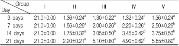
자하거와 전침 처치를 실시한 3일, 7일, 14일, 21일에 BBB 척 도를 검사하였다. 정상군은 차이가 없었으며, Ⅱ군은 손상 후 3 일에 1.36 ± 0.24점에서 21일에는 2.20 ± 0.21점으로 증가되었으 나 유의한 차이가 없었다. Ⅲ군은 3일째 1.30 ± 0.22점에서 21일 째 5.10 ± 0.80점으로, 실험Ⅳ군에서 1.32 ± 0.24점에서 4.90 ± 0.62점, 실험군 Ⅴ는 1.36 ± 0.24점에서 5.65 ± 0.80점으로 유의한 증가를 보였다. 통계학적 유의성을 검정하기 위해 일원배치 분산 분석을 실시한 결과 14일과 21일에서는 실험군 Ⅲ, Ⅳ, Ⅴ에서 유 의한 차이를 보였다(p<0.05)(Table 3).
Table 3. The change of BBB locomotor rating scale in each groups(score)
Group
Day Ⅰ Ⅱ Ⅲ Ⅳ Ⅴ
3 days 21.0±0.00 1.36±0.24 a 1.30±0.22 a 1.32±0.24 a 1.36±0.24 a 7 days 21.0±0.00 1.56±0.26 a 2.00±0.26 b 2.20±0.26 a 2.50±0.26 b 14 days 21.0±0.00 1.75±0.32 a 3.05±0.50 b 3.45±0.42 b 3.75±0.50 b 21 days 21.0±0.00 2.20±0.21 a 5.10±0.80 c 4.90±0.62 c 5.65±0.80 c
All value are showed mean±S.D. Value with different superscripts in the same column are significant(p<.05) by Duncan's multiple range test. Ⅰ: Normal control rat, Ⅱ:Non-treatment after SCI induction, Ⅲ: Hominis placenta pharmacopuncture treatment after SCI induction, Ⅳ: Electroacupuncture therapy after SCI induction, Ⅴ: Hominis placenta pharmacopuncture treatment and electroacupuncture therapy after SCI induction
2) 격자보행 검사(Grid walk test)
자하거와 전침 처치를 실시한 3일, 7일, 14일, 21일에 격자보 행 검사를 실시하였다. Ⅰ군과 Ⅱ군은 유의한 차이가 없었으며,
Ⅲ군은 손상 후 3일에 19.50 ± 0.65점에서 21일에는 10.05 ± 1.88 점, Ⅳ군은 3일째 18.88 ± 0.40점에서 21일째 11.23 ± 1.68점, Ⅴ군 에서 19.50 ± 0.60점에서 9.28 ± 1.28점으로 유의한 증가를 보였 다. 통계학적 유의성을 검정하기 위해 일원배치 분산분석을 실시 한 결과 7일과 14일, 14일과 21일에서는 실험군 Ⅲ, Ⅳ, Ⅴ에서 유의한 차이를 보였다(p<0.05)(Table 4).
Table 4. The change of grid walk test in each groups(number) Group
Day Ⅰ Ⅱ Ⅲ Ⅳ Ⅴ
3 days 0.00±0.00 18.24±1.55 a 19.50±0.65 a 18.88±0.40 a 19.50±0.60 a 7 days 0.00±0.00 17.84±1.65 a 16.20±2.50 a 17.05±2.00 a 16.23±2.08 a 14 days 0.00±0.00 16.23±2.08 b 13.00±2.00 b 14.23±3.02 b 12.04±2.18 b 21 days 0.00±0.00 15.50±1.63 a 10.05±1.88 c 11.23±1.68 c 9.28±1.28 c
All value are showed mean±S.D. Value with different superscripts in the same column are significant(p<.05) by Duncan's multiple range test. Ⅰ: Normal control rat, Ⅱ:Non-treatment after SCI induction, Ⅲ: Hominis placenta pharmacopuncture treatment after SCI induction, Ⅳ: Electroacupuncture therapy after SCI induction, Ⅴ: Hominis placenta pharmacopuncture treatment and electroacupuncture therapy after SCI induction
A B
C D
E
Fig. 1. The histological change of spinal cord in each groups (21 days, H & E stain, × 40).
A: Normal control rat, B: Non-treatment after SCI induction, C: Hominis placenta pharmacopuncture treatment after SCI induction, D:Electroacupuncture therapy after SCI induction, E: Hominis placenta pharmacopuncture treatment and electroacupuncture therapy after SCI induction.
3. 조직학적 관찰
척수 손상 후 척수의 형태학적 변화를 관찰하기 위해 척수 의 H & E 염색을 실시하였다. 실험 종료 후 척수 조직 염색 소 견에서는 Ⅰ군에 비해 Ⅱ군은 척수 조직 손상부위에 광범위한 조직 괴사가 있었으며, 백질과 회백질 부위에 공동화 현상이 관 찰되었다. 정상군(Fig. 1A)에 비하여 SCI 유도된 군에서 손상된 주변부에 공포화(cavitation)가 많이 진행되어 있었다(Fig. 1B). 자 하거 처치와 전침을 처지한 Ⅲ군(Fig. 1C)과 Ⅳ군(Fig. 1D)에서는 괴사 및 공동화 부분이 SCI 유도 군에 비하여 현저히 작아졌다.
Ⅴ군에서는 척수 손상부위는 백질 부분에 공동화 혹은 조직결손 이 일부 구간 남아 있었으며, 척수의 회백질에는 신경섬유들이 대부분 정상적인 신경원 및 축삭의 형태를 갖추고 있지 않아 형
태학적으로 큰 변화는 없었다(Fig. 1E).
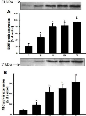
4. BDNF(Brain-derived neurotrophic factor) 및 NT-3 (neurotrophin-3)의 단백질 발현변화
실험 처치가 종료된 후 척수 조직을 적출하여 손상부위를 포함한 척수 조직에 BDNF와 NT-3의 단백질 합성 정도를 관찰 하였다. 각 실험군에서 BDNF의 합성량은 척수손상 후 자하거 약침을 한 Ⅲ군은 80.54 ± 10.67 %, Ⅳ군은 84.01 ± 9.48 %로 대 조군 48.51 ± 9.82 %에 비하여 유의한 증가를 보였으며, 자하거 약침과 전침을 병행하여 실시한 Ⅴ군 93.50 ± 11.26 %로 유의한 단백질 발현의 증가를 보였다(Fig. 2A). NT-3의 합성량은 척수손 상 후 자하거 약침을 한 Ⅲ군은 43.58 ± 9.50 %, Ⅳ군은 50.24 ± 8.36 %로 대조군 18.94 ± 5.68 %에 비하여 유의한 증가를 보였으 며, 자하거 약침과 전침을 병행하여 실시한 Ⅴ군 62.36 ± 12.50 % 로 유의한 단백질 발현의 증가를 보였다(Fig. 2B).
Fig. 2. The protein expression of BDNF(A) and NT-3(B) in rat spinal cord after Hominis placenta pharmacopuncture treatment and electroacupuncture therapy.
All values are means±S.E. of three independent experiments. Value with different superscripts in the same column are significant(p<.05) by Duncan's multiple range test. The autoradiographs are representative of two independent experiments. Ⅰ: Normal control rat, Ⅱ:Non-treatment after SCI induction, Ⅲ: Hominis placenta pharmacopuncture treatment after SCI induction, Ⅳ: Electroacupuncture therapy after SCI induction,
Ⅴ: Hominis placenta pharmacopuncture treatment and electroacupuncture therapy after SCI induction.
고 찰
척수손상 초기에는 신경원과 아교세포의 파괴뿐 아니라 척
수 상행로와 하행로가 손상되어진다. 이는 점진적으로 2차적 흥
분파를 발생하고, 더욱더 신경변성이 가속화 되어진다. 외상 후
이러한 척수손상 후 2차손상의 중요한 요인으로 자가고사
(apoptosis)는 glutamatergic 흥분성, 자유라디칼 손상, 사이토카 인과 염증성 손상의 다양한 요인에 의하여 발생한다 30) . 지질과산 화물(lipid peroxidation)은 외상 후 신경 변성 과정 시 병리적 상 태를 보여주는 중요한 인자이며, 척수손상 직후 빠르게 농도가 상승하며, 1) 이러한 염증은 손상이후 2차적 변성을 더욱 가속화 하는데 기여하는 것으로 알려져 있다 31) . 이러한 신경세포 자가고 사 과정을 통해 척수 신경조직의 소실과 운동장애의 병리학적 과정이 더욱 진행되게 된다. 그래서 현재까지 척수 손상환자의 치료는 이러한 손상의 확대를 최소화 하는데 있다 32) . 본 연구는 자하거 약침과 전침 자극이 신경성장인자의 후지 기능 증진과 신경성장인자 중 특히 BDNF와 NT-3의 단백질 합성에 어떠한 영향을 미치는 지를 알아보고자 하였다.
수정된 Tarlov 평가 방법을 사용하여 척수의 신경학적 손상 정도를 알아보았다 26) . 척수 손상 후 1에서 3일 동안 초기 운동행 동 반응을 측정한 결과 각 군간 행동학적 점수 범위가 통계학적 으로 유의성이 없어, 척수손상 유도가 잘 되어 실험동물의 배치 에 문제가 없었다. 척수 손상 후 운동 기능에 대한 1차적인 회복 은 최근 까지 후지 기능 회복에 초점이 맞춰지고 있으며, 흉부 척수 손상 흰쥐에서 운동을 적용한 결과 기능적 회복과 분자적 인 변화를 가져와 재활능력을 증진한다고 보고되었다 33,34) .
자하거 약침과 전침 자극을 통한 척수 손상 쥐의 후지 운동 기능 회복을 평가하기 위하여 척수 손상 후에 BBB(Basso Beattie Bresnahan) 점수 및 Grid walk 검사를 통해 후지 운동조절 기능 정도를 측정하였다. BBB 평가는 척수손상 흰쥐의 후지 운동 기 능을 평가하기 위해 가장 적합한 척도로 알려져 있으며, 이 검사 는 열린 평면의 공간 위를 보행하게 한 후 보행하는 흰쥐 후지의 운동행동을 관찰하는 것이 목적이다 27) . 척수손상 후 3일, 7일, 14 일, 21일에 BBB 평가를 한 결과 Ⅲ군과 Ⅳ군에서 Ⅱ군에 비하여 21일에 모두 유의하게 점수가 감소하였고, 특히 자하거 약침과 전침을 병행 실시한 Ⅴ군에서 다른 실험군에 비하여 더 많은 감 소를 볼 수 있었다.
Grid walk 검사를 실시한 결과 자하거 약침 처치 및 전침을 처치한 군에서 걷기 실수가 척수손상만 유도한 대조군에 비하여 유의하게 적었고, 병행처치 시에도 통계적으로 다른 실험군과 차 이는 없었으나, 걷기 시 실수하는 횟수가 감소하였다. Bareyre 등
35) 의 연구에서 흉추 척수손상 이후에 척수 내 신경의 재형성을 조사한 결과 척수고유 게재뉴런이 새로 발아함에 따라 새로운 연결 회로가 만들어져 회복 된다고 하였다. 또한 심각한 척수 손 상 이후에도 고유척수 신경원의 회복은 척수 손상환자의 기능 회복을 증진한다고 하였다 36) .
신경영양인자(neurotrophin)는 신경계의 발달, 유지, 가소성 에 매우 중요한 역할을 한다. 신경성장인자(nerve growth factor;
NGF), 뇌유래신경영양인자(brain-derived neurotrophic factor;
BDNF), neurotrophin-3(NT-3)와 neurotrophin-4/5 (NT-4/5)는 4 가지 주요 성장영양인자이며, 이들은 피질 37) , 척수 38) , 후근신경절 신경원 39) 등에 포함되어 있다. 이들 영양인자는 표적기관과 아교 세포(슈반세포, 미세아교세포, 성상세포, 희돌기아교세포)에 의해 생성되어진다. 신경영양인자의 분비는 모든 원시세포에서 손상
이나 질병 과정에 특징적으로 분비되는 것으로 알려져 있으며, 특히 척수 손상 후 손상된 축삭의 성장을 증진하기 위해 신경줄 기세포에서 신경성장인자(nerve growth factor), 뇌유래신경영양 인자(BDNF), 아교유래 신경영양인자(glial-derived neurotrophic factor) 등이 많이 분비되게 된다 40) . 축삭절단과 같은 신경계손상 모델에서 신경경양인자는 신경세포 고사 및 특정 세포사멸을 감 소시키는 능력이 뛰어나다고 알려져 있다 41) .
본 실험 결과에서도 BDNF와 NT-3는 정상군인 Ⅰ군에서는 발현되지 않았으며, 척수손상을 유도한 Ⅱ군은 미약하게 발현되 었다. 척수 손상을 유도하고 자하거 처치를 한 Ⅲ군과 전침을 한
Ⅳ군, 병행 처리한 Ⅴ군은 21일째 BDNF와 NT-3 단백질 함량이 뚜렷하게 증가되어 손상부위의 BDNF와 NT-3의 증가와 함께 척 수조직의 공동화 및 공포화의 구조적 변화를 억제 하는 효과가 있는 것으로 사료된다.
본 연구결과는 자하거와 전침자극이 척수 손상조직 내에 신 경성장인자의 발현을 촉진하여 척수신경의 세포고사와 신경괴사 를 감소와 함께 신경 변성을 지연시키는 것으로 보여지며, 이러 한 신경괴사의 감소를 통해 후지 운동기능의 회복을 일으킨 것 으로 사료된다.
위의 결과를 종합하여 볼 때 척수손상 이후에 자하거 약침 과 전침 자극에 대한 효과를 평가한 결과 자하거 약침과 전침 처 치한 군이 처치하지 않은 군에 비하여 행동 기능학적 소견이 개 선되었으며, 생화학적, 병리조직학적 결과에서 신경보호 효과가 있음이 관찰되었다.
결 론
본 연구는 실험적 척수손상 흰쥐를 대상으로 3주간의 자하 거 약침 처치 및 전침 전기 자극을 적용하여 후지의 운동 기능 회복 및 척수 조직의 변화, 신경영양인자 중 BDNF, NT-3의 단백 질 발현 정도를 관찰하여 다음과 같은 결과를 얻었다.
BBB test에서 Ⅲ군, Ⅳ군, Ⅴ은 손상 후 14일과 21일에 Ⅱ군 과 비해 유의한 차이를 보였으나(p<.05), 실험군 간에서는 유의 한 차이를 보이지 않았다(p>.05).
Grid Walk 평가에서 Ⅲ군, Ⅳ군은 손상 후 14일과 21일에
Ⅱ군에 비해 유의한 차이를 보였으나(p<.05), Ⅲ군, Ⅳ군, Ⅴ은 손 상 후 14일과 21일에 Ⅱ군과 비해 유의한 차이를 보였다(p<.05).
또한 조직학적 평가에서 척수 조직에서는 Ⅱ군과 비교하여 공동화 현상은 남아있었으나 대조군에 비교하여 신경원의 변성 정도 및 염증 소견이 감소하였다.
Western blot을 통한 BDNF와 NT-3 단백질 발현량 변화에 서 각 실험군에서 자하거 약침과 전침 자극한 실험군들에서 신 경성장인자 BDNF와 NT-3의 단백질 합성량이 유의하게 증가함 을 알 수 있었다.
이상의 결과에서처럼 척수손상 흰쥐에서 3주간의 자하거 약
침과 전침 자극은 운동기능의 회복과 척수조직의 염증반응 억제
및 BDNF와 NT-3의 단백질 발현 증가에 영향을 주는 것으로 나
타났다.
참고문헌