Region of Interest Heterogeneity Assessment for Image using Texture Analysis
1)
Yong Sung Park*, Joo Hyun Kang**, Sang Moo Lim***, Sang-Keun Woo****
Abstract
Heterogeneity assessment of tumor in oncology is important for diagnosis of cancer and therapy.
The aim of this study was performed assess heterogeneity tumor region in PET image using texture analysis. For assessment of heterogeneity tumor in PET image, we inserted sphere phantom in torso phantom. Cu-64 labeled radioisotope was administrated by 156.84 MBq in torso phantom. PET/CT image was acquired by PET/CT scanner (Discovery 710, GE Healthcare, Milwaukee, WI). The texture analysis of PET images was calculated using occurrence probability of gray level co-occurrence matrix. Energy and entropy is one of results of texture analysis. We performed the texture analysis in tumor, liver, and background. Assessment textural features of region-of-interest (ROI) in torso phantom used in-house software. We calculated the textural features of torso phantom in PET image using texture analysis. Calculated entropy in tumor, liver, and background were 5.322, 7.639, and 7.818. The further study will perform assessment of heterogeneity using clinical tumor PET image.
▸ Keyword : PET/CT, Tumor heterogeneity, Texture analysis, Gray level co-occurrence matrix, Entropy
I. Introduction
PET/CT는 방사성의약품을 주사하고 체내 생화학적 변화를 관찰하는 양전자 방출 단층촬영기기(Positron Emission Tomography, PET)와 엑스선(X-ray)의 감쇠정보를 이용하여 체내 해부학적 정보를 획득하는 컴퓨터 단층촬영기기 (Computed Tomography, CT)가 융합된 의료영상기기 이다.
PET/CT는 핵의학에서 암 및 난치성 질환을 진단하기 위한 대 표적인 비침습적인 장치로 암의 발생 유무 및 진행 정도를 측 정할 수 있어 필요성이 증대되고 있다[1].
암 환자의 종양조직은 일반조직처럼 유전적 균일성을 갖지 않고 세포간에 유전적으로 이질적인 형태를 나타낸다[2].
종양내 이질성은 동일한 조직내에 서로 다른 특성을 지닌 암 세포들이 존재하는 것으로 유전자 변이에 의해 다양한 세포로
변이되어 항암치료에 내성을 가질 수 있고 환자의 낮은 생존율 과 연관된다[3]. 종양내 이질성 평가는 핵의학 영상기술이 발 달로 영상의 해상도가 증가함에 따라 보다 정확한 분석이 가능 해 졌고 환자의 생존율 예측과 예후 판단에 중요한 예측변수로 활용 가능하다[4]. PET 영상의 텍스처 분석을 통한 종양내 이 질성의 평가는 환자에게 정확하고 효율적인 진단 및 치료계획 을 위해 필요하다[5].
텍스처 분석은 영상이 가지는 질감의 방향성, 대비성, 무늬 의 규칙성을 분석하여 수치화 하는 것으로 구조적 방법, 스펙트 럼 방법, 통계적 방법이 있다. 구조적 방법은 패턴이 규칙적이 고 명확한 경우에 유용하지만 비구조적 텍스처의 분석에 어려 움이 있고 스펙트럼 방법은 비구조적인 텍스처의 분석이 가능 하지만 복잡한 계산 때문에 많은 처리 시간을 필요로 한다. 본
∙First Author: Yong Sung Park, Corresponding Author: Sang-Keun Woo
*Yong Sung Park ([email protected]), Korea Institute of Radiological and Medical Sciences
**Joo Hyun Kang ([email protected]), Korea Institute of Radiological and Medical Sciences
***Sang Moo Lim ([email protected]), Korea Institute of Radiological and Medical Sciences
****Sang-Keun Woo ([email protected]), Korea Institute of Radiological and Medical Sciences
∙Received: 2016. 08. 29, Revised: 2016. 09. 21, Accepted: 2016. 11. 09.
∙This research was supported by a Basic Science Research Program (NRF-2011-0012830) and Nuclear Research &
Development Program (NRF-2012M2A2A7013480) through the NRF of Korea funded by the MEST and by the
National R&D Program through the KIRAMS (No. 1711021908).
연구에서 사용된 방법은 통계적 방법으로 텍스처의 종양과 같 이 규칙이 명확하지 않는 것을 분석하는데 유용하여 다양한 통 계적 평가 지표를 통해 평가 가능한 장점이 있다.
영상의 통계적 텍스처 분석은 영상의 히스토그램 또는 한 개 화소의 발생확률을 사용하는 일차 텍스처 분석(first order texture analysis)과 특정 방향과 거리에 따라 두 개 이상 화소 의 발생 확률을 이용한 고차 텍스처 분석(high order texture analysis)로 분류된다. 일차 텍스처 특징으로는 평균(mean), 표 준편차(standard deviation), 비대칭(skewness), 에너지 (energy), 엔트로피(entropy) 등이 있으며 고차 텍스처 특징은 대비(contrast), 균질성(homogeneity), 에너지, 엔트로피 등이 있으며 텍스처 연산자인 GLCM (gray level co-occurrence matrix)[6]과 GLRLM (gray level run length matrix)[7], 그 리고 GLSZM (gray level size zone matrix)[8] 등을 통하여 계산 된다.
핵의학 영상의 분석이 가능한 프로그램으로는 MATLAB (MathWorks Inc., Natick, MA)에서 구동되는 오픈 소스 코드 인 CGITA (http://code.google.com/p/cgita/)[9]와 C언어 기 반의 OpenGL 라이브러리를 사용한 MaZda (http://www.eletel.p.lodz.pl/programy/cost/)[10]이 있으며 히스토그램(histogram)기반의 통계값, GLCM, GRLM 등의 텍 스처 통계 분석이 가능하다. 본 연구에서는 영상내 이질성 평가 를 위해 종양을 모사한 몸통 팬텀(torso phantom)을 구성하고 PET/CT 스캐너를 이용하여 PET 영상을 획득하고 관심영역의 텍스처 분석을 Visual Studio C++를 이용해 개발된 프로그램 을 이용하여 평가하고자 하였다.
II. Materials and Methods
1. Tumor Model Mimicking
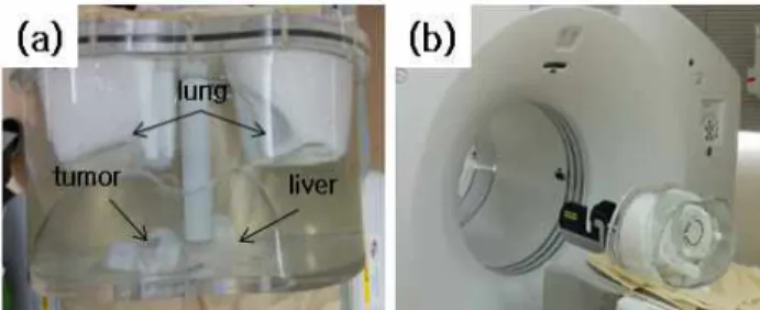
텍스처 분석을 위한 종양 모델은 평균 성인의 해부학적 구조 를 갖는 몸통 팬텀에 종양을 모사하여 Fig. 1(a)와 같이 구현하 였다. 몸통 팬텀은 내부에 폐와 간의 몸통 모양으로 물과 아크 릴로 구성하였으며, 종양을 모사하기 위하여 구 팬텀을 간 부분 에 삽입하여 종양 모델을 구현하였다. 몸통 팬텀의 크기는 26×38 cm
2이고 아크릴의 두께는 9.5 mm 이며 각 영역의 부 피는 폐 2000 cc, 간 1200 cc, 종양 5 cc이다. 종양의 이질성 평가를 위해 Cu-64를 간에 44.4 MBq, 폐에 14.8 MBq, 종양 에 1.44 MBq 배경에 96.2 MBq 주입 하여 제작하였다.
Fig. 1. Imaging of tumor model in torso phantom (a) The torso phantom was composed of body-shaped cylinder lung, liver, and mimic the tumor. (b) PET image of torso
phantom was acquired using PET/CT scanner.
2. Imaging of Tumor Model
종양모사 모델의 PET 영상은 몸통 팬텀에 Cu-64를 주입후 방사성동위원소가 팬텀에 충분히 분포될 수 있도록 1시간 후 Fig. 1(b)와 같이 Discovery 710 PET/CT 스캐너를 이용하여 측정하였다. CT 영상은 같은 시스템의 16-slice helical CT scanner를 이용하여 200 mA, 140 kV, 5.0 mm단위로 획득하 였다. 프레임 당 3분으로 2프레임 획득한 PET 영상의 재구성 은 CT 데이터를 이용하여 감쇠보정하고 3D OSEM (ordered subsets expectation maximization) 알고리즘을 이용하여 영 상화 하였다.
3. Texture Analysis Method
텍스처 분석 프로그램은 영상내 특성을 분석하기위하여 개 발되었으며, 개발환경은 Intel Xeon E5620 (2.4 GHz, 8 core) 의 CPU와 24 GB의 메모리, 그리고 NVIDIA GeForce GTX 760의 그래픽 카드를 사용하였다. 소프트웨어 개발 환경은 Windows7 64Bit의 운영 체제에서 Visual Studio 2010을 사 용하여 OpenCV (v2.4.9, http://www.opencv.org/)를 라이브 러리로 개발하였다. 분석 프로그램은 Analyze 형식의 영상화 파일의 헤더 정보를 이용하여 raw data를 읽고 가시화하였으 며 관심영역(Region of interest, ROI)에 대한 특징정보를 계산 하기 위하여 원하는 영역을 설정할 수 있는 기능을 추가하였다.
관심영역의 일차 텍스처 분석은 영상을 로딩하고 관심영역에 대하여 히스토그램을 계산하여 가시화 할 수 있도록 하였으며 영상의 최대값, 최소값, 평균값, 표준편차, 그리고 히스토그램 을 정규화(normalized)하여 엔트로피를 계산하였다. 고차 텍스 처 분석은 로딩된 영상의 GLCM 변환을 위해 기준 화소와 비 교 화소를 확인하고 선언된 2차배열에 입력되도록 코드를 작성 하였다. GLCM는 0도 45도 90도 135도 방향에 대해 각각 계 산되도록 하였으며 GLCM의 textural feature 계산은 정규화 된 GLCM를 이용하여 계산된다[11].
대비는 인접한 화소의 명암도 차이를 나타내며 다음 식을 이 용하였다.
(1)
식 (1)에서 는 기준이 되는 화소의 명암도(gray level)를 의미 하고 는 인접해있는 화소의 명암도를 나타내며
는 인접한 두 개의 화소가 각각의 명암도로 발생될 확률이며 영상의 명암 도 차이가 클수록 대비는 큰 값을 갖는다.
균질성은 인접한 화소의 명암도 차이가 작은 빈도 분포수를 나타내는 척도로 다음 식을 이용하여 계산하였다.
(2)
식 (2)에서 인접한 화소가 같은 명암도를 갖거나 명암도 차이 가 작을수록 균질성은 큰 값을 나타내고 명암도가 일정한 영상 의 경우 1이 된다.
에너지는 영상의 균일성(uniformity)을 나타내는 척도로 다 음 식을 이용하여 계산하였다.
(3)
식 (3)에서 동일한 명암도 차이를 갖는 화소 쌍이 많을수록 큰 값을 나타내며 명암도가 일정한 영상은 1을 나타낸다.
엔트로피는 영상의 임의성 및 이질성을 나타내는 척도로 명 암도의 변화가 많을수록 큰 값을 나타내며 다음 식을 이용하여 계산된다.
log
(4)
4. Texture Analysis of Tumor Model
몸통 팬텀 텍스처 분석은 PET/CT 스캐너의 측정 파일을 PiView (Infinitt Co. Ltd., Seoul, Korea) 프로그램을 이용하 여 dicom 파일로 변환하고 개발된 프로그램에 입력 data로 사 용하여 분석하였다. 관심영역의 설정은 CT 영상의 해부학적 정보를 이용하여 장기와 종양의 영역을 확인하였고, PET영상 을 분할(segmentation)하여 분석하였다.
III. Result
1. PET/CT Imaging of Tumor Model
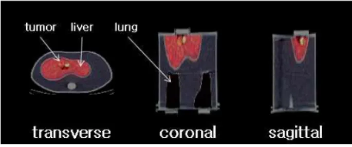
텍스처 분석을 위한 몸통 팬텀의 영상화는 Cu-64를 156.84 MBq 주입후 PET/CT 스캐너를 이용하여 Fig. 2와 같이 간 영 역 내에 종양을 포함하는 영상을 획득하였다. 획득된 PET/CT 정합 영상은 종양 및 간 영역의 경계가 잘 구분되어 영상화 되 었다. 간 및 종양 영역의 경계는 CT 영상의 구조학적 정보를
이용하여 확인할 수 있었으며 주입된 Cu-64의 분포는 PET 영 상을 이용하여 영상화됨을 확인하였다.
Fig. 2. Cu-64 PET/CT torso phantom imaging with tumor region. Torso phantom was consisted by lung, liver and
tumor.
2. Segmentation of ROI
몸통 팬텀의 텍스처 분석을 위한 관심영역의 설정은 Fig. 3 과 같이 종양, 간, 주변영역으로 나누어 설정하였고 영역경계의 영향을 줄이기 위해 관심영역의 중앙부위로 설정 하였다.
CGITA 프로그램은 복셀(voxel)에 기반하여 관심영역을 설정 하기 때문에 Fig. 3(a)에서와 같이 계단(aliasing)현상을 보였 다. 개발된 프로그램에서의 영역설정은 수식을 이용하여 Fig.
3(b)와 같이 계단현상을 줄여 CGITA 프로그램보다 종양영역 을 보다 정확하게 설정할 수 있었다.
Fig. 3. ROI definition of tumor, liver, and background in PET image (a) CGITA program, (b) in-house program 3. Texture Analysis using CGITA
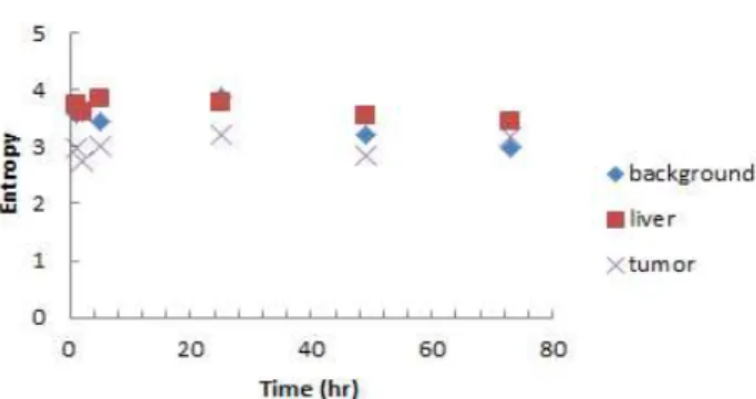
CGITA를 이용한 계산결과는 시간 변화에 따라 엔트로피 변
화를 분석하여 Fig. 4에 나타내었다. CGITA로 계산된 종양 영
역의 엔트로피는 1시간후 4.682, 2시간후 4.527, 5시간후
4.588 이었으며 25시간후 4.741 으로 계산되었다. 간 영역의
엔트로피는 1시간후 6.511, 2시간후 6.406, 5시간후 6.625 이
었으며 25시간후 6.578으로 계산 되었다. 분석된 관심영역의
엔트로피는 몸톰 팬텀을 PET/CT 스캐너에 설치 위치 및 측정
오차를 고려하였을 때 방사성동위원소의 방사능(activity) 변화
가 일정한 것으로 확인되었다.
Fig. 4. CGITA texture analysis graph in liver, tumor and background. Calculated entropy of torso phantom with the
variation of time distribution
4. Texture Analysis using In-house Program 개발된 프로그램은 영상내 텍스처 분석을 위해 영상을 로딩 하고 영상내 값들을 기반으로 히스토그램화하여 일차 텍스처 특징인 최대값, 최소값, 평균값, 표준편차, 엔트로피를 계산 하 였다. 고차 텍스처 특징은 GLCM 연산자를 이용하여 대비, 균 질성, 에너지, 엔트로피를 분석하여 Table 1과 같이 계산되었 다. 종양의 엔트로피는 5.322, 간의 엔트로피는 7.639였으며 배경의 엔트로피는 7.818로 계산되었다. 종양의 에너지는 0.025, 간의 에너지는 0.006이었으며 배경의 에너지는 0.005 로 계산되었다.
관심영역의 설정은 CGITA와 같은 영역을 원형으로 Fig.
3(b)와 같이 설정하였고 동일한 위치를 평가하여 개발된 프로 그램의 적용 가능성을 확인하였다. 분석 결과 CGITA로 계산 된 종양의 엔트로피는 5.193으로 2.48%의 차이를 보였고 간의 경우 6.702로 13.98%의 차이를 보였다. 텍스처 분석결과의 차 이는 관심영역의 설정방법에 의한 것으로 CGITA에서 설정된 영역보다 정확한 영역이 설정되어 엔트로피의 차이를 보였으며 간의 경우 종양 영역보다 보다 확장된 영역을 포함하고 있어 더욱더 큰 차이를 보였다.
Table 1. GLCM based texture analysis parameters in torso phantom using in-house program
Textural
feature Tumor Liver Background
Entropy 5.322 7.639 7.818
Energy 0.025 0.006 0.005
Homogeneity 0.078 0.249 0.196
IV. Conclusion
본 연구에서는 영상내 이질성 평가를 위해 종양을 모사한 몸 통 팬텀에 Cu-64 방사성동위원소를 주입하고 PET/CT 스캐너
를 이용하여 영상화 하였으며 획득된 영상의 관심영역의 특징 을 텍스처 분석을 이용하여 계산하였다. 관심영역의 설정은 CT 영상의 해부학적 정보를 이용하여 영역을 분할하였고 PET 영 상의 분할된 영역을 텍스처 분석하였다. CGITA를 통해 계산된 몸통 팬텀의 텍스처 분석 결과는 Cu-64의 방사능 변화에도 일 정한 결과를 보였고 몸통 펜텀의 구성 및 관심 영역의 설정이 잘 수행되었음을 확인하였다. 개발된 텍스처 분석프로그램은 Visual Studio C++을 이용하여 작성되었고 일차 텍스처 특징 과 고차 텍스처 특징으로 몸통 팬텀의 종양, 간, 주변영역에 대 하여 계산하였다.
개발된 프로그램의 적용 가능성은 CGITA의 텍스처 분석 결 과와 비교하였을 때 관심영역의 설정에서 차이를 보였다.
CGITA는 복셀에 기반하여 최소 영역으로 설정되어 분석되었 고 개발된 프로그램은 정밀한 영역 설정을 위해 수학적 모델을 기반으로 적용되어 보다 정확한 관심영역을 설정하여 분석이 가능하였다. 개발된 프로그램의 관심영역 설정방법은 발병 위 치, 모양 및 크기가 다양한 종양의 관심영역 설정에 탁월할 것 으로 판단되며 CGITA보다 정확한 결과를 도출할 수 있을 것으 로 판단된다. 이 연구를 바탕으로 종양의 이질성평가의 가능성 을 확인하였으며 환자 진단에 정량적인 영상 평가 지표를 제공 함으로써 정확하고 효율적인 진단 및 예측이 가능할 것으로 기 대된다.
REFERENCES
[1] Bar-Shalom, R., Yefremov, N., Guralnik, L., Gaitini, D., Frenkel, A., Kuten, A., Altman, H., Keidar, Z., and Israel, O., "Clinical performance of PET/CT in evaluation of cancer: additional value for diagnostic imaging and patient management." Journal of nuclear medicine Vol. 44, No.
8, pp. 1200-1209, Aug. 2003.
[2] Burrell, R. A., McGranahan, N., Bartek, J., and Swanton, C., "The causes and consequences of genetic heterogeneity in cancer evolution." Nature, Vol. 501, No.
7467, pp. 338-345, July 2013.
[3] Burrell, R. A., and Swanton, C., "Tumour heterogeneity and the evolution of polyclonal drug resistance."
Molecular oncology, Vol. 8, No. 6, pp. 1095-1111, Sep.
2014.
[4] Hyun, S.H., Kim, H.S., Choi, S.H., Choi, D.W., Lee, J.K., Lee, K.H., Park, J.O., Lee, K.H., Kim, B.T. and Choi, J.Y.,
"Intratumoral heterogeneity of 18F-FDG uptake predicts
survival in patients with pancreatic ductal
adenocarcinoma." European journal of nuclear medicine
and molecular imaging, pp. 1-8, Feb. 2016.
Yong Sung Park received the B.S., M.S.
and Ph.D. degrees in Physics and Applied Optics from Dankook University, Korea, in 2008, 2010 and 2016, respectively. He is currently a postdoctoral research associate in Division of RI-convergence Research from KIRAMS. His current research interests include internal dosimetry, texture analysis, and multi-functional fusion image research with PET/CT and MRI.
Joo Hyun Kang received the Pharm. B.
degree in department of pharmacy from Seoul National University and M.S. and Ph.D. degrees in Molecular Biology from KAIST, Korea, in 1985, 1987 and 1991, respectively. She worked as a postdoctoral fellow in KRIBB and associate professor in department of nuclear medicine in Seoul National University. She is currently a principal researcher from 2006 in KIRAMS. Her current research interests include development of new radiopharmaceuticals and molecular imaging methods in small animal disease models with nuclear medicine, optical and MR images.
Sang Moo Lim received the M.D & Ph.D.
degrees in Nuclear medicine from Seoul National University in 1988. He worked a research fellow in USA National Institutes of Health from 1990 to 1991, and University of California, Davis from 1995 to 1996. Dr. Lim joined the Department of Nuclear medicine at KIRAMS, Seoul in 1987. He is currently director of Department of Nuclear medicine in KIRAMS.
He is interested in radionuclide therapy with
131I and
166