http://crossmark.crossref.org/dialog/?doi=10.14474/ptrs.2019.8.3.146&domain=pdf&date_stamp=2019-9-25
Received: 29 August, 2019 Revised: 17 September, 2019 Accepted: 18 September, 2019 Corresponding author: Byoung Hee Lee (ORCID https://orcid.org/0000-0001-9766-6068)
Department of Physical Therapy, College of Health Science and Social Welfare, Sahmyook University, 815 Hwarang-ro, Nowon-gu, Seoul 01795, Republic of Korea
Tel: 82-2-3399-1634 Fax: 82-2-3399-1639 E-mail: [email protected]
This is an Open-Access article distributed under the terms of the Creative Commons Attribution Non-Commercial License (http://creativecommons.org/licenses/
by-nc/4.0) which permits unrestricted non-commercial use, distribution, and reproduction in any medium, provided the original work is properly cited.
Copyright © 2019 Korean Academy of Physical Therapy Rehabilitation Science https://doi.org/10.14474/ptrs.2019.8.3.146
pISSN 2287-7576 eISSN 2287-7584
Phys Ther Rehabil Sci 2019, 8 (3), 146-151 www.jptrs.org
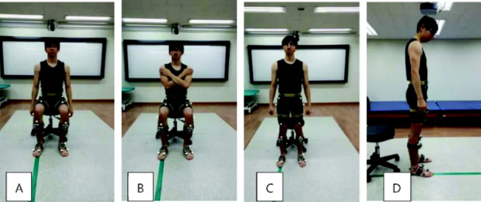
Reliability of joint angle during sit-to-stand movements in persons with stroke using portable gait analysis system based wearable sensors
Jung-Ae An a , Byoung-Hee Lee b
a
Department of Physical Therapy, Seoul Now Hospital, Seongnam, Republic of Korea
b