• 검색 결과가 없습니다.

The evaluation of useful on the additional PET/CT Liver scan

N/A
N/A
Protected

Academic year: 2021

Share "The evaluation of useful on the additional PET/CT Liver scan"

Copied!
3
0
0

로드 중.... (전체 텍스트 보기)

전체 글

(1)

46

핵의학기술 제20권 제2호 2016 J Nucl Med Technol

Vol. 20, No. 2, October 2016

Original Article PET/CT 검사에서 Gastrointestinal Cancer 환자의

Liver 추가촬영에 대한 유용성 평가

부산대학교병원 핵의학과

박세윤·이화진·이무석·김정욱·지혜인

The evaluation of useful on the additional PET/CT Liver scan

Se Youn Park, Hwa Jin Lee, Mu Seok Lee, Jung Uk Kim and Hye In Ji Department of Nuclear Medicine, Pusan National Hospital, Pusan, Korea

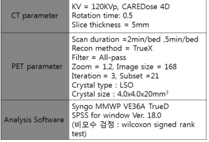
Purpose The liver one of the most common site for distant metastasis for a variety of tumor, especially of gastrointestinal origin. the purpose of this study was to analyze image quality between standard scan and additional liver scan.

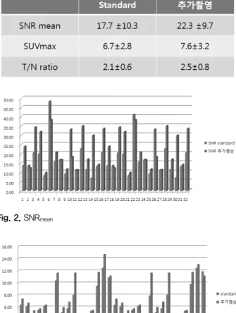
Materials and Methods From September 2015 to February 2016. 152 patients were examined undergo gastrointestinal cancer. 32 patients confirmed liver metastasis analyzed same liver ROI level and check the SNR, SUV and T/N ratio Results The SNR mean of standard was 17.7±10.3; addition was 22.3±9.7 (p<0.05). In SUV max of standard was 6.7±2.8;

addition was 7.6±3.2 ( P <0.05). and the T/N ratio of standard was 2.1±0.6; addition was 2.5±0.8 ( P <0.05).

Conclusion The SNR mean , SUV max and T/N ratio were higher than those on the first scan ( P <0.05). The SNRmean showed the highest change rate among the parameters. A additional liver scan is more favorable for the detection of gastrointestinal cancer patients.

Key Words SNR, SUV, T/N ratio

9) 서론

서 론

Gastrointestinal cancer에서 전이가 가장 흔하게 발견되는 장기는 간이다. 1) Gastrointestinal cancer 환자의 약 20%가 진 단 당시에 이미 간에 전이가 된 상태에서 발견되며, 25%는 치료경과 중 간 전이가 발생하는 것으로 알려져 있다. 2) Gastrointestinal cancer 환자의 간전이 유무를 조기에 정확히 진단하는 것은 치료방침을 결정하는데 있어 매우 중요하 다. 3) 이에 본 연구는 Gastrointestinal cancer 환자에서 PET/CT 검사 시 Liver 추가 촬영을 함으로써 영상의 질적 변 화에 대한 유용성을 평가하고자 하였다.

∙ Received: September 28, 2016 Accepted: October 6, 2016

∙ Correspoding author : Se Youn Park

Address for correspondence : Department of Nuclear Medicine, Pusan National Hospital, 179 Gudeok-ro, Seogu, Pusan 49241, Korea

Tel : +82-10-9422-4590 Fax : +82-51-247-3214 E-mail : ssyy0126@naver.com

연구방법 및 대상

1. 실험방법

검사 장비는 Siemens사의 Biograph Truepoint 40을 이용 하였고 CT parameter는 KV = 120 KVp, CAREDose 4D, Rotation time: 0.5, Slice thickness = 5 ㎜를 사용 하였고 PET parameter에서 스캔 시간은 standard 에서는 2min/bed, 추가 영상에선 5min/bed 으로 검사하였다(table 1).

2. 연구대상

검사대상은 2015년 9월부터 2016년 2월까지 본원에 내원

하여 Gastrointestinal cancer 진단을 받은 152명을 대상으로

PET/CT 검사 후 Liver 부위를 추가를 촬영하였다. 판독을 통

해서 Liver에 전이가 확인된 환자 32명을 대상으로 Standard

영상과 추가 영상에서 같은 Level의 관심영역을 설정하여

Signal to Noise Ratio (SNR), Standardized Uptake Value

(2)

47 박세윤·이화진·이무석·김정욱·지혜인 PET/CT 검사에서 Gastrointestinal Cancer 환자의 Liver 추가촬영에 대한 유용성 평가



  

   

   ×

      ′  

      

          

   

Fig. 1. PET/CT standard Liver scan and additional Liver scan.

Table 1. Specifications of the Siemens Biograph Truepoint 40

(SUV), Tumor/Normal(T/N) ratio의 변화를 통한 영상의 질적 변화를 평균값을 산출하여 비교 평가하였다. 환자는 6시간 이상 금식 하게 하였고 충분한 수분을 섭취하게 하고 심한 운 동은 금지하도록 하였다.

3. 검사방법

18 F-FDG 주사 60분 뒤 Skull base부터 Thigh까지 Bed당 2 분씩 Standard 검사를 시행하고 끝나면 Liver 부위만 추가로 Bed당 5분씩 촬영하였다.

4. 영상분석

SNR은 Normal uptake된 Liver ROI 1과 추가영상의 1’ 에 대해서 구하였는데 Trans axial 이미지의 같은 Pixel영역에 대해서 SUV mean 을 5회 측정 하여 이를 Standard deviation 으 로 나눠주었다. 4)

SUV max 는 region 영역에 대하여 standard의 ROI 2와 추가 영상의 ROI 2’ 의 SUV max 을 5회 측정 하여 평균하여 비교 하 였다(Fig. 1).

결 과

SNR은 Standard 17.7±10.3, 추가영상 22.3±9.7으로 산출 되었으며(P<0.05) 4명의 환자에 대해서는 Standard의 영상 이 더 높게 나왔고 나머지 28명은 추가촬영을 하였을 때가 더 높게 나왔고 통계적으로 유의한 차이가 있었다.

SUV max 는 각각 6.7±2.8와 7.6±3.2이었으며(P<0.05)로 나

타났고 1명을 제외한 모든 환자에서 추가영상에서의

Region 영역 SUV max 가 높게 나타났다. 이는 통계적으로 유

의한 차이가 있는 것으로 나타났다. T/N ratio는 Standard는

2.1±0.6, 추가촬영에서는 2.5±0.8로 분석되었고 이는 통계적

으로 유의한 차이가 있는 것으로 나타났다. 2명을 제외한 나

머지 환자들에게서 추가촬영일 때의 Ratio가 더 높은 것으로

나타났다(table 2).

(3)

48

핵의학기술 제20권 제2호 2016

Table 2. Comparison of SNR mean , SUV max and T/N ratio

Fig. 2. SNR mean

Fig. 3. SUV max

Fig. 4. T/N ratio

결 론

Gastrointestinal cancer 환자에서 Liver 추가 촬영에 따른 SNR과 SUV max , T/N ratio는 상대적으로 standard 보다 높게 나타났다. 이미지 Quality를 나타내는 인자들 중에서 SNR의 변화가 가장 컸다. Gastrointestinal cancer 환자에서 Liver 추 가 촬영을 통한 영상의 질적 향상은 간 전이가 있는 암환자를

판독 하는데 있어서 유용한 검사 방법이라고 사료된다.

요 약

Gastrointestinal cancer 환자의 약 20%가 진단 당시에 이미 간에 전이가 된 상태에서 발견된다. Gastrointestinal cancer 환자에서 PET/CT 검사 시 Liver 추가 촬영을 함으로써 영상 의 질적 변화에 대한 유용성을 평가하고자 하였다. 검사대상 은 2015년 9월부터 2016년 2월까지 본원에 내원하여 Gastrointestinal cancer 진단을 받은 152명을 대상으로 PET/CT 검사 후 Liver 부위를 추가를 촬영하였다. Standard 영상과 추가 영상에서 같은 Level의 관심영역을 설정하여 Signal to Noise Ratio(SNR), Standardized Uptake Value (SUV), Tumor/Normal(T/N) ratio의 변화를 통한 영상의 질적 변화를 평균값을 산출하여 비교 평가한 결과 SNR은 standard 17.7±10.3, 추가영상 22.3±9.7, SUV max 는 각각 6.7±2.8와 7.6±3.2, T/N ratio는 standard는 2.1±0.6, 추가촬영 에서는 2.5±0.8로 분석되었다. 이는 통계적으로 유의한 차이 가 있는 것으로 나타났다. SNR의 변화량이 가장 큰 것으로 나타났고 Liver 추가 촬영에 따른 SNR과 SUV max , T/N ratio 는 상대적으로 Standard 보다 높게 나왔고 Liver 추가 촬영을 통한 영상의 질적 향상은 간 전이가 있는 암 환자를 판독 하 는데 있어서 유용한 검사 방법이라고 사료된다.

REFERENCES

1. JW Lee, SK Kim, SM Lee, SH Moon, TS Kim. Detection of hepatic metastases using dual-time-point FDG PET/CT scan in patients with colorectal cancer. Molecular imaging and Biology 2011;13:565-572.

2. Russ A Kuker, Geraldine Mesoloras, Seza A Gulec.

Optimization of FDG-PET/CT imaging protocol for evaluation of patients with primary and metastatic liver disease. Surgical Oncology;2007, 4:17.

3. D. Fuster, S. Lafuente, X. Setoain, I. Navales, A.

Perissinotti, J. Pavia, P. Paredes, F. Lome˜na, F. Pons.

Dual-time point images of the liver with 18F-FDG PET/CT in suspected recurrence from colorectal cancer. Rev Esp Med Nucl Imagen Mol. 2012;31(3):111116.

4. Halper BS, Dahlbom M, Auerbach MA et (2005)

Optimizing imaging protocols for overweight and obese

patients: a lutetium orthosilicate PET/CT study. J Nucl Med

46:603-607.

수치

Fig. 1. PET/CT standard Liver scan and additional Liver scan.
Table 2. Comparison of SNR mean ,  SUV max and T/N ratio

참조

관련 문서

편의점 간편식 인식도 구간화에 따른 인구통계학적 특성 분포 결과, 성별 분포로 남 학생에서는 D군이 높게 나타났고, 여학생에서는 C군이 높게 나타났으며,

Carmustine (BCNU) Marrow (delayed nadir) GI, liver (high

Nephrotic syndrome Renal insufficiency Heart

1 John Owen, Justification by Faith Alone, in The Works of John Owen, ed. John Bolt, trans. Scott Clark, &#34;Do This and Live: Christ's Active Obedience as the

The histological course of nonalcoholic fatty liver disease: a longitudinal study of 103 patients with sequential liver biopsies.. McPherson S, Hardy T, Henderson

본고에서는 간 질환에서 흔히 사용되는 생화학 검사 또는 간기능 검사 (liver function test) 항목에 대해서 기술하고자 한다..

만 성 B형 간염의 치료 가이드라인은 1990년대 말부터 아시아- 태평양간학회 (Asian Pacific Association for the Study of the Liver, APASL), 미국간학회(American Association

Effect of caloric restriction on the expression of PGC-1 and PPARs mRNA in liver of Otsuka Long-Evans Tokushima Fatty Rats.. Long-lived growth hormone