• 검색 결과가 없습니다.

Serious Complications after Self-expandable Metallic Stent Insertion in a Patient with Malignant Lymphoma

N/A
N/A
Protected

Academic year: 2021

Share "Serious Complications after Self-expandable Metallic Stent Insertion in a Patient with Malignant Lymphoma"

Copied!
5
0
0

로드 중.... (전체 텍스트 보기)

전체 글

(1)

Serious Complications after Self-expandable Metallic Stent Insertion in a Patient with

Malignant Lymphoma

Sung Bae Cho, M.D.

1

, Seon Ah Cha, M.D.

1

, Joon Young Choi, M.D.

1

, Jong Min Lee, M.D.

1

, Hyeon Hui Kang, M.D.

1

, Hwa Sik Moon, M.D., Ph.D.

1

, Sei Won Kim, M.D.

2

, Chang Dong Yeo, M.D.

3

and Sang Haak Lee, M.D., Ph.D.

1

1

Department of Internal Medicine, St. Paul’s Hospital, College of Medicine, The Catholic University of Korea, Seoul,

2

Department of Internal Medicine, Yeouido St. Mary’s Hospital, College of Medicine, The Catholic University of Korea, Seoul,

3

Department of Internal Medicine, Uijeongbu St. Mary’s Hospital, College of Medicine, The Catholic University of Korea, Uijeongbu, Korea

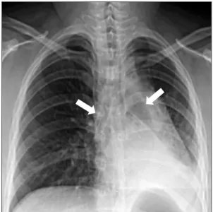
An 18-year-old woman was evaluated for a chronic productive cough and dyspnea. She was subsequently diagnosed with mediastinal non-Hodgkin lymphoma (NHL). A covered self-expandable metallic stent (SEMS) was implanted to relieve narrowing in for both main bronchi. The NHL went into complete remission after six chemotherapy cycles, but atelectasis developed in the left lower lobe 18 months after SEMS insertion. The left main bronchus was completely occluded by granulation tissue. However, the right main bronchus and intermedius bronchus were patent. Granulation tissue was observed adjacent to the SEMS. The granulation tissue and the SEMS were excised, and a silicone stent was successfully implanted using a rigid bronchoscope. SEMS is advantageous owing to its easy implantation, but there are considerable potential complications such as severe reactive granulation, stent rupture, and ventilation failure in serious cases. Therefore, SEMS should be avoided whenever possible in patients with benign airway disease. This case highlights that SEMS implantation should be avoided even in malignant airway obstruction cases if the underlying malignancy is curable.

Keywords: Airway Obstruction; Stents; Bronchoscopy; Granulation Tissue

Introduction

Airway obstruction has numerous causes and can induce serious clinical symptoms and disease such as dyspnea, hy- poxemia, obstructive pneumonia, or respiratory failure. Air- way stenting is a well-established method that immediately relieves symptoms and maintains airway patency in cases of inoperable airway obstruction

1-6

. Currently, two stent types are used therapeutically, a self-expandable metallic stent (SEMS) and a silicone stent. SEMS is used to treat malignant airway obstructions because it can be easily placed by a flexible bron- choscope. However, SEMS may cause long-term complica- tions such as granulation tissue formation, and the incidence of complications increase over time

7,8

. If the stent becomes obstructed by granulation tissue, then stent removal or other interventions are required, which themselves may cause seri- ous complications

9

.

Copyright © 2015

The Korean Academy of Tuberculosis and Respiratory Diseases.

All rights reserved.

Address for correspondence: Sang Haak Lee, M.D., Ph.D.

Department of Internal Medicine, St. Paul’s Hospital, College of Medicine, The Catholic University of Korea, 180 Wangsan-ro, Dongdaemun-gu, Seoul 130-709, Korea

Phone: 82-2-961-4500, Fax: 82-2-968-7250 E-mail: agmante@gmail.com

Received: Aug. 6, 2014 Revised: Sep. 24, 2014 Accepted: Sep. 24, 2014

cc

It is identical to the Creative Commons Attribution Non-Commercial

License (http://creativecommons.org/licenses/by-nc/3.0/).

(2)

Many studies recommend against implanting an SEMS in a patient with benign airway disease and instead only recom- mend it for use in malignant airway disorders after thoroughly exploring all other therapeutic options

2,7-9

.

We describe a case of bilateral main bronchial restenosis caused by granulation tissue in-growth after implanting a cov- ered-SEMS to treat a malignant airway disorder. The patient was successfully treated by removing the obstructed covered- SEMS and replacing it with a silicone stent by using a rigid bronchoscope, which helped maintain airway patency and stability.

Case Report

An 18-year-old woman was evaluated for a 7-month history of chronic purulent sputum and dyspnea. She was diagnosed with non-Hodgkin lymphoma with bilateral bronchial ob- struction 18 months previously. At that time, a covered-SEMS was implanted on emergency and was followed by six chemo- therapy cycles. She eventually achieved complete remission.

Eleven months after implanting the stents, the chronic pro-

Figure 1. Initial chest radiography shows atelectasis of left lower lung field and metallic stents are placed at both main bronchus (ar- rows).

Figure 2. The stents are deployed at both

main bronchi, the left bronchial stent

(arrow) is obstructed and left lower lobe

is collapsed as a result (A, C). The lumen

of the right stent (arrow) is intact (B, D).

(3)

ductive cough with dyspnea began. An initial bronchoscopy and computed tomography revealed stent obstruction with granulation tissue. Despite repeated excision of the granula- tion tissue and other medical therapies, the obstructive lesions continued to deteriorate, and after 3 months, she was referred to our hospital for evaluation.

On initial examination, the blood pressure was 120/70 mm Hg, heart rate 80 beats per minute, respiratory rate 29 breaths per minute, and body temperature was 36.5

o

C. The lung sounds were decreased, and wheezing was auscultated at the left lower lung; thoracic radiography showed atelectasis in the left lower lung field (Figure 1). A chest computed tomography revealed obstruction in the left main bronchus and left lower lobe collapse (Figure 2A).

Leukocyte count was 9,900 cells/mm

3

(71.7% neutrophils, 19.9% lymphocytes, 5.5% monocytes, 1.1% eosinophils, and

1.8% basophils), hemoglobin concentration was 12.3 g/dL, and platelet count was 284,000 cells/mm

3

. Arterial blood gas analysis, which was performed in ambient air, revealed the following: pH, 7.414; Pa CO

2

, 34.7 mm Hg; Pa O

2

, 111.2 mm Hg;

HCO

3

, 21.7 mmol/L; and Sa O

2

, 99.7%. On pulmonary function tests, the forced expiratory volume in one second (FEV

1

) was 1.17 L (41% of predictive value), forced vital capacity (FVC) was 1.69 L (56% of predictive value), and FEV

1

/FVC was 69%.

Flexible bronchoscopy showed an obstruction in the left main bronchus, and the bronchoscope was unable to proceed beyond the bronchus mid portion due to the excessive granu- lation tissue (Figure 2C). The right main bronchus was rela- tively patent (Figure 2B), but excessive granulation tissue was noted (Figure 2D). After excising the granulation tissue using a rigid bronchoscope, the covered-SEMS was visualized distally.

The implant was overgrown with granulation tissue, and the metallic wires were pressed against the bronchial wall. The implant removed from the left bronchus in fragments using alligator forceps (Figure 3), and once removed, bronchoma- lacia and excessive granulation was observed in the left main bronchus. There were no complications after removing the covered-SEMS and attached granulation tissue.

Afterwards, a balloon dilation and silicone stent insertion were performed to maintain airway patency (Figure 4). The right bronchial covered-SEMS was also removed as a single unit to prevent similar SEMS-related complications, especially excessive granulation. Stenosis was present, and a silicone stent was similarly implanted at the right bronchus interme- dius. The patient has reported no complications in 2 years of follow-up examinations.

Discussion

Airway stenting is a highly effective procedure to relieve airway obstruction

1-4

. Generally, an airway stent is indicated in any airway compression condition causing debilitating respi-

Figure 4. (A) The silicone stents are placed at both main bronchus. (B) Left lower lobe collapse is improved after silicone stent insertion.

Figure 3. The covered-self expandable metallic stent is retrieved in

fragments.

(4)

ratory symptoms (dyspnea, obstructive pneumonia, hemor- rhage) and to seal an airway fistula

2,4,10

. The only contraindica- tion is external airway compression from a vessel

4

.

The first airway stent, described in 1964 by Montgomery, was silastic and required insertion using a rigid bronchoscope under general anesthesia

2

. The silicone airway stent was first introduced by Dumon in 1987

11

. Two stent types are currently used, metal and silicone. SEMS was introduced later and is advantageous owing to its easy placement under conscious sedation using a flexible bronchoscope, lower incidence of migration, and improved adaption to irregular airways

3,7

. However, SEMS is disadvantaged by high reactivity to granula- tion tissue, stent obstruction, fracture, erosion of surrounding tissue, and bacterial colonization

2,3,7

. Covered-SEMS was sub- sequently developed to decrease the risks of tumor and gran- ulation tissue in-growth. However, granulation tissue may still form at the stent margins (anchor), leading to obstruction

10

.

In the present case, a covered-SEMS was implanted to restore airway patency. The patient was severely debilitated by the malignant disease and required immediate therapy.

However, her medical condition was not favorable for under- going general anesthesia. Fifteen months later, the mediastinal malignant lymphoma improved, but the airway was still ob- structed by granulation tissue in-growth.

In a retrospective review on the use of metallic stents, Breit- enbucher et al.

12

reported complications in 23 of 172 patients (tumor in-growths in nine, granulation tissue in seven, stent migration in five, and external compression in two). Similarly, Lemaire et al.

13

retrospectively reviewed the use of SEMS in a single institution and reported complications in 23 of 166 patients (tumor in-growths in nine, excessive granulation in seven, stent migration in five, and external compression in two). In a review of benign airway disease, Chung et al.

14

identified 35 out of 75 cases (47.8%) developed SEMS-related obstructive granulation tissue. These complications require stent removal, which is difficult and hazardous to the patient;

removal can induce further serious complications such as mucosal tear, hemorrhage, retained stent fragments, pneumo- thorax, re-obstruction, and ventilation failure in severe cas- es

7,8,15

. Noppen et al.

8

retrospectively analyzed cases of benign airway disease treated with covered-SEMS in a single institu- tion. They reported that 10 of 39 patients experienced com- plications requiring stent removal. In this study, granulomas formed in 17 patients and required repeated intervention; five patients required stent removal because of refractory granula- tion

8

. Several studies of SEMS complications in benign airway disease indicate that complications increase the longer SEMS is in place

7,8,15

. In 2007, the US Food and Drug Administration recommended metallic tracheal stents should be implanted in patients with benign airway disorders only after thoroughly exploring all other treatment options

9

. These risks have not significantly improved in the latest generation of prosthesis covered-SEMS

15

.

The silicone stent is disadvantaged because it can only be inserted using a rigid bronchoscope under general anesthesia.

The silicone stent is difficult to implant in a complex airway.

Mucociliary clearance may also be impaired, and there is higher incidence of stent migration than SEMS. However, these disadvantages typically do not cause any serious com- plication. Furthermore, the silicone stent has a low granulation tissue reactivity and is easily removed should complications arise or following treatment of the underlying disease

2,5,6,9

. In this patient, a silicone stent was the superior initial choice because her disease was potentially curable, and the stent should have been removed immediately after chemotherapy.

In conclusion, the reversibility of the underlying pathology and the potential for stent removal should be considered at the time of stent placement. SEMS must not be used in benign airway disease and even malignant disease if the condition is potentially curable or the patient has a long life expectancy.

This is a rare case of obstructed SEMS removal and high- lights that the silicone stent should be used preferentially in benign or potentially curable malignant airway disease.

Conflicts of Interest

No potential conflict of interest relevant to this article was reported.

References

1. Bolliger CT, Sutedja TG, Strausz J, Freitag L. Therapeutic bron- choscopy with immediate effect: laser, electrocautery, argon plasma coagulation and stents. Eur Respir J 2006;27:1258-71.

2. Casal RF. Update in airway stents. Curr Opin Pulm Med 2010;16:321-8.

3. Ernst A, Feller-Kopman D, Becker HD, Mehta AC. Central air- way obstruction. Am J Respir Crit Care Med 2004;169:1278- 97.

4. Chin CS, Litle V, Yun J, Weiser T, Swanson SJ. Airway stents.

Ann Thorac Surg 2008;85:S792-6.

5. Ryu YJ, Kim H, Yu CM, Choi JC, Kwon YS, Kwon OJ. Use of silicone stents for the management of post-tuberculosis tra- cheobronchial stenosis. Eur Respir J 2006;28:1029-35.

6. Rafanan AL, Mehta AC. Stenting of the tracheobronchial tree.

Radiol Clin North Am 2000;38:395-408.

7. Lunn W, Feller-Kopman D, Wahidi M, Ashiku S, Thurer R, Ernst A. Endoscopic removal of metallic airway stents. Chest 2005;127:2106-12.

8. Noppen M, Stratakos G, D’Haese J, Meysman M, Vinken W.

Removal of covered self-expandable metallic airway stents in benign disorders: indications, technique, and outcomes.

Chest 2005;127:482-7.

9. Lund ME, Force S. Airway stenting for patients with benign

(5)

airway disease and the Food and Drug Administration advi- sory: a call for restraint. Chest 2007;132:1107-8.

10. Dutau H, Musani AI, Plojoux J, Laroumagne S, Astoul P. The use of self-expandable metallic stents in the airways in the adult population. Expert Rev Respir Med 2014;8:179-90.

11. Dumon JF. A dedicated tracheobronchial stent. Chest 1990;97:328-32.

12. Breitenbucher A, Chhajed PN, Brutsche MH, Mordasini C, Schilter D, Tamm M. Long-term follow-up and survival after Ultraflex stent insertion in the management of complex ma- lignant airway stenoses. Respiration 2008;75:443-9.

13. Lemaire A, Burfeind WR, Toloza E, Balderson S, Petersen RP,

Harpole DH Jr, et al. Outcomes of tracheobronchial stents in patients with malignant airway disease. Ann Thorac Surg 2005;80:434-7.

14. Chung FT, Lin SM, Chou CL, Chen HC, Liu CY, Yu CT, et al.

Factors leading to obstructive granulation tissue formation after ultraflex stenting in benign tracheal narrowing. Thorac Cardiovasc Surg 2010;58:102-7.

15. Gaissert HA, Grillo HC, Wright CD, Donahue DM, Wain JC,

Mathisen DJ. Complication of benign tracheobronchial stric-

tures by self-expanding metal stents. J Thorac Cardiovasc

Surg 2003;126:744-7.

수치

Figure 1. Initial chest radiography shows atelectasis of left lower  lung field and metallic stents are placed at both main bronchus  (ar-rows).
Figure 4. (A) The silicone stents are  placed at both main bronchus. (B) Left  lower lobe collapse is improved after  silicone stent insertion.

참조

관련 문서

Based on the research described above, a work expressing the formative beauty of lines, which is the most basic in calligraphy, was produced by using

ventilation unit at the top and a hydrogen charging station with a ventilation unit at the bottom by using it, the flame and temperature generated inside the

The feed is commonly a solution in a solvent like ethanol or t-butanol, and the nonsolvent is water..

The PIG3 was interacted with the CTSB in human prostate cancer cell line PC3, which was verified by co-immunoprecipitation analysis using specific

In collaboration with CGC (C. elegans Genomic Center), the proper mutants were selected for the system and a preliminary module was developed.. The preliminary module was

The structure in (b) corresponds to the part indicated by a dotted circle in the network structure in (a), (c) the molecular model constructed with four buckybowl units

A specimen atom, excited by the incident electron, emits some of the energy by one of the higher level electrons coming down, by emitting a second electron with

A specimen atom, excited by the incident electron, emits some of the energy by one of the higher level electrons coming down, by emitting a second electron with