55
http://dx.doi.org/10.4046/trd.2012.72.1.55 ISSN: 1738-3536(Print)/2005-6184(Online) Tuberc Respir Dis 2012;72:55-58
CopyrightⒸ2012. The Korean Academy of Tuberculosis and Respiratory Diseases. All rights reserved.
완치된 결핵환자에서 발생한
Mycobacterium szulgai
폐질환 1예경희대학교 의과대학 강동경희대학교병원
1호흡기내과학교실,
2병리학교실,
3제주대학교 의학전문대학원 제주대병원 호흡기내과학교실
이은정1, 박지영1, 김은영1, 최재호1, 김현수1, 정상완1, 유지홍1, 최천웅1, 김교영2, 이종후3, 김이형1
A Case of Mycobacterium szulgai Lung Disease in Patient with Healed Tuberculosis
Eun Jung Lee, M.D.
1, Ji Young Park, M.D.
1, Eun Young Kim, M.D.
1, Jaeho Choi, M.D.
1, Hyun Soo Kim, M.D.
1, Sang Wan Chung, M.D.
1, Jee-Hong Yoo, M.D., Ph.D.
1, Cheon Woong Choi, M.D., Ph.D.
1, Gou Young Kim, M.D., Ph.D.
2, Jong Hoo Lee, M.D.
3, Yee Hyung Kim, M.D.
1Departments of
1Pulmonary and Critical Care Medicine, and
2Pathology, Kyung Hee University Hospital at Gangdong, Kyung Hee University School of Medicine, Seoul,
3Department of Pulmonary and Critical Care Medicine, Jeju National University Hospital, Jeju National University School of Medicine, Jeju, Korea
Mycobacterium szulgai is a rare nontuberculous mycobacterium found in Korea. It is an opportunistic pathogen and is usually isolated from patients with a history of alcoholism, chronic pulmonary disease, or an immuno- compromising condition. We present here a case of M. szulgai isolated from a patient with a history of pulmonary tuberculosis. A 54-year-old man was admitted with dyspnea and febrile sensation. He had a history of pulmonary tuberculosis which occurred 30 years earlier and treatment with anti-tuberculosis medication. His chest computed tomography scan showed cavitary consolidation in both upper lungs. A sputum acid-fast bacilli (AFB) smear was positive and anti-tuberculous medication was started. However, a polymerase chain reaction for mycobacterium tuberculosis was negative and anti-tuberculous medication was stopped. M. szulgai was isolated on 3 separate sputum and bronchial wash fluid AFB cultures. He was treated with clarithromycin, rifampicin, and ethambutol.
After 1 month, a sputum AFB smear and culture became negative and no additional M. szulgai were isolated during a 16-month treatment.
Key Words: Nontuberculous Mycobacteria; Chronic Necrotizing Pulmonary Aspergillosis
Address for correspondence: Yee Hyung Kim, M.D.
Department of Pulmonary and Critical Care Medicine, Kyung Hee University Hospital at Gangdong, Kyung Hee University School of Medicine, 149, Sangil-dong, Gangdong- gu, Seoul 134-727, Korea
Phone: 82-2-440-8149, Fax: 82-2-440-8150 E-mail: [email protected]
Received: Jul. 22, 2011 Revised: Jul. 29, 2011 Accepted: Aug. 19, 2011
서 론
국내에서 비결핵항산균(Non-tuberculous mycobac- teria, NTM)의 동정률은 결핵유병률의 감소에도 불구하고 지속적으로 상승하고 있으며, 국내 3차병원의 연구자료에 의하면 비결핵항산균이 동정된 환자의 약 30%가 비결핵
항산균 폐질환으로 진단되고 있는 것으로 알려져 있다1.
Mycobacterium szulgai
는 비결핵항산균 중에서도 드 물게 발견되는 균종으로 자연계에 널리 퍼져있으나 다른 비결핵항산균 균주와 마찬가지로 정상적인 면역능력을 가진 사람에서의 감염은 드문 것으로 알려져 있다2.최근 국내에서도 비결핵항산균에 의한 폐감염 및 폐질 환에 대한 관심이 증가하고 있으며 여러 보고가 이루어지 고 있으나3, 지금까지
M. szulgai
에 대한 증례는 국내에서 단독으로 보고된 바가 없었다.이에 본 저자들은 30년 전에 폐결핵에서 완치된 병력의 환자로 객담 항산균 배양 검사에서 동정된
M. szulgai
로 인한 폐질환을 치료한 경험을 보고하는 바이다.Case Report
EJ Lee et al: A case of Mycobacterium szulgai lung disease
56
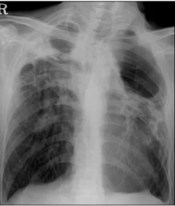
Figure 1. The initial chest X-ray showing destroyed lung lesions. Multifocal cystic lesions and consolidations are also seen.
Figure 2. The initial chest CT showing wall thickening of cavitary lesion in left upper lobe and peribronchial air space consolidation in right lung. CT: computed tomography.
증 례
환 자: 백○○, 남자, 54세
주 소: 내원 5개월 전부터 지속되는 기침, 객담 및 호흡 곤란
현병력: 30년 전 폐결핵에 감염되어 약물치료 후 완치 되었으나 그 후 6년 전, 2년 전에 결핵 재발로 진단받고 완치 판정을 받은 자로 내원 5개월 전부터 기침, 호흡곤란 증상이 지속되고 내원 2일 전 기침, 객담 및 호흡곤란의 악화와 더불어 발열이 동반되어 외래를 경유하여 입원함.
과거력: 30년 전, 6년 전, 2년 전 결핵으로 치료받은 후 완치 판정을 받았고, 1년 전에는 폐쇄성 폐기능 장애로 진단받고 개인의원에서 기관지확장제 등을 복용하고 있 었음. 매일 소주 1∼2병씩 마시는 음주력과 30갑년의 흡
연이력 있었음.
흡연력: 하루에 한 갑씩 30년간 흡연(1갑/1일, 30갑년) 가족력: 특이 사항 없음.
신체검사 소견: 입원 당시 혈압 146/92 mm Hg, 맥박수 110회/분, 호흡수 30회/분, 체온은 38.9oC였으며 산소포 화도는 93%였다. 환자는 급성 병색을 띠고 있었고, 중등 도의 호흡곤란을 호소하였으며, 흉부 진찰상 우상엽에서 악설음이 청진되었고 좌상엽 청음이 감소한 소견이 보였 다. 기타 두경부 검사 및 복부 이학적 검사상 이상 소견은 없었다. 신경학적 검사에서도 특이 소견은 없었다.
검사 소견: 입원 시 백혈구 22,300/μL, 호중구는 86.3%였으며, CRP는 17.34 mg/dL로 측정되었고 전해질 및 기타 생화학 검사에서 이상은 없었으며 혈청학적 검사 상 VDRL, HBs항원, HCV항체 및 HIV항체는 모두 음성이 었다. 심전도는 특이 소견이 없었으며 흉부 단순촬영에서 파괴된 폐와 우상엽과 좌상엽의 경화 소견이 관찰되었고 (Figure 1) 흉부 컴퓨터촬영에서 좌상엽의 공동성 병변과 우상엽의 기관지 주변으로 발생한 경화 소견이 관찰되었 다(Figure 2). 결핵재발 및 지역사회획득 폐렴의 가능성을 고려하여 객담 항산균 도말 검사와 더불어 경험적 항생제 (ceftriaxone 2 g/day, clarithromycin 500 mg/day)를 투여 하였다. 또한 입원 시 시행한 항산균 도말 검사에서 +4 소견으로 1차 항결핵약제를 투여하였고, 반복되는 결핵의 발생 및 과거 치료력 등을 바탕으로 내성 결핵 및 비결핵 항산균 질환의 가능성을 고려해 기관지 세척액에 대한 결
핵균
rpoB
유전자 변이에 대한 검사를 계획하고 시행하였다. 기관지내시경 검사에서는 다량의 화농성 분비물 소견 이 관찰되었으나 기관지 내 병변은 없었다. 항생제치료로 발열 및 흉부 영상학적 소견 및 백혈구 증가 소견은 호전 되었으나 기침과 객담이 지속되고 입원 이후 시행한 3회 의 객담 및 기관지 세척액 항산균 도말(Ziehl-Neelsen
Tuberculosis and Respiratory Diseases Vol. 72. No. 1, Jan. 2012
57
Figure 3. The follow-up chest X-ray after 16 months oftreatment (treatment completion). Decreased extent of consolidation and cavitary lesion are seen.
method)에서 4+로 강양성 소견이 관찰되었으나 동일한 객담 및 기관지 세척액의 TB-PCR 검사는 모두 음성으로, 비결핵항산균 폐질환으로 잠정 진단되었다. 이후 항결핵 치료를 중단하고 항산균 배양 결과를 기다리며 경과관찰 하였다. 1달 후 Ogawa 배지에서 항산균 배양 양성 소견 이었고, 이 배양균에 대한 TB-PCR 결과는 음성이었다. 또 한 이 검체에 대해 시행한 비결핵항산균 동정 검사에서
Mycobacterium szulgai
가 3회 분리, 동정되었다. 최종적 으로Mycobacterium szulgai
에 의한 폐질환으로 진단되 었다.임상 경과 및 치료: 환자에게 clarithromycin 1,000 mg/day, rifampicin 450 mg/day, ethambutol 1,200 mg/day 투여하였으며 치료 시작 1달 후 시행한 객담 항산 균 도말 및 배양 검사가 음전되었다. 16개월간의 약물치 료 기간 동안 환자의 균 배양은 음전된 상태를 유지하였 다. 1년 간의 흉부 영상 및 객담 검사를 통한 추적에서 재발은 없는 상태로 유지되었으며 흉부 단순촬영에서도 예전에 비해 호전된 소견이 관찰되었다(Figure 3). 그러 나, 완치 판정 이후 좌상엽에 공동성 병변 및 경화 소견이 발생하였고 이는 경험적 항생제 투여에도 호전되지 않았 다. 항산균 도말 및 배양 검사에서 비결핵항산균이 동정 되지 않았으나 Aspergillus 항원 양성 소견을 보였고 환자 의 임상 소견 및 영상의학적 소견을 바탕으로 chronic ne- crotizing pulmonary aspergillosis 최종 진단되었다. Itra- conazole을 비롯한 항진균제 투여에도 불구하고 환자는
다량의 객혈로 인한 호흡부전으로 사망하였다.
고 찰
M. szulgai
는 slow growing nontuberculous myco- bacterium (NTM)으로 1972년도에 Marks 등4에 의해 처음 기술되었으며, 이후 이 NTM이 새로운 균종인 것을 밝혀 낸 T. Szulga의 이름을 따서 명명되었다. 달팽이, 수조, 수영장 물, 열대물고기 등 자연계에 분포되어 있고5 특별 한 시기나 지역에 편중 없이 나타나며 주로 폐감염을 일으 키고 전신 파종성 감염은 주로 면역기능이 저하된 환자에 서 보고되고 있다6. 최근의 보고에 의하면, 예전에 알려진 바와 달리 결핵의 유병률이 높은 국가에서도 비결핵항산 균 감염이 드물지 않은 것으로 알려졌으며7 개발도상국의 비결핵항산균에 의한 감염 및 질환의 유병률은 증가하고 있다8. 우리나라에서는 1980년대 초까지는 임상 검체에서 분리되는 mycobacteria의 97∼98% 이상이 결핵균이었으 나, 1990년대 이후 비결핵항산균이 분리되는 비율이 증가 하는 추세에 있다9.비결핵항산균감염증은 폐질환, 림프절염, 피부·연조 직·골감염증, 파종성 질환 등 네 가지 임상증후군으로 분류되며10, 이 중 폐질환은 비결핵항산균 질환의 90% 이 상을 차지하는 가장 흔한 형태이다. 호흡기 검체에서 비 결핵항산균이 분리되었을 때 오염균이나 집락균과 병원 균과의 구별을 위해 정확한 균 동정과 함께 적절한 기준에 따른 진단이 중요한데, 본 증례는 2007년 미국흉부학회 (American Thoracic Society)의 진단기준에10 따라 호흡기 증상과 함께 방사선학적으로 공동성 병변, 객담 및 기관지 세척액 항산균 도말(Ziehl-Neelsen method)에서 4+로 강양성을 보였으며, Ogawa 배지에서 항산균 배양에서 Mycobacterium szulgai가 3회 동정되어 최종적으로
M.
szulgai
에 의한 폐질환으로 진단할 수 있었다. 본 증례에서 치료 시작 후 한번도
M. szulgai
가 동정되지 않은 점에 서 실질적으로 동정된 균만으로 비결핵항산균 폐질환이 아닐 가능성을 고려해 볼 수 있다. 그러나 최근 미국흉부 학회지침10에 따르면 역학적으로M. szulgai
는 환경오염 균일 가능성이 매우 낮으며, 배양된 경우 대부분 임상적 및 병리학적 중요성을 갖는다고 알려져 있고,M. kansassi
폐질환의 경우처럼 적절한 임상적 상황에서는 한 번의 배 양만으로M. szulgai
질환으로 고려될 수 있다고 제시하고 있다. 또한, 장기간의 지속적인 비결핵항산균치료로 임상 증상의 호전과 더불어 환자의 흉부 방사선 소견이 완만하EJ Lee et al: A case of Mycobacterium szulgai lung disease
58
게 호전되었다는 점에서 본 증례가
M. szulgai
에 의한 것 이라 보는 것이 타당하리라 판단된다.한편, 이 증례의 환자는 과거 2차례의 결핵 약제를 복 용한 경력이 있었는데, 최근 결핵치료 시 시행된 객담 배 양 검사에서는 항산균 양성으로만 표기되어 배양균주가 결핵인지 비결핵항산균인지 확인할 수 없었다. 따라서 최 소한 2년 전부터 이 질환을 가지고 있었을 가능성을 완전 히 배제할 수 없다고 판단된다.
M. szulgai
는 국내에서 드물게 분리되는 균종으로 Kim 등11이 2004년도에 NTM 10예의 분리를 보고하면서M.
szulgai
8예를 보고한 바 있으나 당시 반복 배양하여 확인 하는 절차를 거치지 못했고 대부분 환자들이 기존 질환으 로 일찍 사망하거나 특이한 임상 증상이 없어서 병원성 균주가 아니거나 오염균의 가능성을 완전히 배제할 수 없 었다. 그 외에는 아직까지 국내에서M. szulgai
단독으로 보고되었던 예는 없었다. 본 증례는 객담 및 기관지 세척 액에서 동일 균주가 반복 동정되었으며 이에 대한 macro- lide계 항생제를 기반으로 한 16개월 간의 병합화학요법 후 객담 항산균 도말 및 배양 검사가 음전 및 유지되었고 호흡기 및 전신 증상의 호전과 더불어 영상학적 소견이 호전되어 이는M. szulgai
에 의한 폐질환이 약물치료로 완치가 확인된 증례이다. 따라서 본 저자들은M. szulgai
폐질환이 진단되고 약물치료 후 완치되었던 사례가 국내 에서 보고된 바가 없어 이에 본 병원에서 경험한 증례를 보고하는 바이다.참 고 문 헌
1. Park YS, Lee CH, Lee SM, Yang SC, Yoo CG, Kim YW, et al. Rapid increase of non-tuberculous mycobacterial lung diseases at a tertiary referral hospital in South
Korea. Int J Tuberc Lung Dis 2010;14:1069-71.
2. van Ingen J, Boeree MJ, de Lange WC, de Haas PE, Dekhuijzen PN, van Soolingen D. Clinical relevance of Mycobacterium szulgai in the Netherlands. Clin Infect Dis 2008;46:1200-5.
3. Koh WJ, Kwon OJ, Lee KS. Diagnosis and treatment of nontuberculous Mycobacterial pulmonary diseases:
a Korean perspective. J Korean Med Sci 2005;20:
913-25.
4. Marks J, Jenkins PA, Tsukamura M. Mycobacterium szulgai--a new pathogen. Tubercle 1972;53:210-4.
5. Benator DA, Kan V, Gordin FM. Mycobacterium szul- gai infection of the lung: case report and review of an unusual pathogen. Am J Med Sci 1997;313:346-51.
6. Maloney JM, Gregg CR, Stephens DS, Manian FA, Rimland D. Infections caused by Mycobacterium szul- gai in humans. Rev Infect Dis 1987;9:1120-6.
7. Shenai S, Rodrigues C, Mehta A. Time to identify and define non-tuberculous mycobacteria in a tuberculo- sis-endemic region. Int J Tuberc Lung Dis 2010;14:
1001-8.
8. Alvarez-Uria G. Lung disease caused by nontuber- culous mycobacteria. Curr Opin Pulm Med 2010;16:
251-6.
9. Koh WJ, Kwon OJ, Yu CM, Jeon KM, Suh GY, Chung MP, et al. Recovery rate of nontuberculous Mycobac- teria from acid-fast-bacilli smear-positive sputum spe- cimens. Tuberc Respir Dis 2003;54:22-32.
10. Griffith DE, Aksamit T, Brown-Elliott BA, Catanzaro A, Daley C, Gordin F, et al. An official ATS/IDSA state- ment: diagnosis, treatment, and prevention of non- tuberculous mycobacterial diseases. Am J Respir Crit Care Med 2007;175:367-416.
11. Kim JH, Suh JT, Park SY, Lee HJ, Lee WI. Clinical eval- uation of 10 cases of nontuberculous Mycobacteria iso- lated from sputum. Korean J Lab Med 2004;24:49-52.