Keratinocyte-derived Laminin-332 Protein Promotes Melanin
Synthesis via Regulation of Tyrosine Uptake
*
Received for publication, December 6, 2013, and in revised form, June 9, 2014Published, JBC Papers in Press, June 20, 2014, DOI 10.1074/jbc.M113.541177
Heesung Chung‡, Hyejung Jung‡, Jung-hyun Lee‡, Hye Yun Oh§, and Ok Bin Kim§, Inn-Oc Han¶, and Eok-Soo Oh‡1
From the‡Department of Life Sciences, Research Center for Cellular Homeostasis and§Department of Life Sciences,
Interdisciplinary Program of EcoCreative, Ewha Womans University, Seoul 120-750, Korea and the¶College of Medicine,
Department of Physiology and Biophysics, Inha University, Incheon 402-751, Korea
Background:Laminin-332 derived from keratinocytes plays a critical role in adhesion-related cell functions in melanocytes.
Results:Keratinocyte-derived laminin-332 promotes the uptake of extracellular tyrosine and subsequent melanin synthesis in melanoma cells and melanocytes.
Conclusion: Keratinocyte-derived laminin-332 promotes melanogenesis by controlling the uptake of tyrosine into melanocytes.
Significance:Our finding reports novel means for regulating melanogenesis by the insoluble extracellular protein laminin-332.
Melanocytes, which produce the pigment melanin, are known to be closely regulated by neighboring keratinocytes. However, how keratinocytes regulate melanin production is unclear. Here we report that melanin production in melanoma cells (B16F10 and MNT-1) was increased markedly on a keratinocyte-derived extracellular matrix compared with a melanoma cell-derived extracellular matrix. siRNA-mediated reduction of keratino-cyte-derived laminin-332 expression decreased melanin synthe-sis in melanoma cells, and laminin-332, but not fibronectin, enhanced melanin content and␣-melanocyte-stimulating hor-mone-regulated melanin production in melanoma cells. Similar effects were observed in human melanocytes. Interestingly, however, laminin-332 did not affect the expression or activity of tyrosinase. Instead, laminin-332 promoted the uptake of extra-cellular tyrosine and, subsequently, increased intraextra-cellular lev-els of tyrosine in both melanocytes and melanoma cells. Taken together, these data strongly suggest that keratinocyte-derived laminin-332 contributes to melanin production by regulating tyrosine uptake.
Melanocytes, which are present in the skin, hair, eyes, and ears, synthesize melanin via a process known as melanogenesis in the melanosome, a specialized organelle of melanocytes (1). Because melanin is an essential component of the pigmentary system of the skin, melanogenesis is a crucial and special step for the regulation of melanocyte functions such as photopro-tection (2). Therefore, many pigmentary skin diseases are closely correlated with failure of the regulation of melanin syn-thesis. Albinism is induced by the complete or partial absence
of pigmentation in the skin, hair, and eyes (3). On the other hand, vitiligo is caused by depigmentation in the skin when melanocytes die or loss of function because of autoimmune, genetic, oxidative stress, or viral causes (4). Genetic disorder in melanocytes also causes Waardenburg syndrome, which has various symptoms, including pale or blue eyes, white hair, and white patches on the skin (5).
Melanogenesis is critically regulated by the expression of the representative enzymes for promoting melanogenesis. Those include tyrosinase, tyrosinase-related protein 1 (TRP-1), and tyrosinase-related protein 2 (TRP-2). Tyrosinase stimulates the rate-limiting hydroxylation of L-tyrosine to L -3,4-dihydroxy-phenylalanine (L-DOPA).2Sequentially,L-DOPA is converted rapidly intoL-dopaquinone by tyrosinase. On the other hand, TRP-2 stimulates keto-enol tautomerization of dopachrome into 5,6-dihydroxyindole-2-carboxylic acid. Because of this function, TRP-2 is also called dopachrome tautomerase. Although the role of TRP-1 is controversial, TRP-1 has been reported to be involved in tyrosinase activation and stability (6). To serve a photoprotective role, melanin produced in mela-nocytes needs to transfer to neighbor keratimela-nocytes, the pre-dominant cell type in the skin epidermis, to protect keratino-cytes from UV light (7). Therefore, keratinokeratino-cytes are actively involved in the regulation of melanin synthesis. They produce various regulatory soluble factors such as ␣-melanocyte-stim-ulating hormone (␣-MSH), adrenocorticotropic hormone, stem cell factor, and endothelin 1 (8). ␣-MSH, for instance, binds to melanocortin receptor 1 (MC1R) (9) and increases the cAMP level by stimulating adenylyl cyclase. cAMP subse-quently activates PKA and cAMP-responsive element-binding protein. cAMP-responsive element-binding protein works as a transcription factor for microphthalmia-related transcription factor (MITF), a critical regulator of tyrosinase, TRP-1, and TRP-2 (10, 11).
*This work was supported by the Basic Science Research Program through the National Research Foundation of Korea funded by Ministry of Educa-tion, Science, and Technology Grant 2010-0007336; by National Research Foundation of Korea Grant 2012R1A5A1048236 funded by the Korean government; and by Korean Health Technology R&D Project, Ministry of Health and Welfare, Republic of Korea Grant HI12C0050.
1To whom correspondence should be addressed: Dept. of Life Sciences, Ewha Womans University, 52, Ewhayeodae-gil, Seodaemoon-Gu, Seoul 120-750, Korea. Tel.: 82-2-3277-3761; Fax: 82-2-3277-3760; E-mail: [email protected].
2The abbreviations used are:
L-DOPA,L-3,4-dihydroxyphenylalanine;␣-MSH, ␣-melanocyte-stimulating hormone; MITF, microphthalmia-related tran-scription factor; ECM, extracellular matrix/matrices; MEM, minimal essen-tial medium; REF, rat embryonic fibroblast; TRP, tyrosinase-related protein; FN, fibronectin.
at Ewha Medical Library on September 8, 2016
http://www.jbc.org/
In addition to soluble factors, keratinocytes also produce extracellular proteins to regulate the cellular behaviors of mela-nocytes. It has been known that keratinocytes produce various extracellular matrix (ECM) proteins, such as fibronectin, laminin, and collagen (12, 13). These ECM proteins have been commonly reported to regulate cell adhesion, proliferation, and migration (14 –16). One of the ECM proteins produced by kera-tinocytes is laminin-332. Laminin-332 is involved in the initia-tion of hemidesmosome formainitia-tion and in the stable structure of the epidermis (17). In addition, laminin-332 is known to stimulate the migration of keratinocytes in wound healing (18), tumor growth and invasion of melanoma cells (19), and lamel-lipodia formation of keratinocytes (20).
Recently, we have shown that keratinocyte-derived laminin-332 plays a crucial role in adhesion-related cell functions of melanocytes and melanoma cells (21). This suggests that laminin-332 might be involved in the regulation of melanin synthesis. Here we examined whether keratinocyte-derived laminin-332 regulates melanogenesis in melanoma and mela-nocytes cells.
EXPERIMENTAL PROCEDURES
Materials and Antibodies—Polyclonal antibodies against laminin ␥2 chain and tyrosinase and monoclonal antibody against phospho-ERK, ERK, and-actin were purchased from Santa Cruz Biotechnology (Santa Cruz, CA). Polyclonal anti-body to phospho-p38 was purchased from Cell Signaling Tech-nology (Danvers, MA). Human plasma fibronectin (purity, ⬃95%) was purchased from Millipore (Billerica, MA). Human laminin-332 purified from human foreskin keratinocytes (purity, ⬎95%) was from Abcam (Cambridge, MA). Human recombinant laminins (111, 332, and laminin-511) were from BioLamina (Sundbyberg, Sweden). ␣-MSH, L-tyrosine, andL-DOPA were purchased from Sigma.L -[ring-3,5-3H]tyrosine was purchased from PerkinElmer Life Sciences (Waltham, MA). PD98059 was purchased from Calbiochem (Darmstadt, Germany).
Cell Culture and Transfection—The B16F10 mouse mela-noma cell line and the HaCaT human keratinocyte cell line were maintained in DMEM (WelGene, Daegu, Korea) supple-mented with 10% FBS (Hyclone) and gentamicin (50g/ml, Sigma). The MNT-1 human melanoma cell line was maintained in minimal essential medium (MEM) supplemented with 20% FBS, 10% DMEM, 20 mMHepes, and gentamicin (50g/ml). The REF cell line was maintained in␣-MEM (Invitrogen) sup-plemented with 5% FBS and gentamicin (50g/ml). Primary human epidermal melanocytes were purchased from Lonza (Basel, Switzerland). Human epidermal melanocytes were maintained in melanocyte growth medium 4 (Lonza) supple-mented with FBS, recombinant human FGF B, recombinant human insulin, gentamicin sulfate amphotericin B (GA-1000), calcium chloride (CaCl2), phorbol 12-myristate 13-acetate, bovine pituitary extract, and hydrocortisone. Cells were cul-tured at 37 °C in 5% CO2in a humidified atmosphere. Tran-sient transfections were carried out using Lipofectamine 2000 (Invitrogen) according to the instructions provided by the manufacturer.
RNA Extraction and RT-PCR—Total RNA extracted from cells was reverse-transcribed, and aliquots of the resulting cDNA were amplified using the following primers: human LAMC2, 5⬘-TGGAGAACGCTGTGATAGGTGTCG-3⬘ (forward) and 5⬘-TGTGTAAGTCTTGGTGAGCCCAC-3 ⬘(reverse);mousetyrosin-ase, 5⬘-CGAGCCTGTGCCTCCTCTAA-3⬘ (forward) and 5⬘-CCA-GGACTCACGGTCATCCA-3⬘ (reverse); mouse MITF, 5⬘-GGAA-CAGCAACGAGCTAAGG-3⬘ (forward) and 5⬘-TGATGATCCG-ATTCACCAGA-3⬘ (reverse);-actin, 5⬘-TGGAATCCTGTGGC-ATCCATGAAA-3 ⬘(forward)and5⬘-TAAAACGCAGCTCAGTA-ACAGTCCG-3 ⬘(reverse);mouseITGA3,5⬘-CCCACCCGGTGT-GACTTCT-3⬘ (forward) and 5⬘-GACTTGGATACGGCACC-CTC-3⬘ (reverse); mouse ITGA6, 5⬘-AAAGAGACATGA AGTC-CGCGCATC-3⬘ (forward) and 5⬘-AACAATGTCTTGCCACC-CATCTGC-3⬘ (reverse); ITGB1, 5⬘-AATGTTTCAGTGCAG-AGC-3⬘ (forward) and 5⬘-TTGGGATGATGTCGGGAC-3⬘ (reverse); and mouse ITGB4, 5 ⬘-AAGAGTGGCTCCTCCTC-CAG-3⬘ (forward) and 5⬘-TCACCTGAGCCTTCTTGCAG-3⬘ (reverse). After an initial denaturation at 94 °C for 5 min, samples were subjected to 30 cycles of denaturation at 94 °C for 30 s, annealing at 52 °C for 30 s, and extension at 72 °C for 60 s.
siRNA—Human LAMC2-and FN-specific siRNA oligonu-cleotides were designed. ITGA3-, ITGA6-, ITGB1-, and ITGB4-specific siRNA oligonucleotides were also designed. The sequences were as follows: LAMC2 siRNA, 5 ⬘-GCAAAG-AGGAUCAAACAAAUU-3⬘ (sense) and 5⬘-UUUGUUUGUA-UCCUCUUUGCUU-3⬘ (antisense); FN siRNA, 5⬘-GCUGAA-GACACAAGGAAAUUU-3⬘ (sense) and 5⬘-AUUUCCUUGU-GUCUUCAGCUU-3⬘ (antisense); ITGA3 siRNA, 5⬘-CCUAC-UACUUCGAGAGGAAUU-3⬘ (sense) and 5⬘-UUCCUCGAA-GUAGUAGGUU-3⬘ (antisense); ITGA6 siRNA, 5⬘-GAGUAU-GAAUUCAGGGUAAUU-3⬘ (sense) and 5⬘-UUACCCUGAA-UUCAUACUCUU-3⬘ (antisense); ITGB1 siRNA, 5⬘-CCACA-GACAUUUACAUUAAUU-3⬘ (sense) and 5⬘-UUAAUGUAA-AUGUCUGUGGUU-3⬘ (antisense); and ITGB4 siRNA, 5⬘-A-AGAACCGGAUGCUGCUUAUUUU-3⬘ (sense) and 5⬘-AAU-AAGCAGCAUCCGGUUCUUUU-3⬘ (antisense). Scrambled siRNA (siGENOME non-targeting siRNA #2) was purchased from Dharmacon (Lafayette, CO) and used as a control.
Immunoblotting—The cells were washed twice with PBS and lysed in radioimmune precipitation assay buffer (50 mMTris (pH 8.0), 150 mMNaCl, 1% Nonidet P-40, 10 mMNaF, and 2 mM Na3VO4) containing protease inhibitors (1g/ml aprotinin, 1 g/ml antipain, 5 g/ml leupeptin, 1 g/ml pepstatin A, and 20 g/ml phenylmethylsulfonyl fluoride). The lysates were clarified by centrifugation at 13,000⫻ g for 15 min at 4 °C, denatured with SDS sample buffer, boiled, and analyzed by SDS-PAGE. The resolved proteins were transferred to PVDF membranes (Millipore) and probed with the appropriate antibodies. The signals were detected by enhanced chemiluminescence (AbClon, Seoul, Korea).
Immunofluorescence Analysis—Cells were plated onto 12-well plates containing coverslips and fixed with 3.5% parafor-maldehyde for 10 min. After being washed with PBS, cells were blocked with 0.5% BSA and incubated overnight with an anti-laminin␥2 antibody at 4 °C. After being washed with PBS, cells were incubated with Texas Red-conjugated goat anti-rabbit antibody (Invitrogen) for 1 h at 25 °C. For F-actin staining, cells
at Ewha Medical Library on September 8, 2016
http://www.jbc.org/
were fixed with 3.5% paraformaldehyde and permeabilized with 0.5% Triton X-100. After blocking with 0.5% BSA, cells were incubated with FITC-conjugated phalloidin antibody for 1 h at 25 °C. Coverslips were then mounted with mounting solution containing DAPI on glass slides and observed by fluorescence microscopy.
Preparation of Tissue Culture Plates Coated with ECM Substrate—ECM proteins were diluted in serum-free medium (laminin-332, 1g/cm2; fibronectin, 0.5g/cm2) added to the plates and incubated at 25 °C for 1 h to allow adsorption onto the plates. After being washed with PBS, plates were blocked with 0.2% heat-inactivated BSA in PBS for 1 h and then washed three times with PBS. For preparing cells, the cells were detached with 0.05% trypsin and 1 mMEDTA, suspended in medium containing 0.5% FBS, harvested, resuspended in medium containing 0.5% FBS, plated onto ECM-coated plates, and incubated for 24 h at 37 °C and 5% CO2.
Preparation of Keratinocyte-derived ECM—The keratino-cyte-derived ECM was prepared according to the method of Rodeck et al. (22). Briefly, HaCaT cells (90⬃100% confluent) grown on culture plates were detached with 0.05% trypsin and 1 mMEDTA in PBS. The detached cells were removed, and the adherent ECM on the culture plate was washed with PBS and treated with 0.1 mg/ml soybean trypsin inhibitor (Invitrogen). The plates were then washed with PBS, blocked with 0.2% heat-inactivated BSA for 1 h, and washed with PBS. Alternatively, HaCaT cells grown on tissue culture plates were removed by sequential extraction with 1% Triton X-100 in PBS, 2Murea in 1MNaCl, and 8Murea in 1MNaCl (23). After removing cells, ECM-deposited plates were washed and blocked with the same method used to remove cells with 0.05% trypsin and 1 mM EDTA. Melanoma cells were plated on HaCaT ECM for 24 h at 37 °C in medium containing 1% FBS.
Melanin Determination—Cells were plated on an ECM-coated 6-well tissue culture dish for 24 h at 37 °C. Cells were detached using 0.05% trypsin and 1 mMEDTA in PBS at 37 °C in 5% CO2. Detached cells were harvested into a 1.5-ml tube and centrifuged at 1000⫻ g for 3 min. After removing the superna-tant, cell pellets were suspended with PBS. Cells were counted using a hemocytometer. Equal numbers of cells (B16F10 and MNT-1 cells, 3.5⫻ 105cells; melanocytes, 2.0⫻ 105cells) were centrifuged at 1000⫻ g for 3 min and solubilized in 50l of 1 N NaOH and 10% dimethyl sulfoxide for 2 h at 80 °C. The dis-solved melanin was assessed by absorbance at 405 nm, and the melanin content was determined using a standard curve gener-ated with synthetic melanin (Sigma). The results were analyzed in percentage terms.
Tyrosinase Activity Assay—Tyrosinase activity was assayed using a modified version of the method described by Ando et al. (24). After incubation on ECM or␣-MSH for 24 h, cells were lysed in tyrosinase assay buffer (50 mMsodium phosphate (pH 6.8), 1% Triton X-100, 1 mMphenylmethylsulfonyl fluoride, 1 g/ml aprotinin, and 10 g/ml leupeptin) The lysates were clarified by centrifugation at 13,000⫻ g for 15 min at 4 °C and denatured with SDS sample buffer without mercaptoethanol and heating. The proteins (20g) were resolved by SDS-PAGE, and the gels were rinsed with 50 mMsodium phosphate buffer (pH 6.8) and equilibrated at room temperature. After 30 min,
each gel was reacted with staining solution (10 mM L-DOPA in 50 mMsodium phosphate buffer) and incubated in the dark for 2 h at 37 °C. Tyrosinase activity was visualized in the gels as dark bands containing L-DOPA-melanin. Alternatively, clarified lysates were reacted withL-DOPA (5 mM) at 37 °C, and the absorbance was measured at 470 nm (25).
Intracellular Tyrosine Determination—Cells were seeded into a 10-mm dish with MEM (1% FBS) and treated withL -ty-rosine (500M). Cells were lysed with hypotonic solution (20 mMTris, 5 mMEGTA, 2 mMEDTA, 2 mM-mercaptoethanol, 5 mMNaF, and 1 mMNa3VO4) containing protease inhibitors (0.5g/ml aprotinin, 0.5 g/ml antipain, 2.5 g/ml leupeptin, 0.5 g/mlpepstatinA,and10g/mlphenylmethylsulfonylfluo-ride). The lysates were clarified by centrifugation at 13,000⫻ g for 15 min at 4 °C. PlasmaL-tyrosine was measured using an L-8800 automatic amino acid analyzer (L-8800A, Technopark, Inchoen, Korea). The concentrations of intracellular tyrosine were normalized with concentrations of total cell lysates.
Tyrosine Uptake Measurement—Tyrosine uptake into B16F10 cells and melanocytes was measured according to the method of Fuller et al. (26). Cells (1 ⫻ 105cells/well) were seeded into a 12-well plate with MEM (1% FBS) and preincu-bated in transport buffer (140 mMNaCl, 5 mMKCI, 5.6 mM, glucose, 0.9 mMCaCl2, 1.0 mMMgCl2and 25 mMHepes (pH 7.4)) at 37 °C for 20 min. The mixture of unlabeledL-tyrosine andL-[ring-3,5-3H]tyrosine was treated to cells (0.2 mM, 4.28⫻ 105dpm/ml). After 20 min, the tyrosine uptake reaction was stopped with LiCl (100 mM), and cells were lysed with 10% TCA. Lysates were mixed with scintillation solution (Insta-Gel plus, PerkinElmer Life Sciences). Liquid scintillation counting (LS6500, Beckmann) was used to determine radioactivity, which was normalized with respect to the total cell number.
Statistical Analysis—Data are represented as the mean⫾ S.D. from three independent experiments. Statistical analysis was performed using one-way analysis of variance. p⬍ 0.01 or 0.05 was considered to be statistically significant.
RESULTS
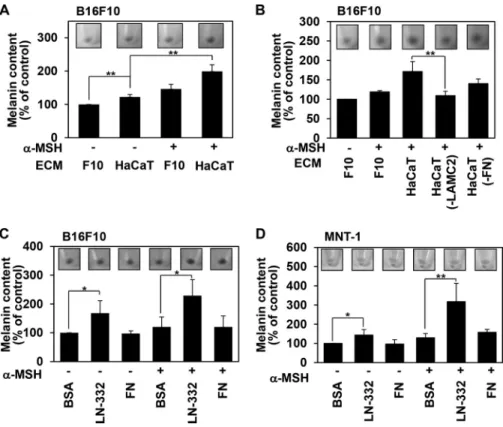
Keratinocyte-derived Laminin-332 Promotes Melanin Syn-thesis of Melanoma Cells—To investigate the effect of keratino-cyte-derived ECM on melanin synthesis, melanoma cells were detached and replated on ECM derived from HaCaT keratino-cytes (HaCaT ECM) or rat embryonic fibroblasts (REF ECM), and the amount of melanin was compared with that of cells on melanoma ECM. Interestingly, B16F10 mouse melanoma cells on HaCaT ECM produced more melanin than cells on B16F10 ECM (F10 ECM) or REF ECM (Fig. 1A). Similarly, MNT-1 human melanoma cells on HaCaT ECM produced more mela-nin than cells on MNT-1 ECM or REF ECM (Fig. 1A). HaCaT ECM increased the amount of melanin from 98.23 pg/cell to 163.2 pg/cell in B16F10 cells and from 153.86 pg/cell to 598.81 pg/cell in MNT-1 cells. We obtained similar results in B16F10 cells on HaCaT ECM that was prepared by removing cells using either trypsin/EDTA or 1% Triton X-100 (Fig. 1B), suggesting that ECM produced specifically by keratinocytes regulates mel-anin synthesis in melanoma cells.
Because laminin-332 is known to regulate adhesion-depen-dent signaling in melanoma and melanocytes (21), we
at Ewha Medical Library on September 8, 2016
http://www.jbc.org/
gated whether laminin-332 regulates melanin synthesis of mel-anoma cells. We found that both B16F10 and MNT-1 cells grown on laminin-332 showed increased production of mela-nin compared with cells grown on either BSA or fibronectin (Fig. 1C). In addition, laminin-332 enhanced melanin synthesis more strongly than other laminins (Fig. 1D).
To further investigate the potential involvement of laminin-332 in the regulation of melanin synthesis, we used unique 21-bp siRNA sequences targeted against the laminin␥2 chain (si-LAMC2) to knock down the expression levels of laminin-332. HaCaT transfected with the siRNA constructs showed decreased mRNA (Fig. 1E, top panel) and protein expression of the targeted proteins (Fig. 1E, center panel). Consistently, our results revealed that B16F10 cells grown on HaCaT ECM derived from laminin-332 knockdown cells showed reduced melanin synthesis (Fig. 1E, bottom panel). Integrins are impor-tant cell surface receptors binding to ECM proteins (27), and integrin ␣61, ␣64, and ␣31 are known receptors for laminin-332 (28). Interestingly, knockdown of integrin ␣6 using unique siRNA sequences targeted against integrin␣6 (si-ITGA6) significantly reduced melanin synthesis in B16F10 cells grown on HaCaT ECM (Fig. 1F). However, those effects were not seen in B16F10 cells transfected with siRNA of integrin␣3 (si-ITGA3), suggesting that integrin ␣6 participates in the laminin-332-mediated melanin synthesis of melanoma cells to keratinocyte-derived ECM. Together, these data strongly sug-gest that laminin-332 enhances melanin synthesis in melanoma cells.
Laminin-332 Potentiates the Melanogenic Response to ␣-MSH in Melanoma Cells—Because ␣-MSH is known to be a key regulator of melanin synthesis in melanocytes (9), we next investigated whether keratinocyte-derived laminin-332 was involved in the regulation of␣-MSH-induced melanin synthe-sis. We found that growth on HaCaT ECM enhanced ␣-MSH-induced melanin synthesis in B16F10 cells (Fig. 2A), whereas growth on HaCaT ECM derived from laminin-332 knockdown cells did not (Fig. 2B). Consistent with these results, growth on laminin-332 directly enhanced␣-MSH-induced melanin syn-thesis in both B16F10 (Fig. 2C) and MNT-1 cells (Fig. 2D). These findings indicate that laminin-332 potentiates ␣-MSH-induced melanin synthesis in melanoma cells and melanocytes. Laminin-332-mediated Melanin Synthesis Is Independent of Tyrosinase—Various enzymes and transcription factors are involved in melanogenesis, including tyrosinase, TRP-1, TRP-2, and MITF (29 –31). Therefore, we investigated whether laminin-332 regulates melanin synthesis by modulating the expression levels of these regulators (Fig. 3). However, the mRNA levels of tyrosinase and MITF in B16F10 cells did not differ significantly in cells grown on B16F10 ECM and those grown on HaCaT ECM (Fig. 3A). In addition, growth on HaCaT ECM failed to affect tyrosinase activity (Fig. 3B). Similarly, growth on laminin-332 did not affect the protein expression level of tyrosinase (Fig. 3C) and the activity of tyrosinase (Fig. 3, D and E). These results suggest that laminin-332 stimulates melanin synthesis through a tyrosin-ase-independent pathway.
FIGURE 1. Keratinocyte-derived laminin-332 promotes melanin synthesis of
melanoma cells. A, melanoma cells were plated on melanoma ECM (B16F10 and
MNT-1 ECM), HaCaT ECM, and REF ECM and incubated at 37 °C. After 24 h, cells were harvested, and cells pellets were photographed with a digital camera (insets). An equal number of cells (3.5⫻ 105cells) was solubilized with 50l of 1 N NaOH-10% dimethyl sulfoxide for 2 h at 80 °C to dissolve melanin. The melanin content was determined by absorbance at 405 nm. Error bars indicate mean⫾ S.D. **, p⬍ 0.01 versus melanoma cell ECM. B, to prepare ECM, B16F10 cells and HaCaT cells grown at confluence in tissue culture plates were detached with 0.05% trypsin and 1 mMEDTA (Trypsin-EDTA) or 1% Triton X-100 (1% Tx-100) as described under “Experimental Procedures.” B16F10 cells were plated on each ECM for 24 h. Cells (3.5⫻ 105cells) were harvested and solubilized, and the mel-anin content was analyzed as described in A. Error bars indicate mean⫾S.D.*,p⬍ 0.05; **, p⬍ 0.01 versus F10 ECM. C, after coating ECM proteins (laminin-332, 1 g/cm2; fibronectin, 0.5g/cm2), cells were plated on either FN or laminin-332 (LN-332) and incubated at 37 °C. After 24 h, cells were harvested, and the melanin content was determined by absorbance at 405 nm. BSA was used as a control.
Error bars indicate mean⫾S.D.*,p⬍0.05;**,p⬍0.01.D,B16F10cellswereplated
on the indicated laminin and incubated at 37 °C. After 24 h, cells (3.5⫻ 105cells) were harvested, and the melanin content was determined by absorbance at 405 nm. Error bars indicate mean⫾ S.D. *, p ⬍ 0.05. E, HaCaT cells were transfected with siRNAs targeting laminin␥-chain (LAMC2). The expression levels of the tar-get mRNAs were analyzed by RT-PCR (top panel). Both HaCaT and B16F10 cells were cultured on cover slips and immunostained with anti-laminin␥2 chain anti-body. The results were visualized with a Texas Red-conjugated goat anti-rabbit antibody. DAPI was used to stain nuclei (center panel). Scale bars⫽ 20m. The cells were removed, the ECM beds were prepared, and B16F10 cells were seeded onto the ECM-deposited plates. After 24 h, the melanin content was determined (bottom panel). Error bars indicate mean⫾S.D.**,p⬍0.01.con,control.F,B16F10 cells were transfected with siRNAs targeting the integrin␣3 (ITGA3), ␣6 (ITGA6), 1 (ITGB1), and 4 (ITGB4) subunits, and the expression levels of the target mRNAs were analyzed by RT-PCR (top panel). These cells were replated on HaCaT ECM and incubated at 37 °C. After 24 h, cells (3.5⫻ 105cells) were harvested, and the melanin content was determined by absorbance at 405 nm (bottom panel).
Error bars indicate mean⫾ S.D. **, p ⬍ 0.01.
at Ewha Medical Library on September 8, 2016
http://www.jbc.org/
Laminin-332 Promotes Tyrosine Uptake into Melanoma Cells—Tyrosine is a precursor of melanin (32, 33), so its avail-ability is a critical regulator of melanogenesis. Because laminin-332 enhanced melanin synthesis without affecting the expres-sion or activity of tyrosinase, we next investigated whether laminin-332 was involved in the regulation of tyrosine uptake. We found that B16F10 cells grown on high-tyrosine medium showed enhanced intracellular tyrosine levels and melanin syn-thesis compared with cells grown on low-tyrosine medium (Fig. 4A), suggesting the possible involvement of intracellular tyro-sine levels in the regulation of melanin synthesis. As expected, in culture media containing tyrosine, B16F10 cells grown on HaCaT ECM showed increased intracellular tyrosine levels in parallel with increased melanin synthesis (Fig. 4B). Consistent with the observation of B16F10 cells grown on HaCaT ECM, growth on laminin-332 directly enhanced both intracellular levels of tyrosine (Fig. 4C) and uptake of extracellular tyrosine (Fig. 4D). Although fibronectin also showed increased tyrosine uptake, laminin-332 showed a much better effect. Interestingly, the expression levels of these transporters, including larger amino acid transporter (LAT1), membrane-associated trans-porter protein, and P-protein, did not appear to be altered in cells grown on HaCaT ECM (data not shown). In addition, although B16F10 cells growth on laminin-332 had a more cylin-drical morphology compared with those incubated on fibronectin, tyrosine did not cause significant morphological changes in both cells (Fig. 4E). Collectively, our data suggest FIGURE 2. Laminin-332 potentiates a melanogenic response to␣-MSH in melanoma cells. A, B16F10 cells plated on B16F10 ECM or HaCaT ECM were incubated at 37 °C for 24 h in the presence or absence of␣-MSH (1 M). The melanin content was determined as described in Fig. 1. Error bars indicate mean⫾ S.D. **, p⬍ 0.01. B, B16F10 cells were plated on B16F10 ECM, HaCaT ECM, and LAMC2 or FN knockdown HaCaT ECM. Cells were incubated with or without ␣-MSH (1 M) for 24 h, and the melanin content was determined. Error bars indicate mean⫾ S.D. **, p ⬍ 0.01. C, B16F10 cells plated on FN or laminin-332 (LN-332) were incubated as described in A, and the melanin content was determined. BSA was used as a control. Error bars indicate mean⫾ S.D. *, p ⬍ 0.05. D, the melanin content of MNT-1 cells plated on the indicated ECM was determined as described in A. Error bars indicate mean⫾ S.D. *, p ⬍ 0.01; **, p ⬍ 0.05. Cells pellets were photographed with a digital camera.
FIGURE 3. Laminin-332 affects neither the expression nor the activation
of tyrosinase. A, B16F10 cells seeded on each ECM and treated with␣-MSH (1
M) for 24 h. The expression levels of tyrosinase (TYR) and MITF were analyzed by RT-PCR.-actin was used as a loading control. B, B16F10 cells plated on either B16F10 ECM or HaCaT ECM were treated with␣-MSH (1 M) for 24 h. Total cell lysate (20g) was subjected to SDS-PAGE and subsequentL-DOPA reaction for tyrosinase. C, B16F10 cells seeded on FN- or laminin-332-coated (LN-332) plates were incubated and treated with␣-MSH (1 M) for 24 h. The total cell lysate was analyzed by Western blotting using anti-tyrosinase (␣-TYR) antibody. Anti--actin (␣--actin) was used as a loading control. D, B16F10 cells plated on the indicated ECM were treated with␣-MSH (1 M). After 24 h, B16F10 cells were lysed with tyrosinase assay buffer. Total cell lysates (20g) were subjected to SDS-PAGE and subsequentL-DOPA reaction for tyrosinase. E, B16F10 cells plated on the indicated ECM were treated with ␣-MSH (1 M). After 24 h, total cell lysates (100g) were reacted withL-DOPA for 2 h at 37 °C, and tyrosinase activity was measured at 470 nm. Error bars indicate mean⫾ S.D. *, p ⬍ 0.05; ns, non-significant.
at Ewha Medical Library on September 8, 2016
http://www.jbc.org/
that laminin-332 regulates melanin synthesis by promoting tyrosine uptake.
Laminin-332 Promotes Tyrosine Uptake into Melanoma Cells via Adhesion-dependent MAP Kinase Activation—Cell adhesion to the ECM is known to activate diverse intracellular signaling molecules, including the MAP kinases, which are involved in melanogenesis. Among the MAP kinases, ERK reg-ulates MITF expression and stability (34), and p38 MAPK has been reported to play a role in pigmentation (35). To investigate the effect of the HaCaT ECM on MAP kinase activation, B16F10 cells and MNT-1 cells were incubated on either their own ECM or HaCaT ECM, and MAP kinase activation was monitored by immunoblotting with the indicated MAP kinase antibodies (Fig. 5). As expected, lysates from B16F10 cells incu-bated on HaCaT ECM showed a clear increase in ERK activa-tion, with a measurable increase in ERK phosphorylation as early as 15 min after replating (data not shown), and HaCaT ECM-mediated ERK activation remained high for up to 24 h when we observed increased melanin production in B16F10 cells on laminin-332 (Fig. 5A). Similarly, lysates from MNT-1 cells incubated on HaCaT ECM showed a clear increase in ERK activation (Fig. 5A). Consistent with these observations,
laminin-332 enhanced HaCaT ECM-mediated ERK phosphor-ylation in both B16F10 and MNT-1 cells (Fig. 5B). These find-ings indicate that laminin-332-induced ERK activation is involved in melanin synthesis. Indeed, PD98059-mediated inhibition of ERK reduced laminin-332-mediated melanin syn-thesis (Fig. 5C). Because cell adhesion and spreading is depen-dent on ERK activation (36), it is possible that laminin-332-mediated ERK activation is involved indirectly in the regulation of melanin synthesis through enhanced cell adhesion. Under our experimental conditions, however, inhibition of ERK activ-ity did not affect the laminin-332-mediated spreading of mela-noma cells (data not shown), suggesting that laminin-332-me-diated ERK activation regulates melanin synthesis. Consistent with these results, laminin-332-mediated tyrosine uptake was reduced significantly when ERK activity was inhibited using the MEK inhibitor PD98059 (Fig. 5D). Therefore, laminin-332 seems to regulate tyrosine uptake into melanoma cells via adhe-sion-dependent MAP kinase activation.
Laminin-332 Regulates Melanin Synthesis in Human Pri-mary Melanocytes—Finally, we used similar experiments to investigate whether laminin-332 regulates melanin synthesis in human melanocytes (Fig. 6). Consistent with our observations in melanoma cells, laminin-332 stimulated melanin synthesis in melanocytes (Fig. 6A), and its effect were synergistic with that FIGURE 4. Laminin-332 promotes tyrosine uptake into melanoma cells. A,
B16F10 cells were cultured in DMEM (high tyrosine) or MEM (low tyrosine). After 24 h, cells (3.5⫻ 105cells) were harvested, and melanin contents were determined. To measured intracellularL-tyrosine levels, cells were lysed with hypotonic solution and analyzed using an L-8800A instrument. Error bars indi-cate mean⫾ S.D. **, p ⬍ 0.01 versus DMEM. B, B16F10 cells plated on the indicated ECM were incubated in MEM containingL-tyrosine (500M). After 24 h, melanin and intracellular tyrosine levels were measured as described in
A. *, p⬍ 0.05 versus F10 ECM(⫹). C, B16F10 cells plated on laminin-332 were
incubated in MEM containingL-tyrosine (500M). Melanin content and intra-cellular tyrosine levels were measured as described in A. Error bars indicate mean⫾ S.D. *, p ⬍ 0.05 versus LN-332(⫺). D, B16F10 cells were plated on the indicated ECM. After 24 h, cells were incubated in tyrosine transport buffer containing 0.2 mM L-tyrosine andL-[ring-3,5-3H]tyrosine. After 20 min, cells were lysed with 10% TCA, and radioactivity was measured by liquid scintilla-tion counting. The results were normalized with total cell numbers and ana-lyzed in percentage terms. Error bars indicate mean⫾ S.D. *, p ⬍ 0.05 versus BSA. E, B16F10 cells plated on either FN or laminin-332 (LN-332) were incu-bated as in C. After 24 h, cells were fixed (3.5% paraformaldehyde) and immu-nostained with FITC-conjugated phalloidin antibody. DAPI was used to stain nuclei. Scale bars⫽ 20m.
FIGURE 5. Laminin-332 promotes tyrosine uptake into melanoma cells
via adhesion-dependent MAP kinase activation. A, B16F10 cells were
plated onto B16F10 ECM or HaCaT ECM, MNT-1 cells were plated onto MNT-1 ECM or HaCaT ECM for 24 h, and phosphorylation of MAP kinase was analyzed by immunoblotting with the indicated antibodies (top panel). The relative density of phospho-ERK (pERK) was quantitated using ImageJ software
(bot-tom panel). Error bars indicate mean⫾ S.D. *, p ⬍ 0.05. B, B16F10 or MNT-1
cells on the indicated ECM were lysed, and ERK phosphorylation was analyzed by immunoblotting with anti-phospho-ERK antibody. The relative density of bands was quantitated using ImageJ software. LN-332, laminin-332. C, B16F10 cells pretreated with PD98059 (1M) were plated on the indicated ECM for 24 h. Cell lysate was analyzed by immunoblotting with anti-phospho-ERK antibody. The relative density of bands was quantitated using ImageJ software (top panel). The melanin content was determined by absorbance at 405 nm (bottom panel). Error bars indicate mean⫾ S.D. *, p ⬍ 0.05 versus PD98059(⫺). D, B16F10 cells preincubated with PD98059 (1M) for 1 h were detached and replated on laminin-332 in MEM containing PD98059 (1M). After 24 h, tyrosine uptake was measured as described in Figure 4D. Error bars indicate mean⫾ S.D.
at Ewha Medical Library on September 8, 2016
http://www.jbc.org/
of␣-MSH (Fig. 6B). Furthermore, laminin-332 did not affect the expression or activity of tyrosinase (Figs. 6, C–E). Like mel-anoma cells,L-tyrosine enhanced melanin synthesis in melano-cytes (Fig. 6F) and laminin-332 stimulated tyrosine uptake into melanocytes (Fig. 6G). Taken together, these results demon-strate that laminin-332 promotes melanin synthesis in melano-cytes by regulating tyrosine uptake.
DISCUSSION
We reported previously that keratinocyte-derived laminin-332 plays important roles in the adhesion, spreading, and migration of melanoma cells and melanocytes (21). Here we further investigated the role of laminin-332 in melanin synthe-sis. As expected, growth on HaCaT ECM enhanced melanin synthesis in both melanoma cells and melanocytes (Figs. 1 and 6). Keratinocyte-derived laminin-332 stimulated melanin syn-thesis in melanoma cells (Fig. 1) and had a synergic effect with ␣-MSH on melanin synthesis (Fig. 2). Therefore, keratinocyte-derived laminin-332 appears to promote melanin synthesis in melanocyte-derived cells.
One of the major regulatory pathways in melanogenesis is the regulation of tyrosinase expression. Even though we observed increased melanin synthesis in cells grown on laminin-332, we failed to detect any altered expression of tyro-sinase (Fig. 3, A and C). Tyrotyro-sinase activity was not regulated by
keratinocyte-derived laminin-332 (Fig. 3, B, D, and E), suggest-ing that laminin-332 promotes melanin synthesis through a tyrosinase-independent pathway. Because melanin is produced from tyrosine, changes in the intracellular concentration of tyrosine form another important regulatory mechanism with increases in tyrosine triggering increases in cellular melanin content (37). Interestingly, increased intracellular tyrosine lev-els correlated with enhanced melanin synthesis (Fig. 4A), kera-tinocyte-derived ECM and laminin-332 caused an increase of intracellular tyrosine levels (Fig. 4, B and C), and laminin-332 enhanced the uptake of extracellular tyrosine (Fig. 4D). Together, these results indicate that laminin-332 regulates mel-anin synthesis by regulating tyrosine uptake. Notably, our results revealed that laminin-332 stimulates melanin synthesis not only in melanoma cells but also in human primary melano-cytes (Fig. 6). As in melanoma cells, melanin synthesis in mela-nocytes was promoted by laminin-332 (Fig. 6, A and B), as was tyrosine uptake (Fig. 6, C–G). Therefore, laminin-332 appears to enhance melanin synthesis via the same mechanism in mela-nocytes and melanoma cells. Several transporters are involved in tyrosine uptake, including LAT1 in the plasma membrane (38) and membrane-associated transporter protein and P-pro-tein in the membrane of the melanosome (39). It appears likely that laminin-332 regulates tyrosine uptake through a still FIGURE 6. Laminin-332 regulates melanin synthesis in human primary melanocytes. A, human primary melanocytes were plated on either FN or laminin-332 (LN-laminin-332) for 24 h. Cells (2⫻ 105cells) were harvested, and the melanin content was determined by absorbance at 405 nm. BSA was used as a control. Error
bars indicate mean⫾ S.D. **, p ⬍ 0.01 versus BSA. B, human primary melanocytes plated on FN or laminin-332 were incubated at 37 °C for 24 h in the presence
or absence of␣-MSH (1 M). The melanin content was determined. Error bars indicate mean⫾ S.D. *, p ⬍ 0.05 versus FN(⫹). C, human primary melanocytes were cultured as described in B. Cell lysates were analyzed by immunoblotting with anti-tyrosinase antibody (␣-TYR). D, total cell lysates (5 g) were reacted with L-DOPA for 20 min at 37 °C, and tyrosinase activity was measured at 470 nm. Error bars indicate mean⫾ S.D. *, p ⬍ 0.05; ns, non-significant. E, cell lysates from human melanocytes on the indicated ECM were analyzed by tyrosinase activity assay (L-DOPA reaction product). F, human primary melanocytes were treated
withL-tyrosine (500M). After 24 h, cells were harvested, and melanin content was determined. Error bars indicate mean⫾ S.D. *, p ⬍ 0.05 versus control. G, melanocytes were incubated on the laminin-332, and tyrosine uptake was measured as described in Fig. 4D. BSA was used as a control. The results were normalized with total cell numbers and analyzed in percentage terms. Error bars indicate mean⫾ S.D. **, p ⬍ 0.01 versus BSA.
at Ewha Medical Library on September 8, 2016
http://www.jbc.org/
unknown mechanism rather than via the regulation of trans-porter expression.
We further found that adhesion-mediated MAPK activation was crucial to melanin synthesis. It has been known that the integrin family, the major receptors involved in mediating cel-lular response to ECM, can activate the focal adhesion kinase and/or Src kinases to promote integrin-mediated intracellular signaling (40). Indeed, we found that siRNA-mediated knock-down of integrin␣6 diminished the ability of laminin-332 to enhance melanin synthesis (Fig. 1F). Therefore, it is likely that integrin ␣61 mediates the laminin-332-mediated melanin synthesis in melanoma cells. Another key regulatory kinase family is the MAP kinase family. Integrin-mediated MAP kinase activation of the Ras pathway can activate cell prolifera-tion (41), and the integrin 1 subunit can activate a MAPK signaling pathway in neural stem cells, contributing to their maintenance (42). The integrin 3 subunit stimulates actin polymerization and migration by activating ERK in human mesangial cells (43), whereas the integrin ␣v subunit induces phosphorylation of p38 for invasion of breast cancer cells (44). Interestingly, the MAPK pathway is crucial to melanin synthe-sis. Activated ERK regulates the activity or stability of MITF, a critical regulator of tyrosinase (45, 34), whereas ERK phosphor-ylates MITF at serine 73 or serine 407, increasing its transcrip-tional activity or ubiquitination, respectively (46). Further-more, p38 MAPK is required for pigmentation (35) and the degradation of tyrosinase (47). Therefore, it is likely that ERK activation plays a role in melanin synthesis. Consistent with this notion, we found that keratinocyte-derived laminin-332 specif-ically increased the phosphorylation of ERK (Fig. 5, A and B), whereas PD98059-mediated inhibition of ERK decreased mel-anin synthesis (Fig. 5C) and laminin-332-mediated tyrosine uptake (Fig. 5D). These results indicate that keratinocyte-de-rived laminin-332 stimulates melanin synthesis via ERK activation.
Keratinocytes produce various soluble regulators for mela-nin synthesis, including ␣-MSH, adrenocorticotropic hor-mone, agouti signaling protein, stem cell factor, and endothelin 1, all of which are involved in mediating the expression of tyro-sinase and TRPs. Interestingly, however, our results indicate that laminin-332 is an insoluble protein that regulates melanin synthesis via a tyrosinase-independent pathway. Instead, laminin-332 enhances the uptake of tyrosine, a substrate of tyrosinase and a precursor of melanin (Figs. 4 – 6). These find-ings suggest that keratinocytes cooperatively regulate melanin synthesis by producing both soluble and insoluble regulators. Our data showing a synergistic effect on melanin synthesis by ␣-MSH and laminin-332 (Fig. 2) strongly support this idea of cooperative regulation.
In sum, here we show the role of keratinocyte-derived laminin-332 in melanin synthesis. Laminin-332 promotes mel-anin synthesis by a tyrosinase-independent pathway. Interest-ingly, laminin-332 stimulates the uptake of tyrosine, which is a precursor of melanin. Although future studies will be required to fully elucidate the mechanism underlying laminin-332-in-duced melanin synthesis in melanocytes, our present findings provide important new insights into the cooperative regulation of melanogenesis by keratinocytes.
REFERENCES
1. Hearing, V. J. (2011) Determination of melanin synthetic pathways. J.
In-vest. Dermatol. 131,E8-E11
2. Brenner, M., and Hearing, V. J. (2008) The protective role of melanin against UV damage in human skin. Photochem. Photobiol. 84, 539 –549 3. Summers, C. G. (2009) Albinism: classification, clinical characteristics,
and recent findings. Optom. Vis. Sci. 86, 659 – 662
4. Ongenae, K., Van Geel, N., and Naeyaert, J. M. (2003) Evidence for an autoimmune pathogenesis of vitiligo. Pigment Cell Res. 16, 90 –100 5. Steingrímsson, E., Copeland, N. G., and Jenkins, N. A. (2004) Melanocytes
and the microphthalmia transcription factor network. Annu. Rev. Genet.
38,365– 411
6. Slominski, A., Tobin, D. J., Shibahara, S., and Wortsman, J. (2004) Melanin pigmentation in mammalian skin and its hormonal regulation. Physiol.
Rev. 84,1155–1228
7. Ando, H., Niki, Y., Ito, M., Akiyama, K., Matsui, M. S., Yarosh, D. B., and Ichihashi, M. (2012) Melanosomes are transferred from melanocytes to keratinocytes through the processes of packaging, release, uptake, and dispersion. J. Invest. Dermatol. 132, 1222–1229
8. Hirobe, T. (2005) Role of keratinocyte-derived factors involved in regulat-ing the proliferation and differentiation of mammalian epidermal mela-nocytes. Pigment Cell Res. 18, 2–12
9. Dores, R. M., and Baron, A. J. (2011) Evolution of POMC: origin, phylog-eny, posttranslational processing, and the melanocortins. Ann. N.Y. Acad.
Sci. 1220,34 – 48
10. Buscà, R., and Ballotti, R. (2000) Cyclic AMP a key messenger in the reg-ulation of skin pigmentation. Pigment Cell Res. 13, 60 – 69
11. Cheli, Y., Ohanna, M., Ballotti, R., and Bertolotto, C. (2010) Fifteen-year quest for microphthalmia-associated transcription factor target genes.
Pigment Cell Melanoma Res. 23,27– 40
12. Scott, G. A., Cassidy, L., Tran, H., Rao, S. K., and Marinkovich, M. P. (1999) Melanocytes adhere to and synthesize laminin-5 in vitro. Exp. Dermatol.
8,212–221
13. O’Toole, E. A. (2001) Extracellular matrix and keratinocyte migration.
Clin. Exp. Dermatol. 26,525–530
14. Lu, P., Weaver, V. M., and Werb, Z. (2012) The extracellular matrix: a dynamic niche in cancer progression. J. Cell Biol. 196, 395– 406 15. Zou, L., Cao, S., Kang, N., Huebert, R. C., and Shah, V. H. (2012)
Fibronec-tin induces endothelial cell migration through1 integrin and Src-depen-dent phosphorylation of fibroblast growth factor receptor-1 at tyrosines 653/654 and 766. J. Biol. Chem. 287, 7190 –7202
16. Kiefer, J. A., and Farach-Carson, M. C. (2001) Type I collagen-mediated proliferation of PC3 prostate carcinoma cell line: implications for en-hanced growth in the bone microenvironment. Matrix Biol. 20, 429 – 437 17. Frijns, E., Sachs, N., Kreft, M., Wilhelmsen, K., and Sonnenberg, A. (2010) EGF-induced MAPK signaling inhibits hemidesmosome forma-tion through phosphorylaforma-tion of the integrin4. J. Biol. Chem. 285, 37650 –37662
18. D’Alessio, S., Gerasi, L., and Blasi, F. (2008) uPAR-deficient mouse kera-tinocytes fail to produce EGFR-dependent laminin-5, affecting migration
in vivoand in vitro. J. Cell Sci. 121, 3922–3932
19. Tsuji, T., Kawada, Y., Kai-Murozono, M., Komatsu, S., Han, S. A., Takeu-chi, K., Mizushima, H., Miyazaki, K., and Irimura, T. (2002) Regulation of melanoma cell migration and invasion by laminin-5 and␣31 integrin (VLA-3). Clin. Exp. Metastasis 19, 127–134
20. Choma, D. P., Pumiglia, K., and DiPersio, C. M. (2004) Integrin␣31 directs the stabilization of a polarized lamellipodium in epithelial cells through activation of Rac1. J. Cell Sci. 117, 3947–3959
21. Chung, H., Suh, E. K., Han, I. O., and Oh, E. S. (2011) Keratinocyte-derived laminin-332 promotes adhesion and migration in melanocytes and mela-noma. J. Biol. Chem. 286, 13438 –13447
22. Rodeck, U., Jost, M., Kari, C., Shih, D. T., Lavker, R. M., Ewert, D. L., and Jensen, P. J. (1997) EGF-R dependent regulation of keratinocyte survival.
J. Cell Sci. 110,113–121
23. Wayner, E. A., Gil, S. G., Murphy, G. F., Wilke, M. S., and Carter, W. G. (1993) Epiligrin, a component of epithelial basement membranes, is an adhesive ligand for␣ 3  1 positive T lymphocytes. J. Cell Biol. 121,
at Ewha Medical Library on September 8, 2016
http://www.jbc.org/
1141–1152
24. Ando, H., Funasaka, Y., Oka, M., Ohashi, A., Furumura, M., Matsunaga, J., Matsunaga, N., Hearing V. J., and Ichihashi, M. (1999) Possible involve-ment of proteolytic degradation of tyrosinase in the regulatory effect of fatty acids on melanogenesis. J. Lipid Res. 40, 1312–1316
25. Hunt, G., Todd, C., Cresswell, J. E., and Thody, A. J. (1994)␣-Melanocyte stimulating hormone and its analogue Nle4DPhe7␣-MSH affect mor-phology, tyrosinase activity and melanogenesis in cultured human mela-nocytes. J. Cell Sci. 107, 205–211
26. Fuller, B. B., Drake, M. A., Spaulding, D.T., and Chaudhry, F. (2000) Downregulation of tyrosinase activity in human melanocyte cell cultures by yohimbine. J. Invest. Dermatol. 114, 268 –276
27. Weber, G. F., Bjerke, M. A., and DeSimone, D. W. (2011) Integrins and cadherins join forces to form adhesive networks. J. Cell Sci. 124, 1183–1193
28. Marinkovich, M. P. (2007) Tumour microenvironment: laminin 332 in squamous-cell carcinoma. Nat. Rev. Cancer 7, 370 –380
29. Olivares, C., and Solano, F. (2009) New insights into the active site struc-ture and catalytic mechanism of tyrosinase and its related proteins.
Pig-ment Cell Melanoma Res. 22,750 –760
30. Sarangarajan, R., and Boissy, R. E. (2001) Tyrp1 and oculocutaneous albi-nism type 3. Pigment Cell Res. 14, 437– 444
31. Guyonneau, L., Murisier, F., Rossier, A., Moulin, A., and Beermann, F. (2004) Melanocytes and pigmentation are affected in dopachrome tau-tomerase knockout mice. Mol. Cell Biol. 24, 3396 –3403
32. Slominski, A., Zmijewski, M. A., and Pawelek, J. (2012)L-tyrosine and L-dihydroxyphenylalanine as hormone-like regulators of melanocyte functions. Pigment Cell Melanoma Res. 25, 14 –27
33. Potterf, S. B., and Hearing, V. J. (1998) Tyrosine transport into melano-somes is increased following stimulation of melanocyte differentiation.
Biochem. Biophys. Res. Commun. 248,795– 800
34. Primot, A., Mogha, A., Corre, S., Roberts, K., Debbache, J., Adamski, H., Dreno, B., Khammari, A., Lesimple, T., Mereau, A., Goding, C. R., and Galibert, M. D. (2010) ERK-regulated differential expression of the Mitf 6a/b splicing isoforms in melanoma. Pigment Cell Melanoma Res. 23, 93–102
35. Ye, Y., Wang, H., Chu, J. H., Chou, G. X., and Yu, Z. L. (2011) Activation of p38 MAPK pathway contributes to the melanogenic property of apigenin in B16 cells. Exp. Dermatol. 20, 755–757
36. Fincham, V. J., James, M., Frame, M. C., and Winder, S. J. (2000) Active ERK/MAP kinase is targeted to newly forming cell-matrix adhesions by integrin engagement and v-Src. EMBO J. 19, 2911–2923
37. Schwahn, D. J., Xu, W., Herrin, A. B., Bales, E. S., and Medrano, E. E. (2001)
Tyrosine levels regulate the melanogenic response to ␣-melanocyte-stim-ulating hormone in human melanocytes: implications for pigmentation and proliferation. Pigment Cell Res. 14, 32–39
38. Vumma, R., Wiesel, F. A., Flyckt, L., Bjerkenstedt, L., and Venizelos, N. (2008) Functional characterization of tyrosine transport in fibroblast cells from healthy controls. Neurosci. Lett. 434, 56 – 60
39. Cook, A. L., Chen, W., Thurber, A. E., Smit, D. J., Smith, A. G., Bladen, T. G., Brown, D. L., Duffy, D. L., Pastorino, L., Bianchi-Scarra, G., Leonard, J. H., Stow, J. L., and Sturm, R. A. (2009) Analysis of cultured human melanocytes based on polymorphisms within the SLC45A2/MATP, SLC24A5/NCKX5, and OCA2/P loci. J. Invest. Dermatol. 129, 392– 405 40. Mitra, S. K., and Schlaepfer, D. D. (2006) Integrin-regulated FAK-Src
sig-naling in normal and cancer cells. Curr. Opin. Cell Biol. 18, 516 –523 41. Illario, M., Cavallo, A. L., Monaco, S., Di Vito, E., Mueller, F., Marzano,
L. A., Troncone, G., Fenzi, G., Rossi, G., and Vitale, M. (2005) Fibronectin-induced proliferation in thyroid cells is mediated by ␣v3 integrin through Ras/Raf-1/MEK/ERK and calcium/CaMKII signals. J. Clin.
Endo-crinol. Metab. 90,2865–2873
42. Campos, L. S., Leone, D. P., Relvas, J. B., Brakebusch, C., Fässler, R., Suter, U., and ffrench-Constant, C. (2004)1 integrins activate a MAPK signal-ling pathway in neural stem cells that contributes to their maintenance.
Development 131,3433–3444
43. Crean, J. K., Finlay, D., Murphy, M., Moss, C., Godson, C., Martin, F., and Brady, H. R. (2002) The role of p42/44 MAPK and protein kinase B in connective tissue growth factor induced extracellular matrix protein pro-duction, cell migration, and actin cytoskeletal rearrangement in human mesangial cells. J. Biol. Chem. 277, 44187– 44194
44. Chen, J., Baskerville, C., Han, Q., Pan, Z. K., and Huang, S. (2001) Alpha(v) integrin, p38 mitogen-activated protein kinase, and urokinase plasmino-gen activator are functionally linked in invasive breast cancer cells. J. Biol.
Chem. 276,47901– 47905
45. Herraiz, C., Jiménez-Cervantes, C., Zanna, P., and García-Borrón, J. C. (2009) Melanocortin 1 receptor mutations impact differentially on signal-ling to the cAMP and the ERK mitogen-activated protein kinase pathways.
FEBS Lett. 583,3269 –3274
46. Kim, D. S., Park, S. H., Kwon, S. B., Park, E. S., Huh, C. H., Youn, S. W., and Park, K. C. (2006) Sphingosylphosphorylcholine-induced ERK activation inhibits melanin synthesis in human melanocytes. Pigment Cell Res. 19, 146 –153
47. Bellei, B., Maresca, V., Flori, E., Pitisci, A., Larue, L., and Picardo, M. (2010) p38 regulates pigmentation via proteasomal degradation of tyrosinase.
J. Biol. Chem. 285,7288 –7299
at Ewha Medical Library on September 8, 2016
http://www.jbc.org/
and Eok-Soo Oh
Heesung Chung, Hyejung Jung, Jung-hyun Lee, Hye Yun Oh, Ok Bin Kim, Inn-Oc Han
Regulation of Tyrosine Uptake
doi: 10.1074/jbc.M113.541177 originally published online June 20, 2014 2014, 289:21751-21759.
J. Biol. Chem.
10.1074/jbc.M113.541177
Access the most updated version of this article at doi: Alerts:
When a correction for this article is posted
•
When this article is cited
•
to choose from all of JBC's e-mail alerts
Click here
http://www.jbc.org/content/289/31/21751.full.html#ref-list-1
This article cites 47 references, 18 of which can be accessed free at
at Ewha Medical Library on September 8, 2016
http://www.jbc.org/