ABSTRACT
Recently, endoscopic screening systems have enabled the diagnosis of gastric cancer in the early stages. Early gastric cancer (EGC) is typically characterized by a shallow invasion depth and small size, which can hinder localization of EGC tumors during laparoscopic surgery. Here, we review nine recently reported tumor localization methods for the laparoscopic resection of EGCs. Preoperative dye or blood tattooing has the disadvantage of spreading. Preoperative 3-dimensional computed tomography reconstruction is not performed in real time during laparoscopic gastrectomy. Thus, they are considered to have a low accuracy. Intraoperative portable abdominal radiography and intraoperative laparoscopic ultrasonography methods can provide real-time feedback, but these methods require expertise, and it can be difficult to define the clips in some gastric regions. Despite a few limitations, intraoperative gastrofibroscopy provides real-time feedback with high accuracy. The detection system using an endoscopic magnetic marking clip, fluorescent clip, and radio-frequency identification detection system clip is considered highly accurate and provides real-time feedback; we expect a commercial version of this setup to be available in the near future. However, there is not yet an easy method for accurate real-time detection. We hope that improved devices will soon be developed and used in clinical settings.
Keywords: Tumor localization; Early gastric cancers; Laparoscopy; Intraoperative methods
INTRODUCTION
The common types of malignancy in human hollow viscera are stomach and colon cancers. According to recently reported data, stomach and colon cancers are the fifth and third most common malignancies worldwide, and stomach cancer is the second leading cause of cancer-related deaths [1]. Due to their high incidence and mortality rates, extensive efforts have been focused on the treatment of cancers of the hollow viscera [2-5]. One of the most effective ways to treat malignancy is to diagnose the patient as soon as possible before the cancer progresses. The diagnosis of cancer at an early stage can improve treatment effectiveness and prognosis; thus, screening systems for the early detection of cancer are of particular interest [6-11]. With respect to the global incidence of cancer, regional discrepancies in gastric cancer are apparent [12-14]. In East Asia, the incidence of stomach cancer is particularly high. The
Review Article
Received: Mar 14, 2021 Revised: Mar 16, 2021 Accepted: Mar 17, 2021 Correspondence to Jae-Seok MinDepartment of Surgery, Dongnam Institute of Radiological and Medical Sciences, Cancer Center, 40 Jwadong-gil, Jangan-eup, Gijang-gun, Busan 46033, Korea.
E-mail: mdoogy@naver.com
Copyright © 2021. Korean Gastric Cancer Association
This is an Open Access article distributed under the terms of the Creative Commons Attribution Non-Commercial License (https:// creativecommons.org/licenses/by-nc/4.0) which permits unrestricted noncommercial use, distribution, and reproduction in any medium, provided the original work is properly cited.
ORCID iDs Sang-Ho Jeong
https://orcid.org/0000-0001-9061-6236 Kyung Won Seo
https://orcid.org/0000-0002-5771-3832 Jae-Seok Min https://orcid.org/0000-0002-5798-4595 Authors Contributions Conceptualization: M.J.S., J.S.H., S.K.W.; Investigation: M.J.S., J.S.H., S.K.W.; Methodology: M.J.S., J.S.H.; Project administration: M.J.S., J.S.H.; Resources: M.J.S., J.S.H., S.K.W.; Writing - original draft: M.J.S., J.S.H.; Writing - review & editing: M.J.S., J.S.H., S.K.W.
Sang-Ho Jeong 1, Kyung Won Seo 2, Jae-Seok Min 3
1Department of Surgery, Gyeongsang National University School of Medicine, Jinju, Korea 2Department of Surgery, Kosin University Gospel Hospital, Busan, Korea
3 Department of Surgery, Dongnam Institute of Radiological and Medical Sciences, Cancer Center, Busan, Korea
Intraoperative Tumor Localization of
Early Gastric Cancers
Conflict of Interest
No potential conflict of interest relevant to this article was reported.
recent implementation of gastroscopy screening systems in this region has increased the proportion of patients diagnosed with early gastric cancer (EGC) among all patients with gastric cancer [15-18]. EGC is defined as a new growth limited to mucous membranes, regardless of local lymph node metastasis [19].
The thickness of the stomach wall depends on the characteristics of the individual (e.g., race, species) and the longitudinal location from the high body to the antrum, which is approximately 0.3 to 1.0 cm. It is impossible to detect EGC by gross analysis of the serosa layer during surgery with or without tactile sensing. Therefore, endoscopic clipping with a hemoclip is the most popular method for preoperatively marking the location of the tumor through an endoscope before surgery. In the case of open surgery, the EGC tumor can be located during surgery by palpating the premarked endoscopic clip or opening the stomach region, which is expected to be the area of gastrectomy. However, most surgeries for EGC are considered minimally invasive surgery (MIS), particularly laparoscopic surgery [15,20,21]. Laparoscopic gastrectomy is one of the best surgical treatments for EGC due to its various benefits, such as reduced complications and superior postoperative recovery [22-24]. Therefore, the laparoscopic approach for gastrectomy can be considered the first option for the treatment of EGC, except when endoscopic resection is necessary [25,26]. However, it is not possible to directly touch the premarked endoscopic clip during total laparoscopic gastrectomy, wherein surgical resection and reconstruction are performed using a
laparoscopic procedure. Additionally, it is impossible to detect an EGC tumor by laparoscopy with the naked eye, and thus, the direct identification of an EGC tumor's position is difficult during total laparoscopic gastrectomy.
To our knowledge, there is no method for detecting EGC tumors that are both delicate and useful in real time during laparoscopic surgery. However, various detection methods have been applied to patients during laparoscopic surgery, which are reviewed in the current article.
METHODS FOR LOCALIZATION OF EGC TUMORS DURING
LAPAROSCOPIC SURGERY
Preoperative endoscopic dye tattooing (dye-tattoo)
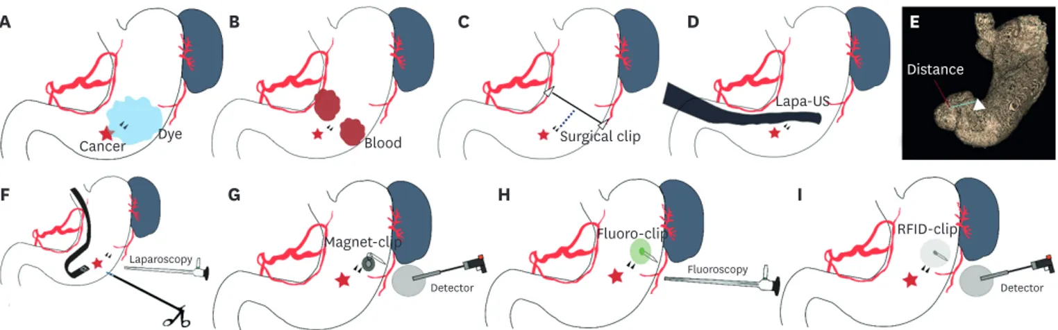
Tattooing with dye before surgery using an endoscope was the first method used for localization and is still commonly used for colon and pancreatic surgery [27]. The dyes currently used are methylene blue, indigo carmine, toluidine blue, isosulfan blue, hematoxylin, eosin, indocyanine green (ICG), and India ink [28]. Considering the nature of laparoscopic surgery, if endoscopy is performed during or immediately before surgery, gas is generated in the intestine; consequently, the intestine will hardly be dilated, which can interfere with the visual field during laparoscopic surgery. Therefore, tattooing is usually performed the day before surgery. Among the eight types of dyes, India ink and ICG are the only dyes that are not absorbed and last more than 24 hours after injection [29] (Fig. 1A). India ink was first used in the form of carbon particles that did not disappear easily. However, India ink can cause severe inflammation and necrosis, such as fat necrosis, inflammatory pseudotumors, abscesses, focal peritonitis, and phlegmonous gastritis [30-32]. These effects are the result of inflammation caused by India ink colloidal suspensions of carbon particles and ethylene glycol, phenol, and shellac, which are used as diluents and stabilizing surfactants, and
gelatin, which is an animal product. A recent report indicated that when diluted 1:100, India ink rarely causes inflammation for several months [33]. Sterile materials have recently been developed, such as SPOT® (GI Supply, Camp Hill, PA, USA), a dye for endoscopic tattooing similar to India ink [27]. SPOT® is approved by the US Food and Drug Administration (FDA) and consists of water, glycerol, polysorbate 80, benzyl alcohol, simethicone, and high-purity carbon black; it does not contain phenol or shellacs. A sterile carbon particle suspension (Black Eye®, The Standard Co., Ltd., Seoul, Korea) has also been developed [34]. These materials can be used safely for tattooing and cause less inflammation than India ink; however, there are no recent reports on their use in laparoscopic gastrectomy surgery.
The ICG remains in the tattoo location for 12 hours or more; however, it later spreads to the lymphatic vessels around the gastric tissue, hindering accurate localization of the tumor. ICG can be visualized only if injected during or immediately before surgery [35,36]. The advantage of this method is that the dye can be visualized directly with the naked eye during laparoscopic surgery; it is also a relatively simple and clinically useful technique. However, the disadvantage is that additional intraoperative endoscopy instruments and endoscopists are required during surgery. Furthermore, the spreading and disappearance of the dye can reduce detection accuracy. If the EGC is located at the posterior wall of the stomach, the location of the dye will be difficult to determine. Since vascularity and lymphatic drainage can be detected with ICG, recent studies have used ICG to confirm blood circulation at the anastomosis site and sentinel lymph node status during surgery [37-40]. Recently, an ICG dye fluorescence marking system for intraoperative localization of EGC tumors during laparoscopic distal gastrectomy was described [37]. However, dye spreading and weak dye uptake remain as limitations that can cause inaccurate resection margins, even when a fluorescence system is applied.
Preoperative endoscopic autologous blood tattooing (blood-tattoo)
Endoscopic autologous blood tattooing involves injecting the patient's own blood into the muscle layer at the 3–4 cm proximal margin of the gastric cancer lesion; this is done using an endoscope the day before surgery [41]. This method has been previously used for localization A
Cancer Dye Blood Surgical clip
Lapa-US Distance RFID-clip Fluoro-clip Magnet-clip Detector Detector Fluoroscopy Laparoscopy B C D E F G H I
Fig. 1. Schematic illustration of intraoperative tumor localization methods for early gastric cancer. (A) Preoperative endoscopic dye tattooing (cancer, red star; endoscopic clip, black triangle; dye injection, blue area). (B) Preoperative endoscopic autologous blood tattooing (blood injection, red area). (C) Intraoperative portable abdominal radiograph. The resection line is measured by the distance (black dotted line) between the endoscopic clip (small black triangle) and surgical clip line (black line) (surgical clip, white triangle). (D) Intraoperative laparoscopic ultrasonography. (E) 3-Dimensional reconstruction utilizing preoperative 3-dimensional computed tomography measurements of the distance from the clip to the pylorus (green line) (endoscopic clip, white triangle). (F) Surgeon indicating the lesion using a laparoscopic device and endoscopist confirming the clip area by intraoperative endoscopy. (G) Magnetic marking clip detection system and Magnet-clip. (H) Endoscopic fluorescent clip detection system and Fluor-clip. (I) Radio-frequency identification detection system and RFID Clip.
during laparoscopic colon resection [42,43]. When injected with an endoscope, the muscle layer is injected as close to the subserosa as much as possible. The closer the blood injection is to the gastric serosa, the better the gross visualization of the blood. Approximately 2–3 mL of blood prepared without heparin was injected (Fig. 1B). To facilitate detection, injections can be performed at two or three sites. The advantage of this method is that, like dye, the injected blood can be viewed immediately and grossly with a laparoscope, without the need for additional instruments and procedures during surgery. However, compared to dye, blood may spread less due to its viscosity. Since the patient's own blood is used, adverse reactions, such as allergic reactions and inflammation, are rare, and complications are mild even if spillage in the peritoneum occurs.
The disadvantages of endoscopic autologous blood tattooing are that it is difficult to find the exact location near the lesser omentum and lymphovascular bundle when the tumor is located in the lesser curvature, and endoscopy is required before surgery. For colon cancers, the removal range is wider than that of gastric cancers; therefore, surgeons only need to know the approximate location. However, in the case of gastric cancer, especially EGC, if the lesions are located in the mid to high body (e.g., an accurate margin is necessary to determine whether subtotal gastrectomy or total gastrectomy should be performed), there is a high possibility of error in estimating the location. A previous study suggested that blood injections at more than two sites can be helpful for more accurate resection, in order to provide guidance for careful gastrectomy; however, in general, detailed manipulation appears to be difficult with this method. Additionally, when used in cancer patients, it is necessary to confirm oncologic safety, that is, whether autologous blood injection can promote cancer progression.
Intraoperative portable abdominal radiography (intraop-X-ray)
The intraoperative portable abdominal radiography method involves endoscopic application of two or more endoscopic clips around the EGC tumor preoperatively, applying a surgical metal clip to the lesser and greater curvatures above the suspected position during laparoscopic surgery, and using a portable X-ray device to determine the distance between the endoscopic clips and the surgical metal clip (Fig. 1C) [44,45]. The resection line is measured as the distance (white dotted line) between the endoscopic clip (black triangle) and surgical metal clip line (black line, Fig. 1C).
A previous retrospective study reported the safe performance of the procedure in 80 patients with gastric cancer [44]. The advantage of this method is that it is intuitive and easy to apply. The disadvantage is that endoscopic clipping is required before surgery, and to perform X-ray imaging without contaminating the operating room during surgery, an operative bed that transmits X-rays is required. Moreover, additional experts are required to acquire portable X-rays, and the procedure is time-consuming. Furthermore, it is not a real-time detection method but an indirect measurement method, and although unlikely, there is an additional risk of radiation exposure for the patients and surgeons.
Intraoperative laparoscopic ultrasonography (lapa-US)
Intraoperative laparoscopic ultrasonography uses a special ultrasound instrument to confirm the location of the tumor or clip during laparoscopic surgery. Two case reports of localization using laparoscopic ultrasonography for gastric submucosal tumors have been reported (Fig. 1D) [46]. Laparoscopic ultrasonography is a technique used to confirm the suspected position during surgery if it is an endophytic mass that is difficult to visualize with laparoscopy. Laparoscopic ultrasonography is used in laparoscopic liver surgery; therefore, it is a good
option if it is performed in the operating room. However, gastric cancer, especially EGC, does not form masses, making it difficult to identify the tumor itself. Therefore, in most gastric cancer surgeries, two or more endoscopic clips are clipped 1–2 cm above the tumor and then located intraoperatively by laparoscopic ultrasonography. However, unless the surgeon has extensive experience with laparoscopic ultrasonography, it will be difficult to locate the endoscopic clip, which is small, during surgery. In particular, if the tumor is located on the posterior wall of the stomach, it may be difficult to detect due to intragastric gasses.
Preoperative 3-dimensional computed tomography (3D-CT) reconstruction
measurements
For computed tomography (CT) reconstruction measurements, a specialist applies an endoscopic clip at the proximal site of the gastric tumor before surgery, performs CT gastroscopy, and measures the distance from the clip to the pylorus or gastroesophageal junction following a 3D reconstruction (Fig. 1E) [47]. Preoperative 3D-CT has become common in recent years and is not a difficult method to perform. However, endoscopic clipping should be performed before CT examination. Unfortunately, ECGs are too small to be detected by CT; therefore, the distance is measured from the clip site instead of the gastric tumor location. For example, it is possible to estimate the location within centimeters of the pylorus during surgery. The advantages are that the gastric tumor location can be predicted by measuring the distance or location in advance before surgery, and no additional examination other than 3D-CT is required. The disadvantages are that endoscopic clipping before the CT scan and cooperation with a radiology specialist for 3D reconstruction are required. In addition, it is difficult to select an accurate location because it is measured in two dimensions during surgery, without real-time feedback.
Intraoperative gastrofibroscopy (IOG)
During laparoscopic gastrectomy, tumor localization can be performed by injecting dye with an endoscope (Fig. 1F) [35,36] or by pressing the suspected site with a laparoscopic device or endoscopic needle (Fig. 1F) [48,49]. In most cases, preoperative endoscopy is performed, and an endoscopic clip is applied 1–2 cm proximal to the EGC tumor. Endoscopic clip preparation is performed because the EGC tumor itself may be difficult to find via intraoperative endoscopy. When the stomach is gripped during surgery, it is difficult to distinguish between mucosal injury and small EGC tumors. Surgical field endoscopy is performed at the beginning of surgery when performed by an endoscopic specialist. After the surgeon creates a pneumoperitoneum after anesthesia, the first specialist holds the intestinal clamp directly under the ligament of Treitz to prevent air from entering the small intestine. Next, the endoscopic procedure is performed; the dye is injected via the endoscope, or the endoscopic specialist checks the area that was pressed by the operator using laparoscopic forceps. If the tumor is located on the posterior wall of the stomach, it is better to dissect the greater omentum in advance for an accurate visual field observation. If the surgeon performs an endoscopic procedure during surgery, considering a lack of manpower, it is more effective to insert an endoscope after dissection of the perigastric lymph nodes and greater curvature omentum than immediately after anesthesia, in order to create a convenient visual field. Additionally, in this situation, the surgeon checks the site by dye injection or presses the mucosal site with laparoscopic forceps to confirm the location of the tumor.
The advantage of intraoperative endoscopy is that it can be visualized directly and in real time during surgery; therefore, it is very useful for malignant small gastric tumors. Additionally, after anastomosis, additional endoscopy can be performed immediately. Fatal complications
can be reduced by checking the area of bleeding and leakage at the anastomosis site during surgery and performing endoscopic or laparoscopic hemostasis, additional suture, and re-anastomosis, if necessary [50]. In recent years, laparoscopic and endoscopic cooperative surgery (LECS), which combines laparoscopic gastric resection with an endoscopic procedure for the local treatment of gastric tumors, has been reported as an emerging modality [51]. The disadvantage of IOG is that expensive endoscopy equipment is required. Furthermore, for endoscopic procedures, an endoscopic specialist or surgeon training is required. During laparoscopic surgery, the location of the tumor in the mucosal layer can be detected with an endoscope while air insufflation is performed in the stomach, and the serosal site should be marked by laparoscopy. When the tumor is located at the lesser curvature or posterior wall of the stomach, visual limitation of the detection field may occur if air fills the stomach while checking all circumferential sites of the stomach by laparoscopy.
Magnetic marking clip detection system (magnet-clip)
A magnetic marking clip is a specially manufactured clip that contains a magnet. The clip is inserted before surgery via preoperative endoscopy and located using a magnetic marking clip detection system (MMCDS) probe during surgery (Fig. 1G) [52]. It was reported that the lesions in 15 patients had an average error of approximately 5.7 mm and a maximum error of approximately 10.1 mm. These results were similar to those obtained using laparoscopic ultrasonography. However, compared with ultrasonography, an advantage of MMCDS is that the clip can be detected easily with only information on the magnetic flux density display units, without any training or learning curves. Among the disadvantages of MMCDS, it is not possible to check the location more precisely than with intraoperative endoscopy, and a novel MMCDS instrument is required. Moreover, no approval or commercialization of the MMCDS has been reported.
Fluorescence detection system
Fluorescence imaging with ICG has been commonly used to detect lymphatic drainage or blood circulation [37]. When fluorescence was used for localization of the tumor, ICG dye injection with fluorescence detection had limitations in dye spreading and weak uptake [53]. To minimize the disadvantage of dye injection at the wall of the hollow viscera, a preoperative endoscopic fluorescent clip (fluor-clip) detection method was developed. In this procedure, a 650-nm diode laser and a digital CCD camera were used during surgery after applying an endoscopic fluorescent clip near the tumor before surgery (Fig. 1H) [54]. Effective localization of lesions in the colon and stomach of experimental animals has been reported. An advantage is that if only fluorescent clipping is performed before surgery, laparoscopy can be performed in real time during surgery. However, if the EGC tumor is located in a thick wall in the stomach, the detection accuracy may decrease if the transmittance of fluorescence is decreased or if the signal is observed to have spread. Additionally, an additional expensive fluorescence laparoscopy detection instrument is required, and only animal experimental results have been reported to date.
Radio-frequency identification detection system
In radio-frequency identification (RFID) detection, a clip containing an RFID tag (RFID clip) is applied using an endoscope before surgery and is detected in real time during laparoscopic surgery with a novel detection system (Fig. 1I) [55-59]. In a previous ex vivo porcine study, the mean detection time was 28 seconds in the colon and 32 seconds in the stomach, and the median detection distance was 6 mm for the colon and 6.5 mm for the stomach. The advantages are that the price of the RFID clip is low, each RFID tag can be recognized by
attaching a unique number, and high detection accuracy is possible. However, this technique is still in the animal experimental stage and has not been commercialized or approved by the FDA. In addition, the purchase of a novel detection system is required.
Comparison of localization methods
Preoperative preparation
All nine methods require preoperative gastrofibroscopy or endoscopic clipping on the day prior to surgery (Table 1). Dye or blood injection is performed using a dye-tattoo or blood-tattoo during preoperative endoscopy. A 3D-CT is performed after the 3D-CT scan, and a 3D reconstruction program is used to calculate the distance between the pylorus and endoscopic clip or gastroesophageal junction and clip.
Intraoperative procedure time
Among the six methods that require localization during surgery, the procedure time of intraoperative portable abdominal radiography is relatively short, but verification requires additional time; however, recently, the digital images can checked immediately after X-ray examination. Lapa-US depends on the physician's expertise, and it is helpful to have a radiologist for diagnosis. The IOG requires an endoscopist to perform endoscopy. Recent techniques using Magnet clips, Fluor-clips, and RFID clips have a short procedure time of several seconds to minutes, but no standard methods or protocols are available.
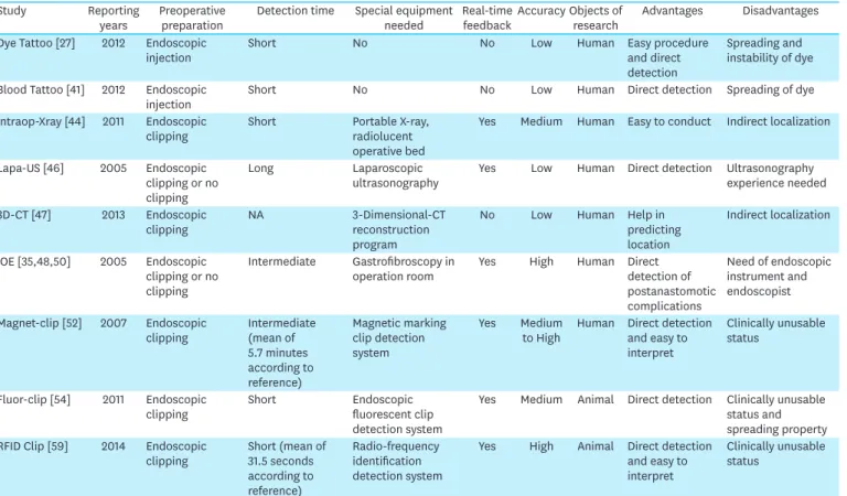
Table 1. Summary of methods for localization of early gastric cancer tumors during laparoscopic surgery Study Reporting
years Preoperative preparation Detection time Special equipment needed Real-time feedbackAccuracy Objects of research Advantages Disadvantages Dye Tattoo [27] 2012 Endoscopic
injection Short No No Low Human Easy procedure and direct
detection
Spreading and instability of dye Blood Tattoo [41] 2012 Endoscopic
injection Short No No Low Human Direct detection Spreading of dye
Intraop-Xray [44] 2011 Endoscopic
clipping Short Portable X-ray, radiolucent operative bed
Yes Medium Human Easy to conduct Indirect localization Lapa-US [46] 2005 Endoscopic
clipping or no clipping
Long Laparoscopic
ultrasonography Yes Low Human Direct detection Ultrasonography experience needed 3D-CT [47] 2013 Endoscopic
clipping NA 3-Dimensional-CT reconstruction program
No Low Human Help in predicting location Indirect localization IOE [35,48,50] 2005 Endoscopic clipping or no clipping Intermediate Gastrofibroscopy in
operation room Yes High Human Direct detection of postanastomotic complications Need of endoscopic instrument and endoscopist Magnet-clip [52] 2007 Endoscopic
clipping Intermediate (mean of 5.7 minutes according to reference) Magnetic marking clip detection system Yes Medium
to High Human Direct detection and easy to interpret
Clinically unusable status
Fluor-clip [54] 2011 Endoscopic
clipping Short Endoscopic fluorescent clip detection system
Yes Medium Animal Direct detection Clinically unusable status and spreading property RFID Clip [59] 2014 Endoscopic
clipping Short (mean of 31.5 seconds according to reference)
Radio-frequency identification detection system
Yes High Animal Direct detection and easy to interpret
Clinically unusable status
Dye tattoo = preoperative endoscopic dye tattooing; blood tattoo = preoperative endoscopic autologous blood tattooing; 3D-CT = preoperative 3-dimensional computed tomography reconstruction measurement; Intraop-Xray = intraoperative portable abdominal radiography; Lapa-US = intraoperative laparoscopic ultrasonography; IOG = intraoperative gastrofibroscopy; Magnet-clip = magnetic marking clip detection system; Fluor-clip = endoscopic fluorescent clip detection system; RFID Clip = radio-frequency identification detection system; NA = not applicable.
Equipment
For methods involving dye, autologous blood is not needed, but other equipment such as an injection needles and dye are needed during the endoscopic procedure. Intraop-X-rays need to be performed on a radiolucent operating room bed to prevent contamination. The Lapa-US method requires laparoscopic ultrasound, and 3D-CT requires a 3D reconstruction program. For IOG, a gastrofibroscopy device is required in the operating room. Magnet clips, Fluor-clips, and RFID clips each require a detection system. Initially, it is better to start with a tattoo or intra-X-ray, which can be performed without additional equipment.
Real-time feedback and accuracy
The methods for dye-tattoos, blood-tattoos, and 3D-CT are considered to have a low accuracy because they do not provide real-time feedback, and these methods are recommended for use with cancers in the antrum or high-body area. Intraop-X-ray and Lapa-US can provide real-time feedback; however, these methods require expertise, and it is difficult to define the clips in some gastric areas. Thus, these methods are considered to have intermediate accuracy. We recommend that IOG, which provides real-time feedback with high accuracy, can be used for the localization of mid-body cancers. The Magnet-clip, Fluor-clip, and RFID Clip methods are considered highly accurate based on the availability of real-time feedback, and we expect commercialized equipment to be available soon.
SUMMARY
More than half of all gastric cancers in some East Asian countries are diagnosed with EGC, highlighting the importance of endoscopic screening in countries with a relatively high incidence of gastric cancer. Additionally, the implementation of endoscopic screening will increase the proportion of EGC among all stages of gastric cancer [15,16]. Laparoscopic surgery is currently performed for the surgical treatment of EGC. Tumor localization may not be necessary in some cases of laparoscopic surgery for gastric cancer; for example, if the EGC is located at a longitudinally lower site, such as the gastric angle or antrum, laparoscopic subtotal gastrectomy can be performed without localization. However, when EGC tumors are located in the longitudinally middle level of the stomach, tumor localization is required in most cases. The number of patients with EGC has gradually increased in some countries, and the number of patients with long-term survival and a very low probability of recurrence after surgery for EGC is increasing. Many attempts have been made to perform function-preserving gastrectomy in patients with a low probability of recurrence, in order to improve their postoperative quality of life. The types of function-preserving gastrectomy include pylorus-preserving gastrectomy, proximal gastrectomy, gastric wedge resection, segmental resection or endoscopic submucosal dissection, and endoscopic full-thickness resection with sentinel node navigation surgery [60-64]. In such minimal operations, the location of the EGC tumor and the extent of tumor removal should be accurately determined before surgery. In addition, since presurgical localization can reduce the operation time and operator fatigue, complications related to the operation can be minimized. Therefore, accurate tumor localization is essential during laparoscopic surgery.
Various methods for tumor localization during surgery have been reported. The historical stages in the development of instruments for accurate EGC localization during laparoscopic surgery are dye, tracer, magnetic metal, luminous fluorescence materials, and the RFID system, which are miniaturized products; devices using next-generation novel materials are
expected to be developed in the near future. However, there is still no easy way to accurately detect EGCs in real time. The development of a new device and its application to patients in the clinic requires considerable time, money, and effort. After development, the new device must be validated by relevant organizations and approved by government regulators, including the FDA. New methods and instruments are indispensable for MIS, and we hope that improved devices will soon be developed and used in clinical settings by surgeons.
REFERENCES
1. Bray F, Ferlay J, Soerjomataram I, Siegel RL, Torre LA, Jemal A. Global cancer statistics 2018: GLOBOCAN estimates of incidence and mortality worldwide for 36 cancers in 185 countries. CA Cancer J Clin 2018;68:394-424.
PUBMED | CROSSREF
2. Choi AH, Kim J, Chao J. Perioperative chemotherapy for resectable gastric cancer: MAGIC and beyond. World J Gastroenterol 2015;21:7343-7348.
PUBMED | CROSSREF
3. Mihmanli M, Ilhan E, Idiz UO, Alemdar A, Demir U. Recent developments and innovations in gastric cancer. World J Gastroenterol 2016;22:4307-4320.
PUBMED | CROSSREF
4. Ilson DH. Advances in the treatment of gastric cancer. Curr Opin Gastroenterol 2017;33:473-476.
PUBMED | CROSSREF
5. Tan Z. Recent advances in the surgical treatment of advanced gastric cancer: a review. Med Sci Monit 2019;25:3537-3541.
PUBMED | CROSSREF
6. Allemani C, Matsuda T, Di Carlo V, Harewood R, Matz M, Nikšić M, et al. Global surveillance of trends in cancer survival 2000-14 (CONCORD-3): analysis of individual records for 37 513 025 patients diagnosed with one of 18 cancers from 322 population-based registries in 71 countries. Lancet 2018;391:1023-1075.
PUBMED | CROSSREF
7. Hamashima C. Current issues and future perspectives of gastric cancer screening. World J Gastroenterol 2014;20:13767-13774.
PUBMED | CROSSREF
8. Rahman R, Asombang AW, Ibdah JA. Characteristics of gastric cancer in Asia. World J Gastroenterol 2014;20:4483-4490.
PUBMED | CROSSREF
9. Hamashima C; Systematic Review Group and Guideline Development Group for Gastric Cancer Screening Guidelines. Update version of the Japanese guidelines for gastric cancer screening. Jpn J Clin Oncol 2018;48:673-683.
PUBMED | CROSSREF
10. Saumoy M, Schneider Y, Shen N, Kahaleh M, Sharaiha RZ, Shah SC. Cost effectiveness of gastric cancer screening according to race and ethnicity. Gastroenterology 2018;155:648-660.
PUBMED | CROSSREF
11. Suh YS, Yang HK. Screening and early detection of gastric cancer: East versus West. Surg Clin North Am 2015;95:1053-1066.
PUBMED | CROSSREF
12. Russo AE, Strong VE. Gastric cancer etiology and management in Asia and the West. Annu Rev Med 2019;70:353-367.
PUBMED | CROSSREF
13. Kim GH, Liang PS, Bang SJ, Hwang JH. Screening and surveillance for gastric cancer in the United States: is it needed? Gastrointest Endosc 2016;84:18-28.
PUBMED | CROSSREF
14. Venerito M, Nardone G, Selgrad M, Rokkas T, Malfertheiner P. Gastric cancer--epidemiologic and clinical aspects. Helicobacter 2014;19 Suppl 1:32-37.
PUBMED | CROSSREF
15. Information Committee of Korean Gastric Cancer Association. Korean Gastric Cancer Association nationwide survey on gastric cancer in 2014. J Gastric Cancer 2016;16:131-140.
16. Katai H, Ishikawa T, Akazawa K, Isobe Y, Miyashiro I, Oda I, et al. Five-year survival analysis of surgically resected gastric cancer cases in Japan: a retrospective analysis of more than 100,000 patients from the nationwide registry of the Japanese Gastric Cancer Association (2001–2007). Gastric Cancer 2018;21:144-154.
PUBMED | CROSSREF
17. Jun JK, Choi KS, Lee HY, Suh M, Park B, Song SH, et al. Effectiveness of the Korean National Cancer Screening Program in reducing gastric cancer mortality. Gastroenterology 2017;152:1319-1328.e7.
PUBMED | CROSSREF
18. Hamashima C, Goto R. Potential capacity of endoscopic screening for gastric cancer in Japan. Cancer Sci 2017;108:101-107.
PUBMED | CROSSREF
19. Costamagna G, Cesaro P. Early gastric cancer: detection and endoscopic treatment. Ann Ital Chir 2012;83:183-191.
PUBMED
20. Huang L, Li TJ. Laparoscopic surgery for gastric cancer: where are we now and where are we going? Expert Rev Anticancer Ther 2018;18:1145-1157.
PUBMED | CROSSREF
21. Santoro R, Ettorre GM, Santoro E. Subtotal gastrectomy for gastric cancer. World J Gastroenterol 2014;20:13667-13680.
PUBMED | CROSSREF
22. Kim W, Kim HH, Han SU, Kim MC, Hyung WJ, Ryu SW, et al. Decreased morbidity of laparoscopic distal gastrectomy compared with open distal gastrectomy for stage I gastric cancer: short-term outcomes from a multicenter randomized controlled trial (KLASS-01). Ann Surg 2016;263:28-35.
PUBMED | CROSSREF
23. Lee HJ, Hyung WJ, Yang HK, Han SU, Park YK, An JY, et al. Short-term outcomes of a multicenter randomized controlled trial comparing laparoscopic distal gastrectomy with D2 lymphadenectomy to open distal gastrectomy for locally advanced gastric cancer (KLASS-02-RCT). Ann Surg 2019;270:983-991.
PUBMED | CROSSREF
24. Song KY, Park CH, Kang HC, Kim JJ, Park SM, Jun KH, et al. Is totally laparoscopic gastrectomy less invasive than laparoscopy-assisted gastrectomy?: prospective, multicenter study. J Gastrointest Surg 2008;12:1015-1021.
PUBMED | CROSSREF
25. Japanese Gastric Cancer Association. Japanese gastric cancer treatment guidelines 2018 (5th edition). Gastric Cancer 2021;24:1-21.
PUBMED | CROSSREF
26. Guideline Committee of the Korean Gastric Cancer Association (KGCA), Development Working Group & Review Panel. Korean practice guideline for gastric cancer 2018: an evidence-based, multi-disciplinary approach. J Gastric Cancer 2019;19:1-48.
PUBMED | CROSSREF
27. Luigiano C, Ferrara F, Morace C, Mangiavillano B, Fabbri C, Cennamo V, et al. Endoscopic tattooing of gastrointestinal and pancreatic lesions. Adv Ther 2012;29:864-873.
PUBMED | CROSSREF
28. Nizam R, Siddiqi N, Landas SK, Kaplan DS, Holtzapple PG. Colonic tattooing with India ink: benefits, risks, and alternatives. Am J Gastroenterol 1996;91:1804-1808.
PUBMED
29. Hammond DC, Lane FR, Welk RA, Madura MJ, Borreson DK, Passinault WJ. Endoscopic tattooing of the colon. An experimental study. Am Surg 1989;55:457-461.
PUBMED
30. Coman E, Brandt LJ, Brenner S, Frank M, Sablay B, Bennett B. Fat necrosis and inflammatory
pseudotumor due to endoscopic tattooing of the colon with india ink. Gastrointest Endosc 1991;37:65-68.
PUBMED | CROSSREF
31. Park SI, Genta RS, Romeo DP, Weesner RE. Colonic abscess and focal peritonitis secondary to India ink tattooing of the colon. Gastrointest Endosc 1991;37:68-71.
PUBMED | CROSSREF
32. Hornig D, Kühn H, Stadelmann O, Bötticher R. Phlegmonous gastritis after Indian ink marking. Endoscopy 1983;15:266-269.
PUBMED | CROSSREF
33. Price N, Gottfried MR, Clary E, Lawson DC, Baillie J, Mergener K, et al. Safety and efficacy of India ink and indocyanine green as colonic tattooing agents. Gastrointest Endosc 2000;51:438-442.
34. Milone M, Vignali A, Manigrasso M, Velotti N, Sarnelli G, Aprea G, et al. Sterile carbon particle suspension vs India ink for endoscopic tattooing of colonic lesions: a randomized controlled trial. Tech Coloproctol 2019;23:1073-1078.
PUBMED | CROSSREF
35. Xuan Y, Hur H, Byun CS, Han SU, Cho YK. Efficacy of intraoperative gastroscopy for tumor localization in totally laparoscopic distal gastrectomy for cancer in the middle third of the stomach. Surg Endosc 2013;27:4364-4370.
PUBMED | CROSSREF
36. Hur H, Son SY, Cho YK, Han SU. Intraoperative gastroscopy for tumor localization in laparoscopic surgery for gastric adenocarcinoma. J Vis Exp 2016;
PUBMED | CROSSREF
37. Namikawa T, Sato T, Hanazaki K. Recent advances in near-infrared fluorescence-guided imaging surgery using indocyanine green. Surg Today 2015;45:1467-1474.
PUBMED | CROSSREF
38. Yukaya T, Saeki H, Kasagi Y, Nakashima Y, Ando K, Imamura Y, et al. Indocyanine green fluorescence angiography for quantitative evaluation of gastric tube perfusion in patients undergoing esophagectomy. J Am Coll Surg 2015;221:e37-e42.
PUBMED | CROSSREF
39. Yoshida M, Kubota K, Kuroda J, Ohta K, Nakamura T, Saito J, et al. Indocyanine green injection for detecting sentinel nodes using color fluorescence camera in the laparoscopy-assisted gastrectomy. J Gastroenterol Hepatol 2012;27 Suppl 3:29-33.
PUBMED | CROSSREF
40. Tajima Y, Murakami M, Yamazaki K, Masuda Y, Kato M, Sato A, et al. Sentinel node mapping guided by indocyanine green fluorescence imaging during laparoscopic surgery in gastric cancer. Ann Surg Oncol 2010;17:1787-1793.
PUBMED | CROSSREF
41. Jeong O, Cho SB, Joo YE, Ryu SY, Park YK. Novel technique for intraoperative tumor localization during totally laparoscopic distal gastrectomy: endoscopic autologous blood tattooing. Surg Endosc 2012;26:1778-1783.
PUBMED | CROSSREF
42. Feingold DL, Addona T, Forde KA, Arnell TD, Carter JJ, Huang EH, et al. Safety and reliability of tattooing colorectal neoplasms prior to laparoscopic resection. J Gastrointest Surg 2004;8:543-546.
PUBMED | CROSSREF
43. Fu KI, Fujii T, Kato S, Sano Y, Koba I, Mera K, et al. A new endoscopic tattooing technique for identifying the location of colonic lesions during laparoscopic surgery: a comparison with the conventional technique. Endoscopy 2001;33:687-691.
PUBMED | CROSSREF
44. Kim HI, Hyung WJ, Lee CR, Lim JS, An JY, Cheong JH, et al. Intraoperative portable abdominal radiograph for tumor localization: a simple and accurate method for laparoscopic gastrectomy. Surg Endosc 2011;25:958-963.
PUBMED | CROSSREF
45. Chung JW, Seo KW, Jung K, Park MI, Kim SE, Park SJ, et al. A promising method for tumor localization during total laparoscopic distal gastrectomy: preoperative endoscopic clipping based on negative biopsy and selective intraoperative radiography findings. J Gastric Cancer 2017;17:220-227.
PUBMED | CROSSREF
46. Hyung WJ, Lim JS, Cheong JH, Kim J, Choi SH, Song SY, et al. Intraoperative tumor localization using laparoscopic ultrasonography in laparoscopic-assisted gastrectomy. Surg Endosc 2005;19:1353-1357.
PUBMED | CROSSREF
47. Jeong SH, Bae K, Ha CY, Lee YJ, Lee OJ, Jung WT, et al. Effectiveness of endoscopic clipping and computed tomography gastroscopy for the preoperative localization of gastric cancer. J Korean Surg Soc 2013;84:80-87.
PUBMED | CROSSREF
48. Park DJ, Lee HJ, Kim SG, Jung HC, Song IS, Lee KU, et al. Intraoperative gastroscopy for gastric surgery. Surg Endosc 2005;19:1358-1361.
PUBMED | CROSSREF
49. Matsui H, Okamoto Y, Nabeshima K, Kondoh Y, Ogoshi K, Makuuchi H. Endoscopy-assisted gastric resection: a safe and reliable procedure for tumor clearance during laparoscopic high distal or proximal gastrectomy. Surg Endosc 2009;23:1146-1149.
50. Park JH, Jeong SH, Lee YJ, Kim TH, Kim JM, Kim DH, et al. Safety and efficacy of post-anastomotic intraoperative endoscopy to avoid early anastomotic complications during gastrectomy for gastric cancer. Surg Endosc 2020;34:5312-5319.
PUBMED | CROSSREF
51. Hiki N, Nunobe S. Laparoscopic endoscopic cooperative surgery (LECS) for the gastrointestinal tract: updated indications. Ann Gastroenterol Surg 2019;3:239-246.
PUBMED | CROSSREF
52. Ohdaira T, Nagai H. Intraoperative localization of early-stage upper gastrointestinal tumors using a magnetic marking clip-detecting system. Surg Endosc 2007;21:810-815.
PUBMED | CROSSREF
53. Ushimaru Y, Omori T, Fujiwara Y, Yanagimoto Y, Sugimura K, Yamamoto K, et al. The feasibility and safety of preoperative fluorescence marking with indocyanine green (ICG) in laparoscopic gastrectomy for gastric cancer. J Gastrointest Surg 2019;23:468-476.
PUBMED | CROSSREF
54. Choi Y, Kim KG, Kim JK, Nam KW, Kim HH, Sohn DK. A novel endoscopic fluorescent clip for the localization of gastrointestinal tumors. Surg Endosc 2011;25:2372-2377.
PUBMED | CROSSREF
55. Choi WJ, Moon JH, Min JS, Song YK, Lee SA, Ahn JW, et al. Real-time detection system for tumor localization during minimally invasive surgery for gastric and colon cancer removal: in vivo feasibility study in a swine model. J Surg Oncol 2018;117:699-706.
PUBMED | CROSSREF
56. Joo HY, Lee BE, Choi CI, Kim DH, Kim GH, Jeon TY, et al. Tumor localization using radio-frequency identification clip marker: experimental results of an ex vivo porcine model. Surg Endosc 2019;33:1441-1450.
PUBMED | CROSSREF
57. Lee KM, Min JS, Choi WJ, Ahn JW, Yoon SW, Kim YJ. An advanced RFID-based system to localize gastric and colon cancers during laparoscopic surgery. Surg Endosc 2021;35:139-147.
PUBMED | CROSSREF
58. Reicher JJ, Reicher MA, Thomas M, Petcavich R. Radiofrequency identification tags for preoperative tumor localization: proof of concept. AJR Am J Roentgenol 2008;191:1359-1365.
PUBMED | CROSSREF
59. Kojima F, Sato T, Tsunoda S, Takahata H, Hamaji M, Komatsu T, et al. Development of a novel marking system for laparoscopic gastrectomy using endoclips with radio frequency identification tags: feasibility study in a canine model. Surg Endosc 2014;28:2752-2759.
PUBMED | CROSSREF
60. Nomura E, Okajima K. Function-preserving gastrectomy for gastric cancer in Japan. World J Gastroenterol 2016;22:5888-5895.
PUBMED | CROSSREF
61. Hiki N, Nunobe S, Kubota T, Jiang X. Function-preserving gastrectomy for early gastric cancer. Ann Surg Oncol 2013;20:2683-2692.
PUBMED | CROSSREF
62. Oh SY, Lee HJ, Yang HK. Pylorus-Preserving Gastrectomy for Gastric Cancer. J Gastric Cancer 2016;16:63-71.
PUBMED | CROSSREF
63. An JY, Min JS, Lee YJ, Jeong SH, Hur H, Han SU, et al. Safety of laparoscopic sentinel basin dissection in patients with gastric cancer: an analysis from the SENORITA prospective multicenter quality control trial. J Gastric Cancer 2018;18:30-36.
PUBMED | CROSSREF
64. An JY, Min JS, Hur H, Lee YJ, Cho GS, Park YK, et al. Laparoscopic sentinel node navigation surgery versus laparoscopic gastrectomy with lymph node dissection for early gastric cancer: short-term outcomes of a multicentre randomized controlled trial (SENORITA). Br J Surg 2020;107:1429-1439.