Introduction
Due to its unknown etiology, pigmented villonodular synovitis (PVNS) had has also been termed synovial fibroendothelioma, chronic hemorrhagic villous synovitis, or fibrohemosideric sarcoma. In 1941, based on its histologic findings, Jaffe, Lich-tenstein and Sutro first used the the term ‘PVNS’, but the path-ogenesis is still not clear.1,2)
The annual incidence of PVNS is 1.8 patients per million in the population, which is why reporting of PVNS was rare in a large scale study.3-5)
Compared to localized PVNS, diffuse-type PVNS permeates
to the bone and surrounding soft tissues, which results in destruction of major joints.2,3,5-8)
The high recurrence rate after surgical re moval has been a major concern.2,3,6-11)
Radiation or intra-articular radioisotope injection has been attempted to solve this problem, but the efficacy and indications of this method are still not established.2,5-7,12-18)
The purpose of this study is to evaluate and analyze the clinical outcomes of diffuse-type PVNS that were treated with open synovectomy and additional electrocautrization. In addition, the value of radiation therapy to prevent recurrence is examined.
Materials and Methods
1. Patient characteristics
From February 1994 to March 2006, 32 patients were diagnosed as PVNS. Histological confirmation was done in all cases. Cases with a follow-up of less than 3 years and localized-type were excluded. In all, 21 cases of diffuse-type PVNS were reviewed.
Received February 11, 2010 Revised May 9, 2010 Accepted June 10, 2010 Correspondence to: Kyoo-Ho Shin, M.D.
Department of Orthopeadic Surgery, Yonsei University College of Medicine, 250, Seongsan-ro, Seodaemun-gu, Seoul 120-752, Korea
TEL: +82-2-2228-2180 FAX: +82-2-363-1139 E-mail: qshin@yuhs.ac
*The abstract of this article was presented for 39th annual congress of the Korean Bone and Joint Tumor Society.
Purpose: Pigmented villonodular synovitis (PVNS) is a rare soft tissue tumor, which usually arises in larger joints, such as the knee. It has a high recurrence rate after surgical treatment. The purpose of this study is to evaluate and analyze the clinical results of diffuse-type pigmented villonodular synovitis cases that were treated with open total synovectomy.
Materials and Methods: Between 1994 and 2006, 21 patients who had diffuse-type pigmented villonodular synovitis were selectively reviewed. Among the 21 cases studied, 14 patients presented at the knee, 5 at the ankle, and 2 at the shoulder and elbow. The mean follow up period was 5.5 years (range, 36-157 months). The average age of the patients was 34 years consist of 7 men and 14 women. Clinical outcomes were analyzed retrospectively, including range of motion and complications.
Results: Open total synovectomy and adjuvant electrocautrization were done in all cases except one. During the regular follow-up period after the surgery, two patients showed symptoms of recurrence. After re-operation, only one case was pathologically confirmed as a recurrence. The patient who had partial synovectomy and the other patient who had second operation due to recur rence received additional radiation therapy. Clinical outcome scores were improved in every aspect (p<0.0001). 2 out of 14 Patients who had pigmented villonodular synovitis at the knee developed stiff knee after the surgery.
Conclusion: After the open total synovectomy with electrocautrization, a low recurrence rate and satisfactory clinical outcome was achieved, observed in a minimum of 3 years of follow-up.
Key words: synovitis, pigmented villonodular, open total synovectomy
Copyrights © 2010 by The Korean Bone and Joint Tumor Society
“This is an Open Access article distributed under the terms of the Creative Commons Attribution Non-Commercial License (http://creativecommons.org/licenses/by-nc/3.0/) which permits unrestricted non-commercial use, distribution, and reproduction in any medium, provided the original work is properly cited.”
대한골관절종양학회지:제16권 제1호 2010
Synovitis (PVNS) after Open Total Synovectomy
Moses Lee, M.D., Soo Hyun Lee, M.D., Jin Suck Suh, M.D.*, Woo-Ik Yang, M.D.
†,
and Kyoo-Ho Shin, M.D.
Department of Orthopeadic Surgery, *Diagnostic Radiology,
†Pathology, Yonsei University College of Medicine, Seoul,
Korea
Table 1. Summary of Cases
No Age/
Sex Site Trauma Previous surgery Surgical method (approach) RTx.
Compli-cation
F/U
(month) Recurrence
1 40/F Left shoulder No Once: arthroscopy 92-11(YUMC)
Open total synovectomy, Electrocautrization (Anterior)
(-) None 43 No
2 38/F Right elbow No Once: arthroscopy 92-07 (YUMC)
Open total synovectomy, Electrocautrization (Anterior+posterior)
(-) None 62 No
3 7/F Left knee No (-) Open total synovectomy,
Electrocautrization (Popliteal fossa)
(-) None 41 No
4 43/F Right knee No *Twice: arthroscopy 92-10 1st op 97-04 2nd op
Open total synovectomy, Electrocautrization (Anterior+popliteal fossa)
(-) Pain swelling
41 No
5 20/M Left knee No (-) Open total synovectomy,
Electrocautrization (Anterior)
(-) Pain 44 No
6 37/F Left knee No (-) Open total synovectomy,
Electrocautrization (Popliteal fossa)
(-) None 54 No
7 38/F Left knee No (-) Open total synovectomy,
Electrocautrization (Anterior)
(-) None 78 No
8 25/F Left knee No (-) Open total synovectomy,
Electrocautrization (Anterior+popliteal fossa)
(-) None 87 Re-operation pathology
(-) 9 47/F Left knee No *Once: open
excision 01-12
Open total synovectomy, Electrocautrization (Anterior+popliteal fossa)
(-) None 75 No
10 31/F Right knee No (-) Open total synovectomy,
Electrocautrization (Anterior+popliteal fossa)
(+) after 2nd op.
LOM 36 Yes
11 44/F Left knee No Once: arthroscopy 04-11 (YUMC)
Open total synovectomy, Electrocautrization (Popliteal fossa)
(-) None 44 No
12 15/M Left knee No *Three times: arthroscopy 03-11 1st op 04-02 2nd op 04-10 3rd op
Open total synovectomy, Electrocautrization (Anterior+popliteal fossa)
(-) Stiff knee (0-45)
38 No
13 42/M Left knee No Twice: open excision (unknown)
Open total synovectomy, Electrocautrization (Anterior)
(-) Stiff knee (15-70)
36 No
14 13/M Right knee No (-) Open total synovectomy,
Electrocautrization (Anterior)
(-) Swelling 41 No
15 39/M Left knee No (-) Open total synovectomy,
Electrocautrization (Popliteal fossa)
(-) None 36 No
16 54/F Right knee No Once: open excision (unknown)
Open partial synovectomy, Electrocautrization (Popliteal fossa)
(+) LOM 37 No
17 42/F Right ankle No *Once: open excision 95-6
Open total synovectomy, Electrocautrization
(Anterolateral+posteromedial)
(-) None 157 No
18 42/F Left ankle No Once: open excision 92-09
(at local hospital)
Open total synovectomy, Electrocautrization
(Anterolateral+posterolateral)
The medical records, age at presentation, sex, initial symptoms and durations, history of trauma, previous operation methods, and the number of operations was reviewed. The duration of follow-up was calculated from the date of surgery to the last follow-follow-up. Through the medical records and the interviews, post-operative complications, recurrence, and treatment details were also collected. The correlation between the initial treatment method and the recurrence was examined. During the follow-up period, the patients who presented clinical suspicion of recurrence, by symptoms, and imaging studies, underwent re-operation. The mean follow-up was 65 months (range, 36-157 months). There were seven men (33%) and fourteen women (67%), with an average age of 34 years (range, 7-54 years). Of the 21 patients, 10 patients were referred for further treatment after a previous operation due to recurrence. 7 patients were referred from other hospitals after recurrences. The other 3 patients were referred from our arthroscopic division, and the patients had PVNS recur after arthroscopic removal. There was one patient who had three incidences of recurrence; at the each recurrence, this patient had arthroscopic removal. Two patients had been referred after two times of recurrence. One patient had arthroscopic removal, and the other had an open procedure. There were a total of 7 patients who had one recurrence before the referral. Among the 7 patients, 3 had a history of arthroscopic removal (Table 1).
2. Surgical method and rehabilitation
All of patients were first treated by an open synovectomy. All of the total synovectomy were performed by only one surgeon (Cor responding author). After removing as much of the syno-vium as possible, additional electrocautrization was done at the base and surrounding soft tissues. There was one referral patient
who had severe postoperative fibrosis and tethering around the neurovascular bundle in the popliteal fossa, which made it impossible to perform total synovectomy. This patient had additional radiation therapy after surgery. A total of 3,000 cGY radiation therapy was done in fifteen rounds of radiation.
The anterior median parapatella approach and the posterior transpopliteal approach were used to treat 14 patients who had a mass at the knee. If the mass was located both anterior and posterior, the posterior mass was removed first, followed by the removal of the anterior one 6 to 8 weeks later. 5 patients received both the anterior and posterior surgeries. For surgery on the anterior, the curvilinear incision was started 1 to 2 cm above the superior pole of the patella, along the medial margin of the patella (Fig. 1B). After incising the anterior capsule, the synovium, including the mass, was exposed. En bloc total synovectomy was done when possible. The anterior cruciate ligament, subpatellar fat pad, medial and lateral gutter, and the menici was the region where careful inspection was done, because those are the places where the small foci of PVNS could be neglected.17,19)
If the patient had a history of arthroscopic surgery, the previous entry portals were also carefully examined. When using the posterior approach, the prone position was used. The incision was began 5 cm above the joint line, at the medial corner of the popliteal area, and extended distally until the proximal portion of the gastrocnemius was exposed, such that the incision formed an ‘S’ shape. The popliteal artery, tibial nerve, and common peroneal nerve were protected during dissection (Fig. 1C). If possible, a branch of the sural nerve was also saved. Anterolateral, posterolateral, posteromedial approach were used to achieve total synovectomy at the ankle joint. After preserving superficial peroneal nerve, extensor digitorum longus tendon Table 1. Continued
No Age/
Sex Site Trauma Previous surgery Surgical method (approach) RTx.
Compli-cation
F/U
(month) Recurrence
19 36M Left ankle Yes (-) Open total synovectomy,
Electrocautrization
(Anterolateral+posterolateral)
(-) LOM 135 No
20 43/F Right ankle No (-) Open total synovectomy,
Electrocautrization
(Anterolateral+posterolateral)
(-) None 38 No
21 19/M Right ankle No (-) Open total synovectomy,
Electrocautrization
(Anterolateral+posteromedial)
(-) Pain 58 No
RTx, radiation therapy; Op, operation; LOM, limitation of motion; YUMC, Yonsei university college of medicine. *Referal from other tertiary medical center.
was retracted medially with neurovascular bundle. By incising proximal portion of extensor digitorum brevis and fat pad at Sinus tarsi, wide exposure of anterior to lateral aspect of ankle joint was possible including calcaneocuboid joint and medial half of talonavicular joint. If needed additional posterolateral or posteromedial approach was done after changing patient to the prone position (Fig. 2). There were two cases on the upper extremity. Anterior approach was used to remove the mass on the shoulder (Fig. 3) and extended posterior and anterior approach was used to perform total synovectomy at the elbow. A posterior splint was applied immediately after operation, and removed 2-4 days later when the drain was removed. Rehabili-tation started with passive range of motion (ROM) exercises and vigorous active assisted ROM exercises were done by a physio-therapist for 1 month. Especially for the patient who had a sur-gery at knee, quadriceps strengthening exercises, while sitting on a bed or chair was emphasized.
3. Assessment
Regular follow-up was done after the surgery at 2 months, 4 months, 6 months, and annually. At every annual follow-up, the
functional clinical outcome was assessed by checking pain, range of motion, symptoms, and activity of daily living (ADL), which was suggested by Ogilvie-Harris,20)
as modified from Laurin’s proposal (Table 2). We compared scores a 3 year follow-up pe-riod to pre-operative scores. Recent symptoms and satisfaction with the surgery were checked by telephone interviews.
SAS v. 9.1 (SAS Inc., North Carolina), the chi-square test, the paired t-test, and one-way ANOVA were used for statistical analysis.
Results
There were 7 male and 14 female patients. Females were pre-dominant in a two to one ratio, but this was not statistically sig-nificant (p=0.126). The knee joint was the most commonly oc-curring site (14 cases), followed by 5 cases at the ankle joint, and 2 cases at the upper extremity. 14 patients (66%) complained of a palpable mass at the presentation, which was the most common symptom (Table 3). Only the one patient had a history of trauma before the initiation of symptoms.
During the follow-up period, which was a mean of 65 months Figure 1. MRI and gross photos representing the diffuse type PVNS located in anterior and posterior aspect of the knee. (A) Pre-operative MRI revealing diffuse mass on anterior and posterior aspect of the knee. (B) Intra-operative findings after an anterior medial para patella approach. (C) Intra-operative findings after a posterior popliteal approach. (D) Gross photo of pathology.
(range, 36-157 months), two patients had a clinical suspicion of recurrence. Both suspected recurrences had symptoms of swelling and pain. Follow-up MRI revealed equivocal findings to the previous operation field, so re-operation was done. Histologic confirmation was also done to identify recurrence after the re-operation. Of the two patients, only one patient showed histologic confirmation of recurrence. This patient had radiation therapy after re-operation. The other patient presented non-specific chronic synovial inflammation, which resolved after 3 months of conservative treatment. The functional clinical outcome assessment, as suggested by Ogilvie-Harris, showed improvement in all aspects after the surgery (Table 4). By comparing scores from 3 years post-operative to pre-operative scores, pain (p=0.0001), swelling (p=0.0009), range of
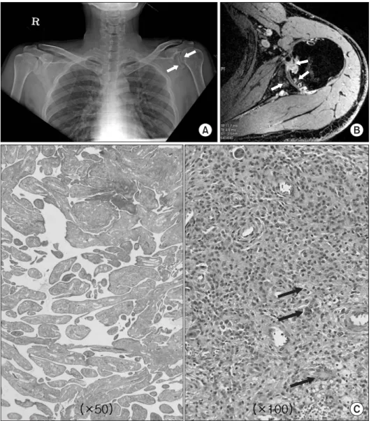
motion (p=0.0004), function (p<0.0001), and total (p<0.0001) scores showed statistically significant improvement. 11 patients (10: referral from other hospitals, 1: received initial surgery by the author) who had re-operation due to recurrence presented overall low scores in all the categories, as compared to the other patients, though the scores were not statistically significant (pain: p=0.844, swelling: p=0.656, range of motion: p=0.662, function: p=0.819, total: p=0.984). In comparing the patients who had recurrences, those who had more than two recurrences showed decreased range of motion (p=0.043). Other categories did not show statistical significance. At the last follow-up, two patients had a range of motion less than 90 degrees, which restrains their daily living. Four other patients complained of intermittent swelling and pain, though the rest of the patients did not have Figure 2. A 36 year old male who had diffuse-type PVNS in his left ankle. (A) Pre-operative x-ray presenting radiolucent lesions on distal fibular and tibia. (B) Pre-operative MRI images showing diffuse mass on anterior and posterior aspect of the ankle. (C) Intra-operative gross findings after a posterolateral approach. (D) Microscopic findings present synovial-like mononuclear cells, hemosiderin laden macrophages, foam cells and giant cells which are consistent findings of PVNS (H&E stain).
any complications that interfered with their living.
Discussion
Pigmented villonodular synovitis (PVNS) generally occurs in patients in the third or fourth decade of life.1-4,10,14)
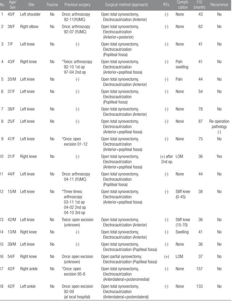
The average age of a patient in this study was 34 years, which matched the Figure 3. Representive plain radiography, MRI and Microscopic findings of diffuse-type PVNS in shoulder joint. (A) Pre-operative plain radiography presenting radiolucent lesion and bony erosions on glenohumeral joint. (B) Pre-operative MRI images (axial) presenting infiltrative diffuse mass on glenohumeral joint with bony erosion and cysts. (C) Microscopic findings present hypertrophied synovium, foam cells, giant cells which are consistent findings of PVNS (H&E stain).
Table 2. Criteria for Assessment Proposed by Ogilvie-Harris
0 1 2 3
Pain Severe Moderate Slight None
Synovitis/swelling Severe Moderate Slight None
ROM More than 20% loss 10% to 20% loss 0% to 10% loss Normal
Function Minimal activity Reduced activity Almost complete activity Complete activity
Table 3. Preoperative Symptoms
Symptom No. of patients
Palpable mass 14 (66%)
Pain 11 (52%)
Swelling 7 (33%)
average age of a previous study. The knee and flexor tendon sheaths of the hand are the most frequent occurring site compare to ankle and shoulder.1,4-6,10,21)
In this study, 66% (14 cases) oc-curred at the knee.
According to Myers and Masi,4)
the distribution of PVNS by sex is close to two to one, with a predominance in male patients. Our study, however, showed a predominance of female cases, by two to one (female: 14 cases, male: 7 cases). Considering that there is also a report of the distribution being equal or opposite, a larger scale study should be done.4,10,14)
Palpable mass was the most common symptom at first presen-tation (66%); only 9% of the patients complained of limipresen-tation of motion as the initial symptom. It should be noted that if the mass locates in the popliteal fossa or ankle, it is easy to palpate. In the
previous report, swelling and pain were the common presenting symptoms.2,6,10)
Even though there is a hypothesis that intra-articular hemor-rhage due to recurrent trauma is the cause of PVNS, many studies report that there is little connection between trauma and etiology of PVNS.2,4,5,10,22) In this study, there was only one patient who
had a history of trauma before the initiation of symptoms. Several authors have insisted on less extensive approaches for the treatment of diffuse PVNS, but there are reasons why an extensive approach or radical synovectomy with a safe margin has been advocated by many authors, treating PVNS like other malignancy.2,5-7,11,13-15,17,19,21,22) Although the pathogenesis of
PVNS is still unclear, Bertoni et al.23)
reported characteristics of a malignant form PVNS in 8 cases, and Oda et al.24) published
Table 4. Result of Clinical Assessment
No Site Pain Swelling ROM Function Total
Pre Post Pre Post Pre Post Pre Post Pre Post
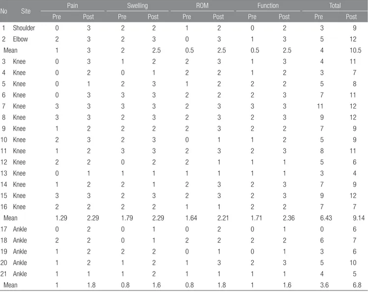
1 Shoulder 0 3 2 2 1 2 0 2 3 9 2 Elbow 2 3 2 3 0 3 1 3 5 12 Mean 1 3 2 2.5 0.5 2.5 0.5 2.5 4 10.5 3 Knee 0 3 1 2 2 3 1 3 4 11 4 Knee 0 2 0 1 2 2 1 2 3 7 5 Knee 0 1 2 3 1 2 2 2 5 8 6 Knee 0 3 3 3 2 2 2 3 7 11 7 Knee 3 3 3 3 2 3 3 3 11 12 8 Knee 3 3 2 3 2 3 2 3 9 12 9 Knee 1 2 2 2 2 3 2 2 7 9 10 Knee 2 3 2 3 0 1 1 2 5 9 11 Knee 1 2 3 3 2 3 2 3 8 11 12 Knee 2 2 0 2 2 1 1 1 5 6 13 Knee 0 1 1 1 1 1 1 1 3 4 14 Knee 1 2 2 1 2 3 2 3 7 9 15 Knee 3 3 2 3 2 3 2 3 9 12 16 Knee 2 2 2 2 1 1 2 2 7 7 Mean 1.29 2.29 1.79 2.29 1.64 2.21 1.71 2.36 6.43 9.14 17 Ankle 0 2 0 1 0 2 0 1 0 6 18 Ankle 2 2 0 1 2 2 2 2 6 7 19 Ankle 1 2 2 2 0 1 0 1 3 6 20 Ankle 1 2 1 2 1 3 2 3 5 10 21 Ankle 1 1 1 2 1 1 1 1 4 5 Mean 1 1.8 0.8 1.6 0.8 1.8 1 1.6 3.6 6.8
a case presenting a malignant transformation of PVNS at the sacrum, following two recurrences. These findings demonstrate that careful attention should be paid to PVNS, due to its potential for malignant behavior. Using a DNA microarray, Finis et al.25)
described that PVNS had a decreased apoptotic cell cycle, like other malignancies. Layfield et al.26)
found trisomies in the fifth and seventh chromosomes within tissue of PVNS. These recent findings about cytogenetic abnormalities, in the form of monoclonalities and chromosomal abnormalities, supports the malignant pathogenesis of PVNS. Addition to that, clinical findings of its local invasiveness, destruction of major joints, and frequent recurrence after the surgery have been the most concerning problems when dealing with PVNS.6-8,11,14,17,19,22) Recurrence rate
after surgery is reported in 8-48% of patients when diffuse-type PVNS occurs at knee.2,3,5,9-11) When PVNS is in the hand, the
reported recurrence rate varies from 7-45%.2,3,16)
There have been several reports of a low recurrence rate when arthroscopy and adjuvant radiation therapy are used, although those methods are generally considered to be appropriate for treating the localized form or biopsy.17,20,27)
Due to the limitation of surgical exposure, arthroscopic removal is not suitable for diffuse-type or extra-articular type PVNS, as in the patients in this study.17,19) 47% (10 cases) of
the patients were referred because of recurrence after primary surgery. Among the 10 cases, 50% had previous arthroscopic removal. Considering that repeated surgery increases the chance of morbidity, such as a stiff knee, a complete resection of the pathologic mass in the initial surgery is the best method to ensure a good prognosis.
The risk that extensive open surgical approaches to the joint will increase post-operative morbidities, such as a stiff joint, or the possibility of infection, has led surgeons to hesitate before performing open procedures, which made them to try minimally invasive techniques for the resection of diffuse-type PVNS, such as arthroscopic removal.20,27) Sharma et al.22)
has reported satisfactory clinical outcomes with low recurrence rate, and preservation of knee joint function, after more than 6 years of follow-up using open synovectomy. Chin et al.17)
has also reported similar clinical outcomes after open surgery. It is thought that insufficient resection leads to recurrence, and that frequent surgery cause functional impairment of the joint. In this study, there were two patients who had a stiff knee after surgery, with a range of motion less than 90 degrees. Both of the patients had more than two re-operations, due to recurrence. The other
patients did not have functional impairment after surgery, and there was improvement in every aspect of the clinical assessment. Additionally, it was determined by telephone interview that if patients were satisfied with the surgical results, they have decreased anxiety about recurrence.
There have been reports of satisfactory results from radia tion therapy as adjuvant therapy, though radiation has drawbacks.12,16)
There is the possibility of sarcomatous change after radiation or post-radiation fibrosis, which might obscure findings of recur-rence. These changes also increase the difficulty of re-operation. Bickels et al.18) reported full thickness necrosis after using the
radio isotope Yttrium-90 in a patient with PVNS of the ankle joint, which led to a cessation of the use of radioisotopes for ad-juvant therapy.
Although it needs demanding surgical skills to perform total synovectomy, there were satisfactory surgical outcomes without radiation therapy with the condition that complete resection with a total synovecmy.5,7,14,15,22)
Atmore et al.13)
and Kotwal et al.16) both reported that compared to surgery alone, adjuvant
radiation therapy is not advantageous. Flandry et al.15)
reported that it is not necessary to perform additional radiation therapy if total synovectomy is done. According to these reports, adjuvant radiation therapy should not be a routine procedure, if a surgeon performs adequate total synovectomy. We limited radiation therapy to patients with recurrence after total synovectomy, or those who had subtotal synovectomy.
We performed total synovectomy on all patients when it was possible, which resulted in all but one having total synovectomy, as for that patient it was impossible to perform total synovectomy due to anatomic reasons. Additional electrocautrization was done at the base and surrounding soft tissues instead of radiation therapy. We expected the thermal effect of electrocautrization to remove remnant pathology. During the follow-up period, of an average of 65 months, there was one (8.3%) recurrence at the knee. It was pathologically confirmed and re-operation was done, and a total of 3,000 cGy of radiation therapy was also applied. Similar to the Chin et al.17) and Sharma et al.22) reports,
our study shows a comparably low recurrence rate without using radiation therapy.
Considering there is no established treatment protocol for diffuse-type PVNS, our surgical outcome supports that complete total synovectomy at initial operation is the key step in successful treatment.
References
1. Jaffe HL, Lichtenstein L, Sutro CJ. Pigmented villonodular synovitis, bursitis and tenosynovitis. A discussion of the synovial and bursal equivalents of the tenosynovial lesion commonly denoted as xanthoma, xanthogranuloma, giant-cell tumor of myeloplasioma of the tendon sheath, with some consideration of this tendon sheath lesion itself. Arch Pathol. 1941;31:731-65.
2. Byers PD, Cotton RE, Deacon OW, et al. The diagnosis and treatment of pigmented villonodular synovitis. J Bone Joint Surg Br. 1968;50:290-305.
3. Granowitz SP, D'Antonio J, Mankin HL. Th e pathogenesis and long-term end results of pigmented villonodular synovitis. Clin Orthop Relat Res. 1976;114:335-51.
4. Myers BW, Masi AT. Pigmented villonodular synovitis and tenosynovitis: a clinical epidemiologic study of 166 cases and literature review. Medicine (Baltimore). 1980;59:223-38. 5. Flandry F, Hughston JC. Pigmented villonodular synovitis. J
Bone Joint Surg Am. 1987;69:942-9.
6. McMaster PE. Pigmented Villonodular Synovitis with Inva-sion of Bone: Report of Six Cases. J Bone Joint Surg Am. 1960; 42:1170-83.
7. Scott PM. Bone lesions in pigmented villonodular synovitis. J Bone Joint Surg Br. 1968;50:306-11.
8. Jergesen HE, Mankin HJ, Schiller AL. Diffuse pigmented villonodular synovitis of the knee mimicking primary bone neoplasms. A report of two cases. J Bone Joint Surg Am. 1978; 60:825-9.
9. Johansson JE, Ajjoub S, Coughlin LP, Wener JA, Cruess RL. Pigmented villonodular synovitis of joints. Clin Orthop Relat Res. 1982:159-66.
10. Rao AS, Vigorita VJ. Pigmented villonodular synovitis (giant-cell tumor of the tendon sheath and synovial membrane). A review of eighty-one cases. J Bone Joint Surg Am. 1984;66:76-94.
11. Schwartz HS, Unni KK, Pritchard DJ. Pigmented villonodular synovitis. a retrospective review of aff ected large joints. Clin Orthop Relat Res. 1989;247:243-55.
12. Wiss DA. Recurrent villonodular synovitis of the knee. Successful treatment with yttrium-90. Clin Orthop Relat Res. 1982;169:139-44.
13. Atmore WG, Dahlin DC, Ghormley RK. Pigmented villono-dular synovitis; a clinical and pathologic study. Minn Med.
1956;39:196-202.
14. Jones FE, Soule EH, Coventry MB. Fibrous xanthoma of synovium (giant-cell tumor of tendon sheath, pigmented no-dular synovitis). A study of one hundred and eighteen cases. J Bone Joint Surg Am. 1969;51:76-86.
15. Flandry FC, Hughston JC, Jacobson KE, Barrack RL, McCann SB, Kurtz DM. Surgical treatment of diffuse pigmented vil-lonodular synovitis of the knee. Clin Orthop Relat Res. 1994; 300:183-92.
16. Kotwal PP, Gupta V, Malhotra R. Giant-cell tumour of the ten-don sheath. Is radiotherapy indicated to prevent recurrence aft er surgery? J Bone Joint Surg Br. 2000;82:571-3.
17. Chin KR, Barr SJ, Winalski C, Zurakowski D, Brick GW. Treatment of advanced primary and recurrent diffuse pig-ment ed villonodular synovitis of the knee. J Bone Joint Surg Am. 2002;84A:2192-202.
18. Bickels J, Isaakov J, Kollender Y, Meller I. Unacceptable com-plications following intra-articular injection of yttrium 90 in the ankle joint for diff use pigmented villonodular synovitis. J Bone Joint Surg Am. 2008;90:326-8.
19. Wu CC, Pritsch T, Bickels J, Wienberg T, Malawer MM. Two incision synovectomy and radiation treatment for diff use pig-mented villonodular synovitis of the knee with extra-articular component. Knee. 2007;14:99-106.
20. Ogilvie-Harris DJ, McLean J, Zarnett ME. Pigmented vil-lonodular synovitis of the knee. The results of total arthros-copic synovectomy, partial, arthrosarthros-copic synovectomy, and arthroscopic local excision. J Bone Joint Surg Am. 1992;74: 119-23.
21. Sharma H, Jane MJ, Reid R. Pigmented villonodular synovitis of the foot and ankle: forty years of experience from the Scottish bone tumor registry. J Foot Ankle Surg. 2006;45:329-36.
22. Sharma H, Rana B, Mahendra A, Jane MJ, Reid R. Outcome of 17 pigmented villonodular synovitis (PVNS) of the knee at 6 years mean follow-up. Knee. 2007;14:390-4.
23. Bertoni F, Unni KK, Beabout JW, Sim FH. Malignant giant cell tumor of the tendon sheaths and joints (malignant pigmented villonodular synovitis). Am J Surg Pathol. 1997;21:153-63. 24. Oda Y, Takahira T, Yokoyama R, Tsuneyoshi M. Diff use-type
giant cell tumor/pigmented villonodular synovitis arising in the sacrum: malignant form. Pathol Int. 2007;57:627-31. 25. Finis K, Sultmann H, Ruschhaupt M, et al. Analysis of
comple-mentary DNA microarray and tissue array technology reveals insight into potential novel therapeutic approaches. Arthritis Rheum. 2006;54:1009-19.
26. Layfi eld LJ, Meloni-Ehrig A, Liu K, Shepard R, Harrelson JM. Malignant giant cell tumor of synovium (malignant pigmented
villonodular synovitis). Arch Pathol Lab Med. 2000;124:1636-41.
27. Beguin J, Locker B, Vielpeau C, Souquieres G. Pigmented villonodular synovitis of the knee: results from 13 cases. Arthroscopy. 1989;5:62-4.