KJTCVS
The Korean Journal of Thoracic and Cardiovascular SurgeryClinical
Research
https://doi.org/10.5090/kjtcs.2020.53.2.41 pISSN: 2233-601X eISSN: 2093-6516
Korean J Thorac Cardiovasc Surg. 2020;53(2):41-48
Chylothorax after Surgery for Congenital Cardiac Disease: A
Prevention and Management Protocol
Yu Rim Shin, M.D., Ha Lee, M.D., Young-Hwan Park, M.D., Han Ki Park, M.D., Ph.D.
Division of Cardiovascular Surgery, Department of Thoracic and Cardiovascular Surgery, Severance Cardiovascular Hospital, Yonsei University College of Medicine, Seoul, Korea
ARTICLE INFO Received September 2, 2019 Revised October 15, 2019 Accepted October 22, 2019 Corresponding author Han Ki Park Tel 82-2-2228-8480 Fax 82-2-313-2992 E-mail hank@yuhs.ac ORCID https://orcid.org/0000-0002-7472-7822 †This article was presented at the 50th Annual Meeting of the Korean Society for Thoracic and Cardiovascular Surgery.
Background: Chylothorax after congenital heart surgery is not an uncommon com-plication, and it is associated with significant morbidity. However, consensus treatment guidelines are lacking. To improve the treatment outcomes of patients with postoperative chylothorax, we implemented a standardized management protocol at Severance Hospi-tal in September 2014.
Methods: A retrospective review of patients treated at a single center was done. All cor-rective and palliative operations for congenital heart disease performed at our institution between January 2008 and April 2018 were reviewed. The incidence and treatment out-comes of postoperative chylothorax were analyzed.
Results: The incidence of chylothorax was 1.9%. Sixty-one percent of the patients could be managed with a low-fat diet, while 28% of the patients required complete restriction of enteral feeding. Thoracic duct embolization was performed in 2 patients and chest tube drainage decreased immediately after the procedure. No patient required thoracic duct ligation or pleurodesis. After implementation of the institutional management protocol, the number of chest tube drainage days decreased (median, 24 vs. 14 days; p=0.45).
Conclusion: Implementing a strategy to reduce postoperative chylothorax resulted in an acceptable incidence of postoperative chylothorax. Instituting a clinical practice proto-col helped to curtail the treatment duration and to decrease the requirement for surgical treatment. Image-guided embolization of the thoracic duct is an effective treatment for postoperative chylothorax.
Keywords: Chylothorax, Congenital heart defects, Postoperative complication, Postop-erative care
Copyright©The Korean Society for Thoracic and Cardiovascular Surgery. 2020. All right reserved.
This is an Open Access article distributed under the terms of the Creative Commons Attribution Non-Commercial License (http://creativecommons.org/licenses/
Introduction
Postoperative chylothorax after surgical procedures for congenital heart disease poses a challenging clinical situa-tion. Its incidence varies across centers, with a reported range from 2% to 5% [1-4]. Postoperative chylothorax is as-sociated with poor nutrition, an electrolyte imbalance, and a higher risk of thrombus development, and its clinical im-pact includes increased mortality, increased length of hos-pital stay, increased length of intensive care unit stay, pro-longed mechanical ventilation, and a higher likelihood of extracorporeal membrane oxygenation support [4].
From a surgical perspective, the mechanism of postoper-ative chylothorax can be categorized as traumatic or
non-traumatic. Traumatic chylothorax is caused by direct injury of the thoracic duct or one of its lymphatic branch-es. Non-traumatic chylothorax is caused by unfavorable hemodynamics, lymphatic malformation, or congenital syndromes such as Noonan syndrome and Turner syn-drome. Traditionally, treatment options included diet modification and octreotide infusion as conservative man-agement, and thoracic duct ligation and pleurodesis as sur-gical management [5-7]. In recent years, new imaging mo-dalities such as magnetic resonance lymphangiography have improved our understanding of chylothorax, and studies on thoracic duct embolization demonstrated an ac-ceptable success rate in patients in whom thoracic duct li-gation failed [8-10].
https://doi.org/10.5090/kjtcs.2020.53.2.41
KJTCVS
Although many treatment options for postoperative chy-lothorax exist, there is no generally accepted standardized treatment guideline. At Severance Hospital, a standardized man agement protocol for postoperative chylothorax was developed and has been implemented since September 2014. The aim of this study was to determine the incidence of postoperative chylothorax after surgery for congenital heart disease and to evaluate the efficacy of implementing a chylothorax management protocol.
Methods
Patients
This is a retrospective review of experiences at a single center. The medical records of all patients who underwent corrective or palliative operations for congenital heart dis-ease at Severance Hospital from January 2008 to April 2018 were reviewed. This study was reviewed and approved by the Institutional Review Board (IRB approval no., 4-2019-0903). The obligation to obtain informed consent was waived by the institutional review board in view of the ret-rospective nature of the study.
Diagnosis of chylothorax
Chylothorax was suspected when persistent drainage from an existing chest tube became cloudy when the pa-tient was fed, or when similar milky fluid was found after placing a tube into a newly discovered effusion. The diag-nosis of chylothorax was confirmed based on the charac-teristics of the pleural fluid recovered from each patient. Chylothorax was diagnosed if any of the following criteria was met: (1) the pleural fluid triglyceride level was >100 mg/dL, (2) the percentage of lymphocytes in the pleural fluid was >80%, or (3) the pleural fluid triglyceride level was higher than that of the serum.
Postoperative chylothorax was defined as chylothorax developing within 30 days of surgery. Based on the maxi-mum cumulative output of all chest tubes for a 24-hour period, chylothorax was classified high (≥20 mL/kg/day) or low (<20 mL/kg/day). The recurrence of chylothorax was diagnosed when a drainage tube was re-inserted for chy-lous effusion. Chylothorax duration was defined as the time interval from the day of diagnosis to the day of chest tube removal.
Institutional policy to reduce postoperative
chylothorax
To minimize the development of postoperative chylotho-rax, we follow our institutional policy. During the opera-tion, we do not remove the thymus routinely. Even in neo-nates or for aortic arch repair, we preserve the thymus whenever possible. If the thymus is large, by splitting its left and right lobes and retracting the lobes with pericardi-al tenting sutures, good exposure of the aortic arch and pulmonary arteries can be obtained. Cannulation of the innominate artery for selective cerebral perfusion can be carried out without thymus resection. Dissection of the aortic arch and superior vena cava (SVC) is minimized if not indicated. We routinely place a SVC drainage cannula through the right atrium and avoid direct cannulation of the SVC. The indications for direct SVC cannulation are superior cavopulmonary anastomosis and anomalous pul-monary venous connection to the SVC.
The postoperative management policies are as follows: (1) early removal of the central venous catheter, (2) avoidance of hyperalimentation, and (3) therapy to lower the SVC pressure in patients with physiologically high SVC pressure (e.g., those who have undergone a Fontan operation or re-ceived bidirectional cavopulmonary shunt), which includes diuresis, pulmonary toileting, supplemental oxygen, pul-monary vasodilator use, nitric oxide inhalation, and after-load reduction.
Management protocol for postoperative
chylothorax
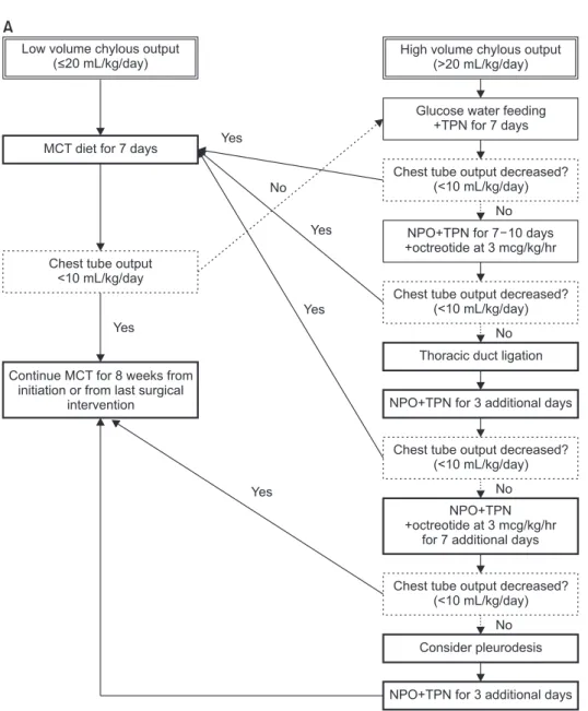
As a quality improvement program, we established a standardized management protocol for postoperative chy-lothorax. The protocol was developed over 3 months, in-cluding a period of literature review and gathering the opinions of multidisciplinary team of experts, after which it was implemented in September 2014. The protocol was then revised in 2017 (Fig. 1).
Based on the chest tube drainage amount, the patients are classified as having low- or high-output chylothorax, according to which a different initial management strategy is applied. The diet of patients with low-output chylothorax is modified to a medium-chain triglyceride (MCT) or low-fat diet. In patients with high-output chylothorax, early adoption of nil per os (NPO) with total parenteral nutri-tion (TPN) treatment is applied. Surgical thoracic duct li-gation, intravenous infusion of octreotide, and pleurodesis are sequentially considered as treatment options for
Ha Lee, et al. Postoperative Chylothorax after Congenital Cardiac Surgery
KJTCVS
non-responders. The version of the protocol revised in 2017 incorporates lymphangiography and thoracic duct embolization as the first choice of treatment modality for adult-sized patients. For the NPO and TPN treatment groups, enteral intake of water, a glucose-water solution, or a balanced ion solution with no fat components is allowed.
Lymphangiography and thoracic duct embolization are performed by interventional radiologists. Inguinal lymph nodes are accessed under real-time ultrasound guidance with a 25-gauge needle, and lymphangiography is per-formed using an oil-based contrast agent (Lipiodol; Guer-bet, Bloomington, IN, USA). After visualization of the tho-racic duct and identification of a target lymphatic vessel, a 21-gauge long Chiba needle is placed in the target
lymphat-ic vessel under fluoroscoplymphat-ic guidance. Next, a mlymphat-icrocathe- microcathe-ter is placed in the lymphatic vessel and contrast dye is in-jected to identify the chylous leak. The thoracic duct is embolized with an embolization coil and/or a mixture of cyanoacrylate (Histoacryl; B. Braun, Barcelona, Spain) and Lipiodol (Guerbet).
Statistical analysis
The incidence of chylothorax was calculated for all cor-rective or palliative surgical procedures for congenital heart disease. Continuous variables were summarized as median with range. Categorical and ordinal variables were presented as frequency and percentage. Group
compari-NPO+TPN +octreotide at 3 mcg/kg/hr
for 7 additional days Continue MCT for 8 weeks from
initiation or from last surgical intervention MCT diet for 7 days
Chest tube output <10 mL/kg/day Low volume chylous output
(<20 mL/kg/day)
High volume chylous output (>20 mL/kg/day)
Glucose water feeding +TPN for 7 days
Chest tube output decreased? (<10 mL/kg/day)
NPO+TPN for 7 10 days +octreotide at 3 mcg/kg/hr
Chest tube output decreased? (<10 mL/kg/day)
Thoracic duct ligation
NPO+TPN for 3 additional days
Chest tube output decreased? (<10 mL/kg/day)
Chest tube output decreased? (<10 mL/kg/day)
Consider pleurodesis
NPO+TPN for 3 additional days Yes No Yes Yes Yes Yes No No No No A
Fig. 1. Management protocol for postoperative chylothorax for chil-dren (A) and adults (B). MCT, medi-um chain triglyceride; NPO, nil per os; TPN, total parenteral nutrition. (Continued on next page).
https://doi.org/10.5090/kjtcs.2020.53.2.41
KJTCVS
sons were made using the Wilcoxon rank sum test. All p-values <0.05 were considered to indicate statistical sig-nificance. Statistical analysis was performed using IBM SPSS ver. 23.0 (IBM Corp., Armonk, NY, USA).
Results
Incidence of postoperative chylothorax
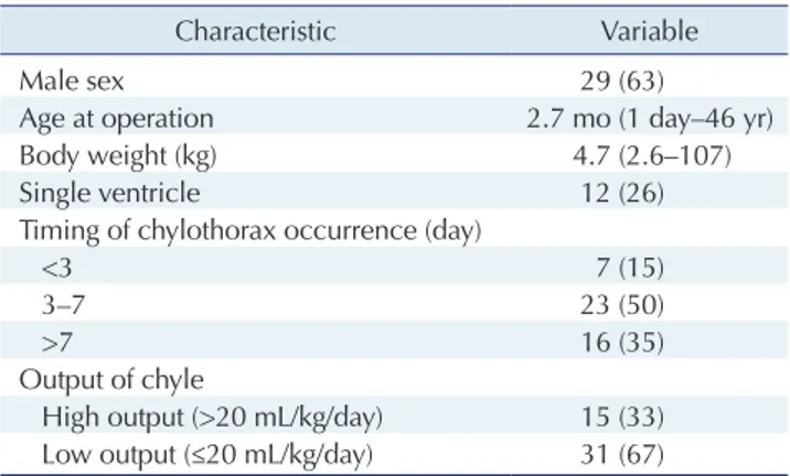
There were 2,465 corrective or palliative surgical proce-dures in the study period, and 46 patients developed post-operative chylothorax. The incidence was 1.9%. In 16 pa-tients (35%), chylothorax developed after postoperative day 7. In 15 patients (33%), the chest tube drainage was classi-fied as high-output (>20 mL/kg/day). The characteristics of the patients are summarized in Table 1.
NPO+TPN +octreotide at 3 mcg/kg/hr
for 7 additional days Chylous output
Thoracic duct embolization
Chest tube output decreased? (<100 mL/day)
NPO+TPN for 7 10 days +octreotide at 3 mcg/kg/hr
Chest tube output decreased? (<100 mL/day)
Thoracic duct ligation
NPO+TPN for 3 additional days
Chest tube output decreased? (<100 mL/day)
Chest tube output decreased? (<100 mL/day)
Consider pleurodesis
NPO+TPN for 3 additional days Continue MCT for 8 weeks
from initiation or from last surgical intervention
MCT diet for 7 days
Chest tube output <100 mL/day Yes No No No No No
Low fat diet MCT diet for 3 days
Chest tube output <100 mL/day Yes Yes Yes Yes No B Yes
Fig. 1. (Continued; caption shown on previous page).
Table 1. Characteristics of patients with postoperative chylothorax (n=46)
Characteristic Variable
Male sex 29 (63)
Age at operation 2.7 mo (1 day–46 yr)
Body weight (kg) 4.7 (2.6–107)
Single ventricle 12 (26)
Timing of chylothorax occurrence (day)
<3 7 (15)
3–7 23 (50)
>7 16 (35)
Output of chyle
High output (>20 mL/kg/day) 15 (33) Low output (≤20 mL/kg/day) 31 (67) Values are presented as number (%) or median (range).
Ha Lee, et al. Postoperative Chylothorax after Congenital Cardiac Surgery
KJTCVS
Twenty-six percent of cases of postoperative chylothoraxoccurred in patients with a functional single ventricle. The incidence of postoperative chylothorax was markedly high-er afthigh-er repair of a vascular ring (3 of 11, 27%), the Fontan operation (5 of 21, 24%), and the Norwood operation (2 of 10, 20%) than after other procedures (Table 2).
Treatment modalities
In 28 patients (61%), the chylothorax could be managed with only an MCT or low-fat diet. Restriction of food ex-cept for water or a glucose-water solution was required in 13 patients (27%). Intravenous somatostatin was adminis-tered in 4 patients (9%). Lymphangiography and thoracic duct embolization were performed in 2 adult patients, in whom chest tube drainage decreased immediately after the procedure. No patient required surgical thoracic duct liga-tion or pleurodesis. Data on the treatment modalities, chy-lothorax duration, and hospital stay are summarized in Table 3.
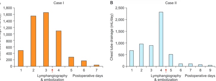
Percutaneous thoracic duct embolization was performed in 2 patients, and it was effective for controlling the chy-lous drainage (Fig. 2). A 46-year-old woman was diagnosed with postoperative chylothorax on postoperative day 16
af-Table 2. Incidence of postoperative chylothorax based on type of surgery and RACHS category
Variable Occurrence of
chylothorax (%) Type of surgery
Ventricular septal defect repair 9 (1) Coarctation of the aorta repair 2 (4) Systemic to pulmonary artery shunt 3 (9) Arterial switch operation 1 (2) Bidirectional cavopulmonary shunt 3 (5)
Fontan operation 5 (24)
Norwood operation 2 (20)
Vascular ring repair 3 (27)
Total anomalous pulmonary venous connection repair
3 (10) Tetralogy of Fallot repair 4 (9)
Others 11 (1) RACHS category I 4 (9) II 19 (41) III 18 (39) IV 2 (4) V 0 IV 2 (4) Not categorized 1 (2)
RACHS, risk adjustment for congenial heart surgery.
Table 3. Treatment modalities for postoperative chylothorax
Treatment modality No. (%) Chylothorax duration (day) Hospital stay (day) MCT or low-fat diet 44 (96)
MCT or low-fat diet only 28 (61) 9 (6–49) 23 (7–331) Nil per os 13 (28) 17 (3–66) 28 (15–93) Octreotide 4 (9) 35 (17–196) 50 (22–196) Lymphangiography and
thoracic duct embolization
2 (4) 6 (5–6) 37 (21–38) Thoracic duct ligation 0
Pleurodesis 0
Protocol based approach 20 (43) 14 (6–39) 21 (9–184) Values are presented as median (range), unless otherwise stated. MCT, medium chain triglyceride.
1,800 1,600 1,400 1,200 1,000 800 600 400 200 7 Chest tube drainage (mL/day) Postoperative days 0 1 2 3 4 5 6 Lymphangiography & embolization Case I Case II 2,500 2,000 1,500 1,000 500 7 8 9 Chest tube drainage (mL/day) Postoperative days 0 1 2 3 4 5 6 Lymphangiography & embolization A B
Fig. 2. (A, B) Chest tube drainage amount of the 2 patients who underwent lymphangiography and thoracic duct embolization for post-operative chylothorax.
https://doi.org/10.5090/kjtcs.2020.53.2.41
KJTCVS
ter the repair of pulmonary valve stenosis. On postopera-tive day 18, lymphangiography and thoracic duct emboli-zation were carried out. Her chest tube drainage decreased from 1,500 mL/day pre-procedure to 200 mL/day on the day after the procedure. The chest tube was removed 3 days after thoracic duct embolization. The other patient was a 20-year-old man who underwent vascular ring re-pair. His chest tube drainage was 2,300 mL/day despite full restriction of enteral intake, but decreased to 510 and 110 mL/day on post-procedure days 1 and 2. There were no procedure-related complications or recurrence of chylotho-rax.
Comparison of clinical outcomes before and after
implementation of the treatment protocol
The median chest tube drainage duration of all chy-lothorax patients was 20 days (range, 6–196 days). In our cohort of 46 patients with postoperative chylothorax, 26 (57%) were diagnosed before September 2014, when our management protocol was implemented. The number of chest tube drainage days decreased after implementation of the protocol (median, 24 versus 14 days; p=0.045). The length of hospital stay also decreased (median, 37 versus 21 days; p=0.067), with marginal statistical significance. Chy-lothorax recurred in 2 patients both before and after im-plementation of the protocol (p=0.78). There were 2 in-hos-pital deaths in the pre-implantation period, whereas no in-hospital mortality occurred in the post-implementation period (p=0.20) (Table 4).
Discussion
In our patient cohort, the incidence of postoperative chy-lothorax for congenital heart surgery was 1.9%. This is
lower than has been reported in other previous reports [1-4]. Therefore, we think that our intraoperative and postop-erative management strategy to reduce postoppostop-erative chy-lothorax is effective. To prevent traumatic damage to lymphatic channels, we minimize the dissection of the structures around the lymphatic pathway. In neonates and infants, the thymus is normally large, which limits the ex-posure of the great vessels. The fissure between the left and right lobes of the thymus can be easily dissected, and later-al retraction of the right and left lobes with pericardilater-al tenting provides good exposure of the aortic arch and the branch pulmonary arteries. In most cases, neonatal aortic arch repair or repair of tetralogy of Fallot can be per-formed without resection of the thymus. Another surgical precaution is dissection around the SVC. To avoid dissec-tion around the SVC, we do not directly cannulate the SVC unless indicated. The SVC is directly cannulated only for repair of an anomalous pulmonary venous connection to the SVC or superior cavopulmonary anastomosis. Creation of a superior cavopulmonary connection or a Fontan pro-cedure leads to elevated SVC pressure and can result in lymph leakage. These operations are known to be high-risk procedures for postoperative chylothorax. In our cohort, 24% of the patients developed chylothorax after the Fontan operation, but the incidence was only 5% after placement of a bidirectional cavopulmonary shunt. This suggests that our strategy of minimal intraoperative dissection and post-operative SVC pressure-lowering management is effective for reducing the incidence of chylothorax. In our cohort, 35% of the cases of chylothorax developed later than post-operative day 7. This suggests that non-traumatic causes are an important factor in chylothorax development. In this regard, postoperative management is also important to prevent postoperative chylothorax. Central venous lines should be removed as early as possible [11] and medical management should have the goal of hemodynamic stabili-ty and lowering the central venous pressure.
Protocol-based management has been proposed to im-prove the outcomes of chylothorax, and studies have shown that implementing such management strategies led to re-ductions in chest tube utilization time and improvements in clinical outcomes [12-14]. We developed a management protocol and implemented it in September 2014. The num-ber of chest tube utilization days decreased (median, 24 to 14 days; p=0.045), and the length of hospital stay also de-creased (median, 37 to 21 days; p=0.067). There was no significant difference in in-hospital mortality or the recur-rence of chylothorax. Even before implementing the proto-col, physicians at our institution treated the patients in a
Table 4. Impact of a protocol-based approach on outcomes
Variable Pre-implementation (n=26) Post-implementation (n=20) p-value Era Before September
2014 After September 2014 Amount (mL/kg/day) 18 (1.0–115) 13 (0.9–79) 0.48 Chylothorax duration (day) 24 (6–196) 14 (6–39) 0.045 Hospital stay (day) 37 (7–331) 21 (9–184) 0.067
In-hospital death 2 0 0.20
Recurrent chylothorax
2 2 0.78
Ha Lee, et al. Postoperative Chylothorax after Congenital Cardiac Surgery
KJTCVS
similar way to that described in the protocol. However, theprotocol more specifically stipulated the relevant details, which helped to decrease the variability in practice among physicians and led to a shortened treatment duration.
Recently, percutaneous lymphatic embolization has been reported as a successful treatment option for patients with postoperative chylothorax [10]. In our cohort, lymphangi-ography and interventional thoracic duct embolization were performed in 2 patients. These patients were adults who underwent vascular ring repair and pulmonary valve replacement. In both patients, the daily chest tube drainage was more than 1,500 mL, but after embolization, the chy-lous drainage decreased immediately and the chest tubes could be removed in 4 days. Based on our experience, we revised our management protocol to consider lymphangi-ography and lymphatic embolization as the first treatment option in adult-sized patients. Our experience is limited to adults, but according to the case series published by Savla et al. [10], lymphatic embolization was successfully per-formed in young infants, suggesting new possibilities for chylothorax treatment, even for younger patients.
The main limitation of this study is that it presents a ret-rospective analysis of a limited number of patients. We compared clinical outcomes between before and after the implementation of the treatment protocol. The protocol helped to decrease variability in practice, but the manage-ment of patients did not always strictly adhere to the pro-tocol. Each patient’s characteristics and medical conditions influenced small details of the management. With the ac-cumulation of more data and advances in medical practice, the protocol should be revised further to obtain the best treatment results for this postoperative problem.
In conclusion, the utilization of a strategy to reduce post-operative chylothorax after surgical procedures for con-genital heart disease resulted in an acceptably low inci-dence. Implementation of a standardized treatment protocol for postoperative chylothorax helped to reduce the treatment duration and to improve treatment outcomes. A strategy focusing on protocol-guided conservative manage-ment and image-guided embolization of the thoracic duct helped to reduce the need for surgical treatment for post-operative chylothorax.
Conflict of interest
No potential conflict of interest relevant to this article was reported.
Acknowledgments
This study was supported by a Grant of the Samsung Vein Clinic Network (Daejeon, Anyang, Cheongju, Cheonan; Fund no. KTCS04-135).
ORCID
Ha Lee: https://orcid.org/0000-0003-3976-8554 Yu Rim Shin: https://orcid.org/0000-0001-7685-0018 Young-Hwan Park: https://orcid.org/0000-0001-9802-8017 Han Ki Park: https://orcid.org/0000-0002-7472-7822
References
1. Milonakis M, Chatzis AC, Giannopoulos NM, et al. Etiology and management of chylothorax following pediatric heart surgery. J Card Surg 2009;24:369-73.
2. Bauman ME, Moher C, Bruce AK, Kuhle S, Kaur S, Massicotte MP. Chylothorax in children with congenital heart disease: incidence of thrombosis. Thromb Res 2013;132:e83-5.
3. Mery CM, Moffett BS, Khan MS, et al. Incidence and treatment of chylothorax after cardiac surgery in children: analysis of a large multi-institution database. J Thorac Cardiovasc Surg 2014;147:678-86.
4. Buckley JR, Graham EM, Gaies M, et al. Clinical epidemiology and centre variation in chylothorax rates after cardiac surgery in children: a report from the Pediatric Cardiac Critical Care Consortium. Cardiol Young 2017;27:1678-85.
5. Nath DS, Savla J, Khemani RG, Nussbaum DP, Greene CL, Wells WJ. Thoracic duct ligation for persistent chylothorax after pediatric cardiothoracic surgery. Ann Thorac Surg 2009;88:246-52.
6. Aljazairi AS, Bhuiyan TA, Alwadai AH, Almehizia RA. Octreotide use in post-cardiac surgery chylothorax: a 12-year perspective. Asian Cardiovasc Thorac Ann 2017;25:6-12.
7. Ok YJ, Kim YH, Park CS. Surgical reconstruction for high-output chylothorax associated with thrombo-occlusion of superior vena cava and left innominate vein in a neonate. Korean J Thorac Cardiovasc Surg 2018;51:202-4.
8. Lee KH, Jung JS, Cho SB, Lee SH, Kim HJ, Son HS. Thoracic duct embolization with lipiodol for chylothorax due to thoracic endovas-cular aortic repair with debranching procedure. Korean J Thorac Cardiovasc Surg 2015;48:74-7.
9. Hur S, Shin JH, Lee IJ, et al. Early experience in the management of postoperative lymphatic leakage using lipiodol lymphangiography and adjunctive glue embolization. J Vasc Interv Radiol 2016;27: 1177-86.
10. Savla JJ, Itkin M, Rossano JW, Dori Y. Post-operative chylothorax in patients with congenital heart disease. J Am Coll Cardiol 2017;69:
https://doi.org/10.5090/kjtcs.2020.53.2.41
KJTCVS
2410-22.
11. Borasino S, Diaz F, El Masri K, Dabal RJ, Alten JA. Central venous lines are a risk factor for chylothorax in infants after cardiac surgery. World J Pediatr Congenit Heart Surg 2014;5:522-6.
12. Yeh J, Brown ER, Kellogg KA, et al. Utility of a clinical practice guideline in treatment of chylothorax in the postoperative congenital heart patient. Ann Thorac Surg 2013;96:930-6.
13. Day TG, Zannino D, Golshevsky D, d’Udekem Y, Brizard C, Cheung MMH. Chylothorax following paediatric cardiac surgery: a case-con-trol study. Cardiol Young 2018;28:222-8.
14. Winder MM, Eckhauser AW, Delgado-Corcoran C, Smout RJ, Mari-etta J, Bailly DK. A protocol to decrease postoperative chylous effu-sion duration in children. Cardiol Young 2018;28:816-25.