16
Background : Aberrant methylation of CpG islands in promoter regions is one of the major
mechanisms for silencing of tumor suppressor genes in various types of human cancers includ-ing non-Hodgkin’s lymphomas (NHL). In this study, we investigated the aberrant promoter methylation status of known or suspected tumor suppressor genes in NHLs and compared the methylation profiles between B-cell and T/NK-cell NHLs. Methods : 54 cases of B-cell NHLs and 16 cases of T/NK-cell NHLs were examined for the methylation status of eight genes using methylation specific PCR. Results : CpG islands methylation was variously found in eight genes as follows; DAPK (71%), MT1G (70%), p16 (53%), CDH1 (53%), THBS1 (56%),
MGMT (27.1%), COX2 (13%), and RUNX3 (11.4%). In six cases (8 %), methylation was not
observed in any of these genes. Overall methylation index of B-cell NHLs (0.48) was signifi-cantly higher than that of T/NK-cell NHLs (0.32). Of eight genes tested, THBS1 and CDH1 methylations were much more prominent in diffuse large B-cell lymphomas than in T/NK-cell NHLs or other B-cell NHLs. Conclusion : This study suggests that aberrant CpG island methy-lation is a frequent event in NHLs, and diffuse large B-cell lymphomas show overlapping but distinct methylation profiles.
Key Words : Lymphoma, non-Hodgkin; DNA methylation; Genes, tumor suppressor
Sun Och Yoon∙∙Young A Kim1
Yoon Kyung Jeon∙∙Ji-Eun Kim1
Gyeong Hoon Kang Chul Woo Kim
16
Diffuse Large B Cell Lymphoma Shows Distinct Methylation Profiles of
the Tumor Suppressor Genes among the Non-Hodgkin’
s Lymphomas
16 16 Corresponding Author
Young A Kim, M.D.
Department of Pathology, Seoul National University Boramae Hospital, 425 Shindaebang 2-dong, Dongjak-gu, Seoul 156-707, Korea Tel: 02-840-2680
Fax: 02-831-0261 E-mail: pathgirl@paran.com
Department of Pathology, Seoul National University College of Medicine, Seoul; 1Department of Pathology, Seoul National University Boramae Hospital, Seoul, Korea
Received : December 3, 2007 Accepted : January 26, 2008
Hypermethylation of CpG islands in the promoter regions is an important mechanism of gene silencing for tumor suppressor genes (TSG).1The aberrant methylation of the CpG islands has
been correlated with loss of gene expression, and DNA methy-lation provides an alternative pathway for gene deletion or muta-tion for the loss of TSG funcmuta-tion.1,2Aberrant promoter
methy-lation has been described in several kinds of malignant tumors, and each type of tumor may have its own distinct pattern of methylation.2-4Hematological neoplasms are also known to have
very different hypermethylation profiles than those of other solid tumors.4To date, there haven’t been extensive studies about
aber-rant promoter methylation of TSGs in non-Hodgkin’s lymphomas (NHL); only a limited numbers of TSGs have been tested and their analysis has been restricted to certain types of NHLs.5-9
In this study, we explored the prevalence of aberrant methy-lation in a selected panel of eight TSGs that are known or rarely known to exist in lymphomas using methylation specific PCR. The selected eight TSGs are known to be involved in cell cycle regulation (p16, COX2),10,11DNA repair (MGMT),7apoptosis
(DAPK, RUNX3),8,12angiogenesis inhibitor (THBS1),13
inva-sion and metastasis (CDH1)11and cell proliferation (MT1G).14
The methylation status was examined in all the enrolled lym-phoma cases and this was analyzed specifically according to the cellular origins (B-cells or T/NK-cells) of the NHLs.
MATERIALS AND METHODS
Tumor samples and DNA preparation
Seventy tumor samples that were diagnosed as NHLs were obtained from the archives of Seoul National University Hospi-tal and Seoul National University Boramae HospiHospi-tal, Seoul, Korea between 1999 and 2003. The tumor samples were for-malin-fixed, paraffin-embedded tissues derived from lymph nodes, gastrointestinal tracts, nasal cavity, tonsils, brain or other involved organs. There were 54 cases of B-cell NHLs and 16 cases of T/NK-cell NHLs. The subclassification of lymphomas,
according to the WHO classification,15is summarized in Table
1. The control samples included the DNAs obtained from periph-eral blood lymphocytes from two healthy adult volunteers and three lymph node tissues diagnosed as reactive hyperplasia. The genomic DNA was isolated using a QIAamp DNA mini kit (Qiagen, Hilden, Germany) according to the manufacturer’s instruction. The institutional review board of Seoul National University Boramae Hospital approved this study.
Bisulphite modifications
DNAs were subjected to sodium bisulphite modification, as described previously.11In brief, 40 Lof DNA (2 g) was
dena-tured at 97℃for 6 min, it was quickly centrifuged and then chilled on ice. Ten microliters of 1 M NaOH was then added and the mixture was stored at room temperature for 15 min. Five hundred fifty microliters of 3.5 M sodium bisulphite and 1 mM hydroquinone mixture was then added to the denatured DNA, which was then stored at 55℃for 16 h. The treated DNA was purified with a JETSORB gel extraction kit (Genomed,
Bad Oeynhausen, Germany) and desulphonated with 0.3 M NaOH at room temperature for 10 min. After adding three volumes of 100% cold ethanol and a two-thirds volume of 7.5 M ammonium acetate and storing at -20℃for 12 h, the pre-cipitated DNA was centrifuged. After washing in 70% ethanol and drying, it was dissolved in 10 mM Tris buffer.
Methylation-specific PCR (MSP)
A panel of eight genes was analyzed for the methylation sta-tus using MSP. The primer sequences of each gene, the product size, the annealing temperature, and references are given in Table 2. All the PCR amplifications were performed using bisulphate-modified DNA (30-50 ng), primers (10 pmol each), dNTPs (1 mM each), 10X standard PCR buffer (Qiagen) and 0.5 U of HotStarTaq Plus DNA polymerase (Qiagen) in a volume of 20
L. The reactions were hot-started at 95℃for 5 min, followed by 35 cycles at 94℃(30 s per cycles), with the annealing temper-ature being specific for each reaction (30 s per cycle), and 72℃
(30 s per cycle), and a final extension step was done at 72℃for
B-cell NHL (n=54) T/NK-cell NHL (n=16)
Burkitt’s lymphoma 2 T-lymphoblastic lymphoma 1
Diffuse large B-cell lymphoma 46 Angioimmunoblastic T-cell lymphoma 2 Follicular lymphoma 1 Anaplastic large cell lymphoma 5 Mantle cell lymphoma 2 Extranodal NK/T-cell lymphoma 4 Extranodal marginal zone B-cell lymphoma 2 Peripheral T-cell lymphoma, unspecified 4 Small lymphocytic lymphoma 1
Table 1. Case summary of non-Hodgkin’s lymphomas (NHL) according to WHO Classification
Primer name Primer sequence (5’-3’) Forward Reverse Product size (bp) Annealing temperature ( °C) Refer-ence CDH1 m TTAGGTTAGAGGGTTATCGCGT TAACTAAAAATTCACCTACCGAC 97 53 11 u TAATTTTAGGTTAGAGGGTTATTGT CACAACCAATCAACAACACA 91 59 COX2 m TTAGATACGGCGGCGGCGGC TCTTTACCCGAACGCTTCCG 161 61 10
u ATAGATTAGATATGGTGGTGG TGGT CACAATCTTTACCCAAACACTTCCA 171 61 DAPK m GGATAGTCGGATCGAGTTAACGTC CCCTCCCAAACGCCGA 98 60 8
u GGAGGATAGTTGGATTGAGTTAATGTT CAAATCCCTCCCAAACACCAA 98 60 MGMT m TTTCGACGTTCGTAGGTTTTCGC GCACTCTTCCGAAAACGAAACG 81 65 7 u TTTGTGTTTTGATGTTTGTAGGTTTTTGT AACTCCACACTCTTCCAAAAACAAAACA 93 59 MT1G m TGCGAAAGGGGTCGTTTTGC GCGATCCCGACCTAAACTATACG 93 59 14 u GTGAGTTGGTGTGAAAGGGGTT CCACACCACCCACAATCCCA 113 59 P16 m TTATTAGAGGGTGGGGCGGATCGC GACCCCGAACCGCGACCGTAA 150 65 11 u TTATTAGAGGGTGGGGTGGATTGT CAACCCCAAACCACAACCATAA 151 60
RUNX3 m TTCGTTTATTTTGTCGTCGT CGCTATTATACGTATTCCCG 100 55 12
u TTTGGGTTTATGGGAATATG TTCTCACAACAACAACAACC 120 55
THBS1 m TGCGAGCGTTTTTTTAAATGC TAAACTCGCAAACCAACTCG 74 62 13
u GTTTGGTTGTTGTTTATTGGTTG CCTAAACTCACAAACCAACTCA 115 62 Table 2. Primer sequences and PCR conditions for methylation-specific PCR analysis
10 min in the PTC200 thermal cycler (MJ research, Waltham, MA, USA). The PCR products (5 L) were electrophoresed on 2% agarose gels and they were visualized after staining with ethidium bromide. For each MSP reaction, we used normal lymphocyte DNA treated with Sss1 methyltransferase (New England Biolabs, Beverly, MA, USA) and distilled water with-out template DNA as a positive and negative control, respec-tively.
Statistical analysis
The frequencies of methylation for the two groups were com-pared using Fisher’s exact test or the 2test. The methylation
index (MI), as a reflection of the methylation status of all of the tested genes, is defined as the total number of genes methylat-ed dividmethylat-ed by the total number of genes analyzmethylat-ed. We calculat-ed the MIs for each case to compare the extent of methylation for the panel of the examined genes16and then we determined
the mean for the different groups. Statistical analysis of MI bet-ween two variables was performed using the Mann-Whitney U nonparametric test. For all tests, p value <0.05 was considered to be statistically significant. All statistical analyses were per-formed using SPSS software (SPSS for windows Release, version 12.0, SpSS, Chicago, IL, USA).
RESULTS
None of the eight genes had methylation detected in the five control samples. However, methylation for these genes was com-mon in NHLs (examples in Fig. 1). Out of the 70 cases of NHLs,
Fig. 1. Methylation specific PCR results for eight genes in non-Hodgkin’s lymphomas. The PCR products in lane U indicate the presence of unmethylated alleles, and the products in lane M indicate the presence of methylated alleles.
L, size marker (100 bp DNA ladder).
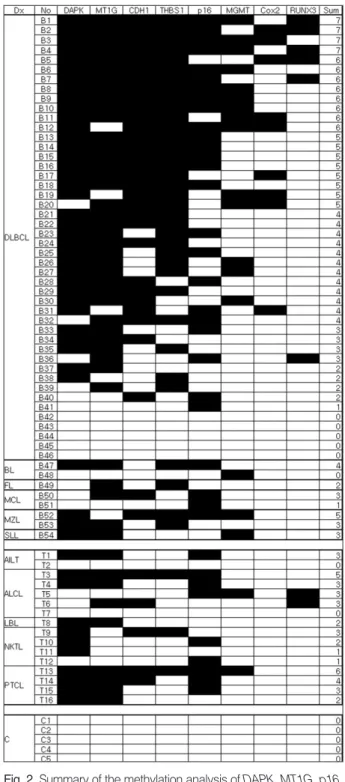
Fig. 2. Summary of the methylation analysis of DAPK, MT1G, p16,
CDH1, THBS1, MGMT, COX2, and RUNX3 in non-Hodgkin’s
lym-phoma samples. The filled boxes indicate the presence of methy-lation and the open boxes indicate the absence of methymethy-lation. Dx, diagnosis; No, case number; Sum, the number of methylated genes; DLBCL, diffuse large B-cell lymphoma; MZL, extranodal marginal zone B-cell lymphoma; BL, Burkitt’s lymphoma; MCL, mantle cell lymphoma; FL, follicular lymphoma; SLL, small lym-phocytic lymphoma; AILT, angioimmunoblastic T-cell lymphoma; ALCL, anaplastic large cell lymphoma; NKTL, Extranodal NK/T-cell lymphoma; PTCL, peripheral T NK/T-cell lymphoma, unspecified; LBL, T-lymphoblastic lymphoma; C, control sample.
COX2 DAPK CDH1 MGMT MT1G P16 RUNX3 THBS1 L U M U M U M U M U M U M U M U M T13 B5 B31 B52 B17 B38 B21 B19
64 cases (91.4%) exhibited aberrant promoter methylation in at least one gene. The detailed results for the methylation of eight genes in each lineage of lymphoma are given in Fig. 2. Each of the eight genes showed methylation of CpG islands in the pro-moter region at frequencies of 11-71%, and the methylation frequencies of each examined gene are summarized in Table 3. In particular, five genes (CDH1, DAPK, MT1G, P16 and THBS1) were frequently methylated (>50%) in all the NHLs.
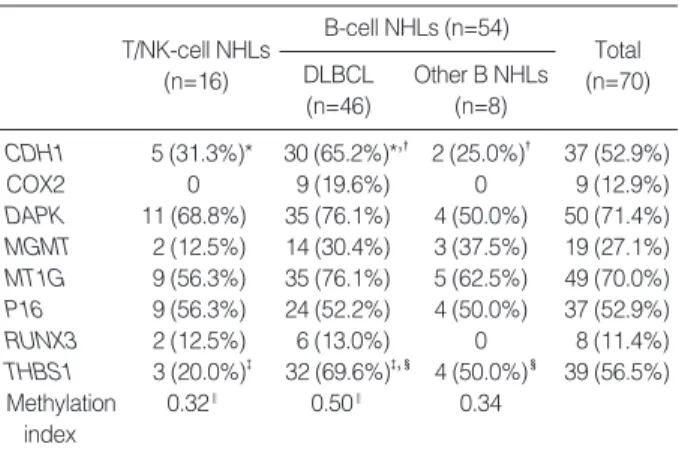
Because most of the B-cell NHLs were diffuse large B cell lymphomas (DLBCL), we analyzed the methylation frequencies of DLBCL compared to those of the T/NK-cell lymphomas or the other B-cell lymphomas. Of the eight genes tested, CDH1 and THBS1 were more methylated in DLBCLs than that in the T/NK-cell lymphomas or the other B-cell lymphomas (Table 3). In addition, the mean MI of the DLBCLs (0.5) was significant-ly higher than that of the T/NK-cell NHLs (0.32; p=0.007). The differences of the mean MI among the B cell lymphomas (0.5 for the DLBCLs and 0.34 for the other B-cell NHLs) were statistically insignificant (p=0.055).
DISCUSSION
In this study, we comprehensively investigated aberrant pro-moter methylation of multiple tumor suppressor genes in NHLs using MSP. Out of these eight genes included in our study, four genes (DAPK, CDH1, p16, and MGMT) have been previously reported to be frequently methylated (>20%) in hematological malignancies,5-9,17and our findings fall within the published
ranges. To the best of our knowledge, the other four genes
(TH-BS1, MT1G, RUNX3, and COX2) we studied have not
previ-ously been investigated in detail in NHLs. THBS1 is a potent inhibitor of angiogenesis and its inactivation by promoter methy-lation was detected in glioblastoma18 as well as many other
tumors.6,12,13MT1G encodes a member of the metallothionein
(MT) heavy metal binding proteins that are active in metal home-ostasis and they have been linked with cell proliferation, and
MT1G gene methylation was observed in esophagus, kidney,
thyroid and prostate carcinomas.14,19-21 RUNX3 is one of the
genes with a RUNT domain, and RUNX3 has been reported to be an important tumor suppressor gene in gastric cancer,22
whereas COX2 is the rate-limiting enzyme for the production of prostanoids (prostaglandins and thromboxanes) from arachi-donic acid, which is known to be causally involved in colorectal carcinogenesis and gastric cancers.10,23Of these four genes, we
discovered that the aberrant methylation of MT1G and THBS1 occurred frequently (70.0% and 56.5%, respectively) in NHLs, which suggested that MT1G and THBS1 may also contribute to the pathogenesis of NHLs. However COX2 and RUNX3 were rarely methylated in NHLs (12.9% and 11.4%, respectively).
Our results showing high methylation frequencies (>50%) in five genes (DAPK, MT1G, THBS1, p16 and CDH1) togeth-er with high MI indicated that the simultaneous inactivation of multiple genes occurred in the lymphoma samples. These find-ings may be of potential significance for creating demethylat-ing therapeutic strategies.24
Most of the B-cell lymphomas in this study were DLBCL, and we found that the mean MI of the DLBCLs was hignificantly higher than that of the T/NK-cell lymphomas, but the differ-ence between the mean MI of the DLBCL and that of the other B-cell lymphomas was not statistically significant. However, the number of cases of other B-cell lymphomas (8 cases) in this study was relatively smaller than that of the other subgroups, so further studies encompassing many other B-cell lymphomas are needed.
Among the eight genes we examined, THBS1 and CDH1 were more frequently methylated in DLBCLs compared to T/NK-cell NHLs or the other B-T/NK-cell NHLs. Methylation of THBS1 in tumor cells will theoretically be expected to produce neovascu-larization or vascular proliferation, but we did not generally observe more prominent vascular components in the DLBCLs than in the other NHLs. There might be some additional path-ways related to angiogenesis rather than THBS1 methylation or there may be other unknown roles for this gene. Further research-es should be done regarding this issue. CDH1 (E-cadherin), as one of the cadherin molecules, may enhance tumor progression and invasion by multiple mechanisms including reduced cell-to-cell T/NK-cell NHLs (n=16) Total (n=70) B-cell NHLs (n=54) DLBCL (n=46) Other B NHLs (n=8) CDH1 5 (31.3%)* 30 (65.2%)*,� 2 (25.0%)� 37 (52.9%) COX2 0 9 (19.6%) 0 9 (12.9%) DAPK 11 (68.8%) 35 (76.1%) 4 (50.0%) 50 (71.4%) MGMT 2 (12.5%) 14 (30.4%) 3 (37.5%) 19 (27.1%) MT1G 9 (56.3%) 35 (76.1%) 5 (62.5%) 49 (70.0%) P16 9 (56.3%) 24 (52.2%) 4 (50.0%) 37 (52.9%) RUNX3 2 (12.5%) 6 (13.0%) 0 8 (11.4%) THBS1 3 (20.0%)� 32 (69.6%)�,� 4 (50.0%)� 39 (56.5%) Methylation 0.32‖ 0.50‖ 0.34 index *p, 0.018; � p, 0.033; � p, 0.001; � p, 0.033; ‖p, 0.007.
Table 3. Methylation frequency of each gene tested in B-cell and T/NK-cell non-Hodgkin’s lymphomas (NHL)
adhesion. The fact that CDH1 was highly methylated in DLB-CLs correlated with the aggressive behavior of DLBDLB-CLs.
In this study, 5 cases of diffuse large B cell lymphoma showed no methylation in any of the eight tested genes. Three of the cases were from lymph nodes, one from brain and the other was from ileum. There were no significant differences between those methylation-nil cases and the other methylated cases regarding the morphology, the immunohistochemical markers (such as CD10, bcl2, bcl6, mum1, etc) and the proliferation indices (data not shown).
Due to the limited number of cases other than the diffuse large B cell lymphomas, we could not perform a subtype spe-cific methylation analyses among the eight genes in this study. However, this study provides insight for understanding the molecular pathogenesis of NHLs. DLBCLs showed a different and distinct methylation pattern compared to T/NK-NHLs or the other B-cell NHLs, which may reflect the different mecha-nisms of lymphomagenesis according to their cellular lineage.
REFERENCES
1. Jones PA, Baylin SB. The fundamental role of epigenetic events in cancer. Nat Rev Genet 2002; 3: 415-28.
2. Costello JF, Fruhwald MC, Smiraglia DJ, et al. Aberrant CpG-island methylation has non-random and tumour-type-specific patterns. Nat Genet 2000; 24: 132-8.
3. Esteller M. CpG island hypermethylation and tumor suppressor genes: a booming present, a brighter future. Oncogene 2002; 21: 5427-40.
4. Esteller M, Corn PG, Baylin SB, Herman JG. A gene hypermethyla-tion profile of human cancer. Cancer Res 2001; 61: 3225-9. 5. Baur AS, Shaw P, Burri N, Delacretaz F, Bosman FT, Chaubert P.
Frequent methylation silencing of p15(INK4b) (MTS2) and p16 (INK4a) (MTS1) in B-cell and T-cell lymphomas. Blood 1999; 94: 1773-81.
6. Chu LC, Eberhart CG, Grossman SA, Herman JG. Epigenetic silenc-ing of multiple genes in primary CNS lymphoma. Int J Cancer 2006; 119: 2487-91.
7. Esteller M, Gaidano G, Goodman SN, et al. Hypermethylation of the DNA repair gene O(6)-methylguanine DNA methyltransferase and survival of patients with diffuse large B-cell lymphoma. J Natl Cancer Inst 2002; 94: 26-32.
8. Katzenellenbogen RA, Baylin SB, Herman JG. Hypermethylation of the DAP-kinase CpG island is a common alteration in B-cell malignancies. Blood 1999; 93: 4347-53.
9. Rossi D, Capello D, Gloghini A, et al. Aberrant promoter methyla-tion of multiple genes throughout the clinico-pathologic spectrum of B-cell neoplasia. Haematologica 2004; 89: 154-64.
10. Akhtar M, Cheng Y, Magno RM, et al. Promoter methylation regu-lates Helicobacter pylori-stimulated cyclooxygenase-2 expression in gastric epithelial cells. Cancer Res 2001; 61: 2399-403.
11. Herman JG, Graff JR, Myohanen S, Nelkin BD, Baylin SB. Methyla-tion-specific PCR: a novel PCR assay for methylation status of CpG islands. Proc Natl Acad Sci USA 1996; 93: 9821-6.
12. Kang GH, Lee S, Lee HJ, Hwang KS. Aberrant CpG island hyper-methylation of multiple genes in prostate cancer and prostatic intraep-ithelial neoplasia. J Pathol 2004; 202: 233-40.
13. Ueki T, Toyota M, Sohn T, et al. Hypermethylation of multiple genes in pancreatic adenocarcinoma. Cancer Res 2000; 60: 1835-9. 14. Morris MR, Hesson LB, Wagner KJ, et al. Multigene methylation
analysis of Wilms’ tumour and adult renal cell carcinoma. Onco-gene 2003; 22: 6794-801.
15. Jaffe ES. Pathology and genetics of tumours of haematopoietic and lymphoid tissues. Lyon: IARC Press; Oxford: Oxford University Press [distributor], 2001; 351 p.
16. Maruyama R, Toyooka S, Toyooka KO, et al. Aberrant promoter methylation profile of bladder cancer and its relationship to clinico-pathological features. Cancer Res 2001; 61: 8659-63.
17. Takahashi T, Shivapurkar N, Reddy J, et al. DNA methylation pro-files of lymphoid and hematopoietic malignancies. Clin Cancer Res 2004; 10: 2928-35.
18. Li Q, Ahuja N, Burger PC, Issa JP. Methylation and silencing of the Thrombospondin-1 promoter in human cancer. Oncogene 1999; 18: 3284-9.
19. Henrique R, Jeronimo C, Hoque MO, et al. MT1G hypermethylation is associated with higher tumor stage in prostate cancer. Cancer Epi-demiol Biomarkers Prev 2005; 14: 1274-8.
20. Huang Y, de la Chapelle A, Pellegata NS. Hypermethylation, but not LOH, is associated with the low expression of MT1G and CRABP1 in papillary thyroid carcinoma. Int J Cancer 2003; 104: 735-44. 21. Roth MJ, Abnet CC, Hu N, et al. p16, MGMT, RARbeta2, CLDN3,
CRBP and MT1G gene methylation in esophageal squamous cell carcinoma and its precursor lesions. Oncol Rep 2006; 15: 1591-7. 22. Li QL, Ito K, Sakakura C, et al. Causal relationship between the loss
of RUNX3 expression and gastric cancer. Cell 2002; 109: 113-24. 23. Toyota M, Shen L, Ohe-Toyota M, Hamilton SR, Sinicrope FA, Issa
JP. Aberrant methylation of the Cyclooxygenase 2 CpG island in colorectal tumors. Cancer Res 2000; 60: 4044-8.
24. Santini V, Kantarjian HM, Issa JP. Changes in DNA methylation in neoplasia: pathophysiology and therapeutic implications. Ann Intern Med 2001; 134: 573-86.