저작자표시-비영리-변경금지 2.0 대한민국 이용자는 아래의 조건을 따르는 경우에 한하여 자유롭게 l 이 저작물을 복제, 배포, 전송, 전시, 공연 및 방송할 수 있습니다. 다음과 같은 조건을 따라야 합니다: l 귀하는, 이 저작물의 재이용이나 배포의 경우, 이 저작물에 적용된 이용허락조건 을 명확하게 나타내어야 합니다. l 저작권자로부터 별도의 허가를 받으면 이러한 조건들은 적용되지 않습니다. 저작권법에 따른 이용자의 권리는 위의 내용에 의하여 영향을 받지 않습니다. 이것은 이용허락규약(Legal Code)을 이해하기 쉽게 요약한 것입니다. Disclaimer 저작자표시. 귀하는 원저작자를 표시하여야 합니다. 비영리. 귀하는 이 저작물을 영리 목적으로 이용할 수 없습니다. 변경금지. 귀하는 이 저작물을 개작, 변형 또는 가공할 수 없습니다.
Effects of irreversible electroporation on
nerves: histologic and functional
evaluation in a rabbit model
Joon Ho Kwon
Department of Medicine
The Graduate School, Yonsei University
[UCI]I804:11046-000000520100
[UCI]I804:11046-000000520100
Effects of irreversible electroporation on
nerves: histologic and functional
evaluation in a rabbit model
Joon Ho Kwon
Department of Medicine
Effects of irreversible electroporation on
nerves: histologic and functional
evaluation in a rabbit model
Directed by Professor Man-Deuk Kim
The Doctoral Dissertation
submitted to the Department of Medicine,
the Graduate School of Yonsei University
in partial fulfillment of the requirements for the degree
of Doctor of Philosophy
Joon Ho Kwon
This certifies that the Doctoral
Dissertation of Joon Ho Kwon is
approved.
---
Thesis Supervisor : Man-Deuk Kim
---
Thesis Committee Member#1 : Se Hoon Kim
---
Thesis Committee Member#2 : Hwan Jun Jae
---
Thesis Committee Member#3: Won Jun Kang
---
Thesis Committee Member#4: Seung Up Kim
The Graduate School
Yonsei University
ACKNOWLEDGEMENTS
I acknowledge my deep gratitude to Professor Man-Deuk Kim,
who is my thesis director, for supporting my efforts with total
commitment and facilitating every step of the process. My
appreciation for his guidance and encouragement is
tremendous. I am also indebted to Professor Se Hoon Kim,
Hwan Jun Jae, Won Jun Kang and Seung Up Kim for their
help for pertinent advice to assure the superior quality of this
paper.
<TABLE OF CONTENTS>
ABSTRACT ··· 1
I. INTRODUCTION ··· 3
II. MATERIALS AND METHODS ··· 4
1. Experimental animals ··· 4
2. IRE procedure ··· 4
3. Funtional assessment ··· 5
4. Histopathologic analysis ··· 5
III. RESULTS ··· 6
1. Functional assessment ··· 6
2. Gross pathologic findings ··· 7
3. Histopathologic findings ··· 7
IV. DISCUSSION ··· 10
V. CONCLUSION ··· 13
REFERENCES ··· 14
LIST OF FIGURES
Figure 1. Functional evaluation of rabbit legs after IRE ··· 7
Figure 2. Effects of IRE on neural and perineural tissues ··· 8
Figure 3. Histopathologic results after IRE ··· 9
1
ABSTRACT
Effects of irreversible electroporation on nerves: functional and
histologic evaluation in a rabbit model
Joon Ho Kwon
Department of Medicine
The Graduate School, Yonsei University
(Directed by Professor Man-Deuk Kim)
Background: Irreversible electroporation (IRE) is a non-thermal ablation technique using pulsatile high-voltage current through electrodes. Recently, IRE is being used to treat various tumors, including pancreas, liver and prostate. The aim of this study was to sequentially evaluate the effects of IRE on nerve during acute to delayed periods in a rabbit model.
Materials and Methods: The study population consisted of 8 male New Zealand White rabbits; 1 rabbit for control subject and 7 for IRE subjects. Ultrasound guided IRE of femoral neurovascular bundles was performed in 7 rabbits. Functional assessment and histopathologic evaluation was performed sequentially at 3 days, 1 week, 2 weeks, 3 weeks, 4 weeks, 6 weeks and 8 weeks after the IRE. Functional assessment was addressed independently immediately after the procedure and then every two days until euthanasia using modified Tarlov scale. All nerves and surrounding tissues were assessed for histopathologic findings consistent with nerve injury and repair, such as axonal swelling, fragmentation, loss, and regeneration, Schwann cell loss and proliferation, ellipsoids, inflammatory cell infiltrates, and fibrosis. The proportion of nerve fiber affected and the area proportion of perineurial inflammation and surrounding tissue injury were recorded.
2
function was damaged before 4 weeks, but then the femoral nerve function recovered gradually to normal. The area of perineural inflammatory infiltration was marked in 3 days after IRE, ranging from 80 to 85% and, was normalized after 8 weeks. Surrounding tissue injury and coagulative necrosis was prominent in 3 days and 1 week after IRE, ranging from 80 to 90%, and recovered after that with fibrous scar. On the other hand, the peripheral nerve fibers were marked destructed in post 1 and 2 weeks with proportion of the affected nerve fiber varied from 80-100%. And then, the nerve fibers became recovered, and normalized after 8 weeks.
Conclusion: The nerve tissues injury with functional impairment can occur after IRE. However, endoneurium and epineurial extracellular matrix are preserved with Schwann cell regeneration, which can lead to regeneration of nerve tissues within 8 weeks. Therefore, IRE may be a potential treatment modality for the tumors that involves a major nerve.
3
Effects of irreversible electroporation on nerves: functional and
histologic evaluation in a rabbit model
Joon Ho Kwon
Department of Medicine
The Graduate School, Yonsei University
(Directed by Professor Man-Deuk Kim)
I. INTRODUCTION
Irreversible electroporation (IRE) is a novel non-thermal ablation technique that uses pulsatile high-voltage current through electrodes placed into
or around the tumor 1. The current creates nanoscale pores in the lipid bilayer of
the cell membrane, disrupting cellular homeostasis and leading to apoptosis 2,3.
As opposed to thermal ablation technique, IRE does not result in inadvertent thermal injury to adjacent major structures, including vessels, bile ducts, urethral or renal collecting systems after IRE for the tumors of pancreas, liver, prostate or kidney 4-6.
In cases of a tumor involves a major nerve, radical resection intended
to complete tumor removal must sacrifice the nerves 7. Although the impressive
development of nerve reconstruction technique, complete recovery and
normalization of nerve function is difficult to achieve 8. The unique
characteristic of IRE, selective cell destruction, overcomes the nerve damage seen with thermal ablation technique or radical resection and allows the preservation of neural function in treating tumors adjacent to the nerves 9.
Few experimental studies have investigated the effects of IRE on nerve
7,9-12
. Schoellnast et al reported that IRE has the potential to damage nerves in acute periods, including axonal swelling, fragmentation, and distal Wallerian
4
degeneration in porcine model 9. However, preservation of endoneurium
architecture and proliferation of Schwann cells enable axonal regeneration as demonstrated in delayed period, after 2months, and Li et al identified similar findings in a rat model 7,9,10.
However, there has been no study of the sequential changes of nerve tissue with functional assessment from acute to delayed periods after the IRE. Thus, the aim of this study was to sequentially evaluate the effects of IRE on nerve during acute to delayed periods in a rabbit model. Furthermore, a comparison of histopathological and functional findings is investigated.
II. MATERIALS AND METHODS 1. Experimental animals
The study was approved by the institutional animal care and use committee. The study population consisted of 8 male New Zealand White rabbits (body weight, 3.0-3.5 kg); 1 rabbit for control subject and 7 for IRE subjects. 1 control subject was euthanasia without IRE to demonstrate the normal histopathologic feature of ablation zone. Anesthesia was induced through intravenous injection of 3mg/kg alfaxalone (Alfaxan) and 5mg/kg 2% xylazine (Rompun; Bayer). After intubation with a 3.0mm endotracheal tube, 1.5%-2% isoflurane was used to maintain general anesthesia. Vercuronium (Vecaron, 0.11mg/kg/h) was injected intravenously to block the muscle contractions that may occur during IRE. Post-procedural pain was managed with intramuscular ketorolac (1mg/kg) and Enrofloxacine (Baytril, 5mg/kg), a fluoroquinolone antibiotic, was injected intramuscularly for 3 days after IRE to prevent wound infection. Euthanasia was performed with an intravenous injection of potassium chloride.
2. IRE procedure
5
Bilateral femoral neurovascular bundles were identified to conduct the experiment using ultrasound (US). The skin overlying the entry position of electrodes was shaved and sterilized using alcohol and Betadine. Two monopolar electrodes (NanoKnife, Angiodynamics, Queenbury, NY, USA) were inserted perpendicular to the neurovascular bundle under US guidance. The neurovascular bundle was positioned at the midpoint of the exposed part of the electrodes. IRE parameters were considered that used in previous studies and to simulate the clinical setting 6,7,9,13. Ablation was performed using an active electrode exposure of 1 cm, an electrode spacing of 1 cm, a voltage of 2000 V/cm, and a pulse length of 90 μs. The pulse number was applied 90 times on the right side and 180 times on the left side to investigate the degree of nerve damage according to pulse number.
3. Functional assessment
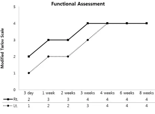
Functional assessment was addressed independently immediately after the procedure and then every two days until euthanasia using modified Tarlov scale. According to modified 5-step Tarlov scale, grade 0 indicates complete paraplegia of hind limb; grade 1, barely detectable movement of hind limbs in response to a hind limb pinch; grade 2, spontaneous movement at all hind limb joints but inability to bear weight of walk; grade 3, able to bear weight and to walk with an abnormal gait on a 1.8-cm wide ledge; grade 4, able to walk with normal gait on a ledge 14.
4. Histopathologic analysis
Pathologic analyses were carried out by a diagnostic pathologist. The pathologic examination was performed sequentially at 3 days, 1 week, 2 weeks, 3 weeks, 4 weeks, 6 weeks and 8 weeks after the IRE to determine the damage and recovery of the nerve. Immediately after euthanasia, a postmortem examination limited to the region of the ablation was performed. The ablation
6
zone including the femoral nerve, vessels, and surrounding muscle and soft tissue was harvested and fixed in 10% neutral buffered formalin. Nerve, vessels, and surrounding tissues were routinely processed, embedded in paraffin, cut into 4-μm-thick sections, and stained with haematoxylin and eosin (H&E). Sections from selected specimens were also stained with Luxol-fast blue for myelin and Bielschowsky silver stain for axon. Immunohistochemial staining (IHC) was performed on selected sections for S100 (for mainly Schwann cell) using rabbit polyclonal antibody S100 protein (dilution 1: 400, NCL-L-S100p, Leica Biosystem, Newcastle, UK), and for neurofilaments (NF, axonal marker) using monoclonal neurofilament protein (dilution 1:400, Clone 2F11, Dako Agilent, CA, USA). IHC was performed according to the manufacturer’s recommended protocol.
All nerves and surrounding tissues were assessed for histopathologic findings consistent with nerve injury and repair, such as axonal swelling, fragmentation, loss, and regeneration, Schwann cell loss and proliferation, ellipsoids, inflammatory cell infiltrates, and fibrosis. The pathologist subjectively measured the area proportion of perineurial inflammation, and surrounding tissue injury and coagulative necrosis. In case of nerve injury, the pathologist counted the damaged nerve fiber proportion. Of the ablation zone, the most severely damaged areas were used as a reference.
III. RESULTS
1. Funtional assessment
Minor bruising had shown at the needle puncture site in all animals, which resolved by days 3 to 5 after IRE. The functional recovery progressed gradually with regard to the animal’s ability to stand without assistance and bear weight on the treated limb. After IRE, the modified Tarlov scale showed that the femoral nerve function was damaged before 4 weeks, but then the femoral nerve function recovered gradually to normal. According to the pulse
7
number, the function of left limb with 180 pulses was decreased short time after the IRE compared to right limb with 90 pulses, but not after 4 weeks (Fig 1).
Figure 1. Functional evaluation of rabbit legs after IRE.
2. Gross pathologic findings
At gross examination, a well-demarcated focal lesion was observed around femoral neurovascular bundle. On 3 days after IRE, the lesions were red and soft, and pale tan and firm after 1 week to 8 weeks. No gross changes were observed in the nerves bundles. Additionally, there was no evidence of thrombosis in the femoral vessels.
3. Histopathologic findings
8
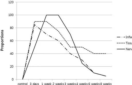
area of perineural inflammatory infiltration was marked in 3 days after IRE, ranging from 80 to 85%. Surrounding tissue injury and coagulative necrosis was prominent in 3 days and 1 week after IRE, ranging from 80 to 90%. Both were decreased and recovered after that, and perineurial inflammation was normalized after 8 weeks after IRE. Some areas of the surrounding tissue were replaced by fibrous scar, approximately 40%.
On the other hand, the peripheral nerve fibers were marked destructed in post 1 and 2 weeks with proportion of the affected nerve fiber varied from 80-100%. And then, the nerve fibers became recovered, and normalized after 8 weeks. There was no difference in pathologic changes between both limbs.
Figure 2. Effects of IRE on neural and perineural tissues. Perineural inflammatory infiltration and surrounding tissue injury was prominent in 3 days and 1 week after IRE and was gradually recovered. The nerve fibers were marked destructed in 1-2 weeks after IRE and normalized after 8 weeks.
9
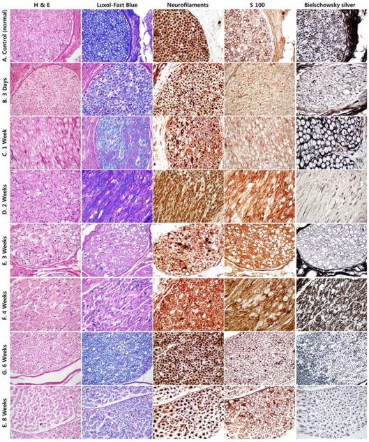
Figure 3. Histopathologic results after IRE. H&E = hematoxylin-eosin stain, S100 = S100 immunohistochemical stain. Original magnification, x400. A. Slides of normal nerve fibers. B, C. Axonal swelling and fragmentation were shown with perineural inflammation and surrounding tissue injury. Endoneurium and Epineurium remained intact and Schwann cell morphology and number are normal. D, E. Significant axonal loss with
10
ellipsoid was seen. F, G. Ellipsoids were gradually resolved and small caliber axons were shown. E. The nerve tissues became normalized and perineural inflammatory infiltrations have resolved.
IV. DISCUSSION
Thermal ablation techniques, including radiofrequency ablation (RFA), microwave ablation (MWA) and cryoablation, have been widely used for the
treatment of various malignancies with promising results 15-18. Although
effective, these techniques can cause inadvertent thermal injury to adjacent hallow viscus, major vessels and endocrine ducts, resulting in high mortality
and mortality 19,20. Nerve also has been experimentally confirmed to have
permanent injuries after thermal ablation, and several clinical reports have been reported 9,21.
IRE is based on a non-thermal mechanism that applies pulsatile high-voltage current through electrodes placed into or around the tumor and the current creates nanoscale pores in the lipid bilayer of the cell membrane, disrupting cellular homeostasis and leading to apoptosis 2,3. So in theory, the supporting extracellular matrix structures with high content of collagenous and elastic fibers are preserved 22. Therefore, adjacent vulnerable tissues, such as vessels, endocrine ducts, urethra and renal collecting system, should remain intact 4. Based on this mechanism of action, IRE is being to treat tumors, including pancreas, liver, kidney and prostate 4-6.
Soft tissue tumors that involves a major nerve, radical resection
intended to complete tumor removal must sacrifice the nerves 7. Unfortunately,
although the impressive development of nerve reconstruction technique, complete function recovery after surgical repair of injured nerves is suboptimal
8
. There were several studies that have investigated the effects of IRE on nerve using animal models. Onik et al demonstrated that the nerves were apparently not affected to IRE 23. On the other hand, Schoellnast et al reported that IRE
11
lead to acute nerve damage including axonal swelling and fragmentation within 14 days in pig models. However, axonal regeneration was demonstrated after 2 months in their study, which was considered to be due to the preservation of external architectures of nerve and proliferation of Schwann cells 9,10. Li et al reported that nerves treated with IRE can attain full recovery after 7 weeks in rat model 7.
In the present study, the histopathologic results were similar to findings of Schoellnast et al and Li et al. The axonal swelling and fragmentation were observed combined with perineural inflammatory infiltration and surrounding tissue injury 3 days and 1 week after IRE. However, the external architectures including endoneurial and epineurial extracellular matrix remained intact and Schwann cell morphology and number are normal. After 2 weeks, a significant axonal loss was observed combined with ellipsoids. The Schwann cell loss was also seen in affected area. However, the regeneration of the Schwann cells was demonstrated at 4 weeks and 6 weeks after IRE that there is potential for axonal regeneration because Schwann cells play an important role in nerve regeneration at the site of injury 24. In addition, most of ellipsoids have resolved and small caliber axons are presented. At 8 weeks after IRE, the nerve tissues became normalized and perineural inflammatory infiltrations have resolved.
To evaluate the function of the femoral nerve after IRE, modified
Tarlov scale was used 25. Both limbs had functional damage immediate after
IRE but gradually recovered. According to pulse numbers, there was a difference in scale until 3 weeks, but both were completely recovered after 4 weeks. In association with histopathologic findings, the period of functional damage and axonal loss was correlated (during 3 weeks), and the function of femoral nerve normalized at 4 weeks after IRE, consistent with the regeneration of the axon. These findings suggest that the functional recovery can be used for predicting regeneration of the damaged nerve.
12
The main parameters affecting the results of IRE ablation are electrical
filed strength, pulse length, interval between pulses, and number of pulses 26. Of
those, pulse number plays an important role in the antitumor effect, and trains of
a large number of short pulses results in the best antitumor effect 27,28. Luo et al
reported that the size of the tumor is more decreased when the number of pulse
was increased without difference in nerve damage 11. However, there was no
difference in nerve damage according to number of pulse in their study 11. Similarly, the present study showed that the degree of nerve damage was not different between 90 times of pulse and 180 times. As mentioned above, IRE is effective in tissues with high density structures, such as cancer, and less effective with collagenous and elastic fibers, potentially avoiding nerve, connective tissue or vessel destruction 7,9. Therefore, the pulse number can be increased sufficiently to increase the antitumor effect until the thermal effect occurs. In most clinical studies, a total 90-180 pulse was applied and 270 pulses were applied in a rare instance 13,29-31.
During the IRE, actual electric field is inhomogeneous and the regions with the largest electric fields occur immediately adjacent to the electrodes 22,32. Therefore, if the histopathologic examination is performed to the regions with small electric field, the degree with nerve damage may be underestimated. There were several previous studies of the effects of IRE on nerves 7,9-11. However, previous studies did not consider the electric field strength when assessing nerve damage. In the present study, the most severely damaged area on the pathological examination of the ablation zone was included in the result. Additionally, most affected area was consistent with the area closest to the electrodes.
This study had several limitations. First, the sample size was small and the results from animal models may not be completely applicable in human. Future studies on human subjects are warranted. Second, there was no control group, such as subjected treated by thermal ablation including radiofrequency
13
ablation and cryoablation. However, nerve injury after thermal ablation has been described on previous studies 33,34. Third, lack of nerve conduction study or electromyography to the clinical assessment of nerve function was other limitation. Nerve conduction study and electromyography can provide objective information of nerve injury and further studies are needed.
V. CONCLUSION
In conclusion, the nerve tissues injury with functional impairment can occur after IRE. However, endoneurium and epineurial extracellular matrix are preserved with Schwann cell regeneration, which can lead to regeneration of nerve tissues within 8 weeks. Therefore, IRE may be a potential treatment modality for the tumors that involves a major nerve. Additionally, future studies of human subject should be preceded.
14
REFERENCES
1. Maor E, Ivorra A, Mitchell JJ, Rubinsky B. Vascular smooth muscle
cells ablation with endovascular nonthermal irreversible electroporation. J Vasc Interv Radiol 2010;21:1708-15.
2. Lee EW, Thai S, Kee ST. Irreversible electroporation: a novel
image-guided cancer therapy. Gut Liver 2010;4 Suppl 1:S99-s104.
3. Lee EW, Wong D, Prikhodko SV, Perez A, Tran C, Loh CT, et al.
Electron microscopic demonstration and evaluation of irreversible
electroporation-induced nanopores on hepatocyte membranes. J Vasc Interv Radiol 2012;23:107-13.
4. Rubinsky B, Onik G, Mikus P. Irreversible electroporation: a new
ablation modality--clinical implications. Technol Cancer Res Treat
2007;6:37-48.
5. Deodhar A, Monette S, Single GW, Jr., Hamilton WC, Jr., Thornton R,
Maybody M, et al. Renal tissue ablation with irreversible electroporation: preliminary results in a porcine model. Urology 2011;77:754-60.
6. Lee EW, Chen C, Prieto VE, Dry SM, Loh CT, Kee ST. Advanced
hepatic ablation technique for creating complete cell death: irreversible electroporation. Radiology 2010;255:426-33.
7. Li W, Fan Q, Ji Z, Qiu X, Li Z. The effects of irreversible
electroporation (IRE) on nerves. PLoS One 2011;6:e18831.
8. Gordon T, Sulaiman OA, Ladak A. Chapter 24: Electrical stimulation
for improving nerve regeneration: where do we stand? Int Rev Neurobiol 2009;87:433-44.
9. Schoellnast H, Monette S, Ezell PC, Deodhar A, Maybody M, Erinjeri
JP, et al. Acute and subacute effects of irreversible electroporation on nerves: experimental study in a pig model. Radiology 2011;260:421-7.
10. Schoellnast H, Monette S, Ezell PC, Maybody M, Erinjeri JP,
15
ablation on nerves. Eur Radiol 2013;23:375-80.
11. Luo X, Qin Z, Tao H, Shi J, Fang G, Li Z, et al. The Safety of
Irreversible Electroporation on Nerves Adjacent to Treated Tumors. World Neurosurg 2017;108:642-9.
12. Wong SS, Hui JW, Chan AW, Chu CM, Rowlands DK, Yu SC.
Irreversible Electroporation of the Femoral Neurovascular Bundle: Imaging and Histologic Evaluation in a Swine Model. J Vasc Interv Radiol 2015;26:1212-20.e1.
13. Martin RC. Irreversible electroporation of locally advanced pancreatic
head adenocarcinoma. J Gastrointest Surg 2013;17:1850-6.
14. Eidelberg E. Letter: Grading animals with spinal cord injury. J
Neurosurg 1975;43:646-7.
15. Aoun HD, Littrup PJ, Jaber M, Memon F, Adam B, Krycia M, et al.
Percutaneous Cryoablation of Renal Tumors: Is It Time for a New Paradigm Shift? J Vasc Interv Radiol 2017;28:1363-70.
16. Song KD. Percutaneous cryoablation for hepatocellular carcinoma.
Clin Mol Hepatol 2016;22:509-15.
17. Cazzato RL, Garnon J, Ramamurthy N, Koch G, Tsoumakidou G,
Caudrelier J, et al. Percutaneous image-guided cryoablation: current applications and results in the oncologic field. Med Oncol 2016;33:140.
18. Facciorusso A, Di Maso M, Muscatiello N. Microwave ablation versus
radiofrequency ablation for the treatment of hepatocellular carcinoma: A systematic review and meta-analysis. Int J Hyperthermia 2016;32:339-44.
19. Casadei R, Ricci C, Pezzilli R, Serra C, Calculli L, Morselli-Labate
AM, et al. A prospective study on radiofrequency ablation locally advanced pancreatic cancer. Hepatobiliary Pancreat Dis Int 2010;9:306-11.
20. Girelli R, Frigerio I, Salvia R, Barbi E, Tinazzi Martini P, Bassi C. Feasibility and safety of radiofrequency ablation for locally advanced pancreatic cancer. Br J Surg 2010;97:220-5.
16
21. Philip A, Gupta S, Ahrar K, Tam AL. A spectrum of nerve injury after
thermal ablation: a report of four cases and review of the literature. Cardiovasc Intervent Radiol 2013;36:1427-35.
22. Davalos RV, Mir IL, Rubinsky B. Tissue ablation with irreversible
electroporation. Ann Biomed Eng 2005;33:223-31.
23. Onik G, Mikus P, Rubinsky B. Irreversible electroporation:
implications for prostate ablation. Technol Cancer Res Treat 2007;6:295-300.
24. Son YJ, Thompson WJ. Schwann cell processes guide regeneration of
peripheral axons. Neuron 1995;14:125-32.
25. Shen J, Zhou CP, Zhong XM, Guo RM, Griffith JF, Cheng LN, et al.
MR neurography: T1 and T2 measurements in acute peripheral nerve traction injury in rabbits. Radiology 2010;254:729-38.
26. Rubinsky J, Onik G, Mikus P, Rubinsky B. Optimal parameters for the
destruction of prostate cancer using irreversible electroporation. J Urol 2008;180:2668-74.
27. Miller L, Leor J, Rubinsky B. Cancer cells ablation with irreversible
electroporation. Technol Cancer Res Treat 2005;4:699-705.
28. Al-Sakere B, Andre F, Bernat C, Connault E, Opolon P, Davalos RV,
et al. Tumor ablation with irreversible electroporation. PLoS One 2007;2:e1135.
29. Narayanan G, Hosein PJ, Arora G, Barbery KJ, Froud T, Livingstone
AS, et al. Percutaneous irreversible electroporation for downstaging and control of unresectable pancreatic adenocarcinoma. J Vasc Interv Radiol 2012;23:1613-21.
30. Martin RC, 2nd, Kwon D, Chalikonda S, Sellers M, Kotz E, Scoggins
C, et al. Treatment of 200 locally advanced (stage III) pancreatic adenocarcinoma patients with irreversible electroporation: safety and efficacy. Ann Surg 2015;262:486-94; discussion 92-4.
31. Martin RC, 2nd, McFarland K, Ellis S, Velanovich V. Irreversible
17
adenocarcinoma. J Am Coll Surg 2012;215:361-9.
32. Edd JF, Davalos RV. Mathematical modeling of irreversible
electroporation for treatment planning. Technol Cancer Res Treat 2007;6:275-86.
33. Yilmaz S, Ozdogan M, Cevener M, Ozluk A, Kargi A, Kendiroglu F,
et al. Use of cryoablation beyond the prostate. Insights Imaging 2016;7:223-32.
34. Bunch TJ, Bruce GK, Mahapatra S, Johnson SB, Miller DV,
Sarabanda AV, et al. Mechanisms of phrenic nerve injury during radiofrequency ablation at the pulmonary vein orifice. J Cardiovasc Electrophysiol 2005;16:1318-25.
18
ABSTRACT(IN KOREAN)
신경에 대한 비가역적 전기천공술의 효과: 토끼 모델을 이용한
병리학적, 기능적 분석
<지도교수 김 만 득>
연세대학교 대학원 의학과
권 준 호
배경: 비가역적 전기 천공술은 전극을 통해 박동성 고전압 전류를 이용하는 비열 절제 기술이다. 최근 비가역적 전기천공술은 췌장, 간 및 전립선을 비롯한 다양한 종양 치료에 사용되고 있다. 본 연구는 토끼 모델에서 신경에 대한 비가역적 전기천공술의 시기에 따른 효과를 평가하고자 하였다. 방법: 연구 집단은 뉴질랜드 수컷 흰 토끼로 대조군 1마리와 실험군 7마리로 총 8마리로 구성되었다. 7마리의 실험군에 대하여 초음파 유도하에 대퇴 신경 혈관 다발에 대하여 비가역적 전기천공술을 시행하였다. 기능적 평가와 병리학적 평가는 비가역적 전기천공술 후 3일, 1주, 2주, 3주, 4주, 6주 및 8주에 순차적으로 시행하였다. 기능적 분석은 modified Tarlov scale을 이용하였다. 모든 신경 조직 및 주변 조직은 신경 손상과 재생을 나타내는 축삭 부종, 분열, 손실 및 재생, Schwann 세포 손실 및 증식, 타원체 및 염증 세포 침윤과 섬유증에 대한 병리적 소견에 대하여 평가하였다. 신경의 손상 정도는 영향을 받은 신경다발의 비율로 분석하였으며 주변 조직의 손상은 영향을 받은 부분의 면적의 비율로 분석하였다.19 손상이 나타났으나, 점차 정상으로 회복되었다. 병리학적 검사상 신경 주변 조직의 염증세포 침윤은 시술 3일 후 가장 심했으며 80-85%로 측정되었다. 신경 주변 조직의 손상 및 응고 괴사는 시술 3일에서 1주일 후에 80-90%로 가장 심하였으며 점차 회복되었고 일부는 섬유성 흉터로 대치되었다. 한편, 신경 섬유는 시술 1-2주 후에 가장 심하게 파괴되었으며, 영향을 받은 신경 섬유의 비율은 80-100% 였다. 그 후 점차 회복되어 시술 8주 후 정상으로 회복되었다. 결론: 비가역적 전기천공술 후 신경의 손상과 함께 기능적 손상이 발생할 수 있다. 하지만, Schwann 세포의 재생과 더불어 신경 내, 외막의 보존은 8주 이내에 신경 조직의 재생을 유도할 수 있다. 따라서 비가역적 전기천공술은 신경에 인접한 종양의 잠재적 치료 방법이 될 수 있다. 핵심되는 말 : 비가역적 전기천공술, 신경 조직, 재생