Analysis of the Cellular Stress Response in MCF10A Cells Exposed
to Combined Radio Frequency Radiation
Han-Na KIM
1†, Na-Kyung HAN
1,2†, Mi-Na HONG
1, Sung-Gil CHI
2, Yun-Sil LEE
3,
Taehong KIM
4, Jeong-Ki PACK
4, Hyung-Do CHOI
5,
Nam KIM
6and Jae-Seon LEE
1*
RF radiation/HSP27/ERK1/2/MCF10A cells.
Exposure to environmental stressors can be measured by monitoring the cellular stress response in target cells. Here, we used the cellular stress response to investigate whether single or combined radio fre-quency (RF) radiation could induce stress response in human cells. Cellular stress responses in MCF10A human breast epithelial cells were characterized after exposure to 4 h of RF radiation [code division mul-tiple access (CDMA) or CDMA plus wideband CDMA (WCDMA)] or 2 h RF radiation on 3 consecutive days. Specific absorption rate (SAR) was 4.0 W/kg for CDMA signal alone exposure and 2.0 W/kg each, 4.0 W/kg in total for combined CDMA plus WCDMA signals. Expression levels and phosphorylation states of specific heat shock proteins (HSPs) and mitogen-activated protein kinases (MAPKs) were ana-lyzed by Western blot. It was found that HSP27 and ERK1/2 phosphorylations are the most sensitive markers of the stress response in MCF10A cells exposed to heat shock or ionizing radiation. Using these markers, we demonstrated that neither one-time nor repeated single (CDMA alone) or combined (CDMA plus WCDMA) RF radiation exposure significantly altered HSP27 and ERK1/2 phosphorylations in MCF10A cells (p > 0.05). The lack of a statistically significant alteration in HSP27 and ERK1/2 phospho-rylations suggests that single or combined RF radiation exposure did not elicit activation of HSP27 and ERK1/2 in MCF10A cells.
INTRODUCTION
Mobile phone use has increased dramatically over the last two decades and is now one of the main sources of environ-mental radiofrequency (RF) radiation. However, it remains unclear whether RF radiation poses a hazard to human health. Although RF radiation may not have sufficient ener-gy to break the covalent bonds of macromolecules, it might be sufficiently energetic to induce the cellular stress
response.1) The situation is complicated further by recent
development of mobile electronic devices that employ dif-ferent frequencies of RF signals. However, the biological effects of simultaneous exposure to multiple RF radiation are unknown.
Environmental stressors such as heat, ethanol, infection, heavy metals, UV, and γ-radiation are known to induce a cas-cade of cellular stress response in exposed cells, a key com-ponent of which are the heat shock proteins (HSPs).2) HSPs
are abundantly expressed and highly conserved molecular chaperones, with diverse functions from bacteria to mam-mals.3) In mammals, HSPs are classified into several families
according to their molecular sizes: HSP90, HSP70, HSP60, and HSP40, and the small HSPs such as HSP27.4)
Phospho-rylation of HSP27 is a rapid response to environmental stres-sors, but it is also observed in unstressed cells upon stimu-lation by a variety of mitogens, cytokines, and inducers of differentiation.5,6)
The mitogen-activated protein kinases (MAPKs) are well known for their pivotal roles in relaying intra- and extra-cel-lular signals in diverse processes such as cell proliferation, differentiation, and cell death.7) Three major classes of
MAPKs are recognized, including the extracellular
signal-*Corresponding author: Phone: +82-2-970-1388, Fax: +82-2-970-1388, E-mail: jaeslee@kcch.re.kr
1Division of Radiation Cancer Research, Korea Institute of Radiological & Medical Sciences, Seoul 139-706, Korea; 2School of Life Sciences and Biotechnology, Korea University, Seoul 136-701, Korea; 3College of Pharmacy & Division of Life Science and Pharmaceuticals, Ewha Womans University, Seoul 120-750, Korea: 4Electromagnetic Research Center, Chungnam National University, Daejeon 305-764, Korea; 5EM Environment Research Team, Electronics and Telecommunications Research Institute, Daejeon 305-700, Korea; 6School of Electrical and Computer Engineering, Chungbuk National University, Cheongju 361-763, Korea.
†HN Kim and NK Han are equally contributed for this work. doi:10.1269/jrr.11048
regulated kinases (ERKs), c-Jun N-terminal kinases (JNKs), and p38.8) MAPKs play an important role in a cascade of
cellular stress response, and they are also implicated in the induction and activation of specific HSPs.9)
In this study, we first characterized the expression and activation of a panel of MAPKs and HSPs in MCF10A human breast epithelial cells stressed by either heat shock or ionizing radiation (IR) to find the most sensitive biomarkers of cellular stress, which were used to investigate stress response to RF radiation. Cells were exposed for 4 h once or 2 h per day for 3 consecutive days to RF radiation at a specific absorption rate (SAR) value of 4 W/kg. Four exper-imental groups included an incubator control group, a sham-exposed group, a code division multiple access (CDMA) RF radiation (836.5 MHz)-exposed group, and a CDMA plus wideband CDMA (WCDMA) RF radiation (1,950 MHz)-exposed group. Following RF exposure, we analyzed the effects of single or combined RF radiation exposure on cel-lular stress response.
MATERIALS AND METHODS
Cell cultures
MCF10A human breast epithelial cell line was purchased from the American Type Culture Collection (ATCC) (Manassas, USA) and maintained in a 1:1 mix of DMEM
and Ham’s F12 media (Invitrogen, Carlsbad, USA) supple-mented with 5% horse serum (Invitrogen), 100 μg/ml of strep-tomycin and 100 U/ml of penicillin (WelGENE, Daegu, Korea), 11.2 μg/ml insulin (Sigma, St. Louis, USA), 21 ng/ml epidermal growth factor (Peprotech, Rocky Hill, USA), 50 ng/ml cholera toxin (Sigma) and 525 ng/ml Hydrocortizone
(Sigma) in a 5% CO2 incubator at 37°C. MCF7 human
breast carcinoma cell line was obtained from the ATCC and grown in DMEM medium (WelGENE) supplemented with 10% fetal bovine serum (FBS) (WelGENE), 100 μg/ml of streptomycin, and 100 U/ml of penicillin in a 5% CO2
incu-bator at 37°C.
RF radiation exposure system
The Radial Transmission Line (RTL) exposure sys-tem10,11) was used as an in vitro multi-frequency radiation
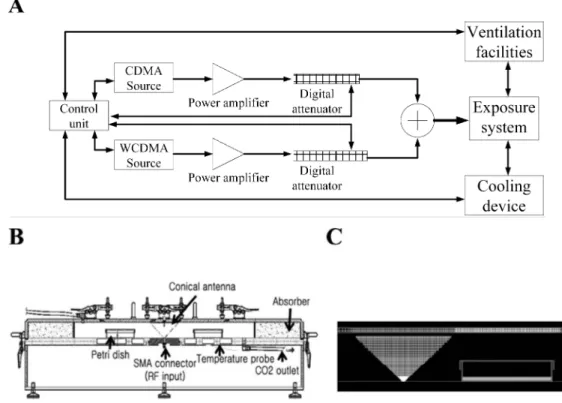
exposure system for this study. The details about exposure system are described previously.12) Block diagram and
cross-sectional view of the entire exposure chamber and of the conical antenna and the culture medium are shown in Fig. 1. CDMA signal at 837 MHz and WCDMA signal at 1,950 MHz were applied to the RTL after amplification.
RF radiation exposure
The exposure system was warmed up for 30 minutes, to equilibrate it prior to RF exposure. The Petri dishes
(diam-Fig. 1. The Radial Transmission Line type RF radiation exposure system for in vitro study. A: Block diagram of the exposure system. B: Cross-sectional view of the entire exposure chamber. C: Cross-sectional view of the conical antenna and the culture medium.
eter 100 mm) were then placed within the exposure cham-ber, at the position shown in Fig. 1B. Cells in petri dishes were exposed either to the CDMA signal alone (4.0 W/kg) or simultaneously to CDMA (2.0 W/kg) and WCDMA (2.0 W/kg) signals for 4 h once or 2 h per day for three consec-utive days. During the exposure period, the temperature in the chamber was maintained within a range of 37 ± 0.3°C, by circulating water within the cavity. The temperature of the culture media was monitored twice per second through-out the exposure period in the sham-exposed and RF radia-tion-exposed groups. Cells were kept for the indicated time for the experiments in a cell culture incubator, immediately after RF radiation exposure. For the sham exposure, the cells were kept in the RF radiation device, but were not exposed to RF radiation.
IR exposure and heat treatment
For irradiation, MCF10A cells exposed to 0.2 Gy of gam-ma radiation from a 137Cs gamma ray source (Atomic Energy
of Canada Ltd., Mississauga, Canada) at a dose rate of 3.81 Gy/min, then recovered at CO2 cell culture incubator at 37°C
for the indicated periods. For heat treatment, MCF10A cells were incubated for 30 min at 39, 41 or 43°C in the temper-ature-controlled water baths, and then recovered at CO2 cell
culture incubator at 37°C for the indicated time periods. MCF7 cells were incubated for 1 h at 43°C in a temperature-controlled water bath, and then immediately harvested to use as a positive control.
Western blotting and image analysis
Cells were washed with phosphate-buffered saline (PBS) (WelGENE), scraped and collected in extraction buffer (25 mM Tris-HCl, pH 7.4, 1 mM EDTA, 1% Triton X-100, 100 mM NaCl, 1 mM phenylmethylsulfonyl fluoride, 10 mM sodium azide, 1 mM orthovanidate, 20 μg/ml aprotinin, and 3 mM dithiothreitol). The collected cells were incubated on ice for 30 min. The lysate was centrifuged and quantitated with a Bradford Assay Reagents (Bio-Rad, Hercules, USA). Equal amounts of proteins (30 μg) were separated on 10% SDS-polyacrylamide gels and then transferred to nitrocellu-lose membranes. The membranes were incubated overnight at 4°C with specific antibodies. Antibodies against HSP90, JNK, ERK1/2, p38, phospho-JNK (Thr183/Tyr185), phos-pho-ERK1/2 (Thr202/Tyr204), and phospho-p38 (Thr180/ Tyr182) were purchased from Cell Signaling (Beverly,
USA). HSP70, HSP27, Phospho-HSP27 (78Ser) (P-HSP27),
α-Tubulin and β-actin were obtained from Santa Cruz Biotech (Santa Cruz, USA). After washing three times in TBST, horseradish peroxidase-conjugated goat anti-mouse, goat anti-rabbit, or donkey anti-goat antibodies from Santa Cruz Biotech were applied. The proteins were visualized using enhanced chemiluminescence (ECL) reagent (Amersham biosciences, Little Chalfont, UK). The signal intensity was quantified with Gel-pro analyzer (http://software.informer.
com/) after normalization with internal control density.
Statistical analysis
All values are expressed as mean ± SEM. The Student’s t test was performed with Origin 6.0 (Origin Lab, Northampton, USA), and statistical significance was set at P values < 0.05.
RESULTS
Phosphorylations of ERK and HSP27 in MCF10A cells
are sensitive to heat shock and IR
We first analyzed the stress response caused by heat shock or IR to identify the most sensitive marker of cellular stress in MCF10A human breast epithelial cells. When we heat shocked MCF10A cells at 43°C, we observed increased phosphorylation of JNK and ERK1/2, but p38 remained unphosphorylated (Fig. 2A, left panel). To determine the sensitivity of this response, we heat shocked MCF10A cells at either 41°C or 39°C (Fig. 2A, right panel). Whereas JNK and ERK1/2 were both phosphorylated after heat shock at 41°C, only ERK1/2 was phosphorylated at 39°C. We next determined the sensitivity of the HSPs to extracellular stim-uli. Three members of the HSP family, HSP27, HSP70, and HSP90, were all increased at the protein level by heat shock at 43°C. In addition, HSP27 became phosphorylated very rapidly after the onset of heat shock. Heat shock at 41°C had less of an effect on the expression levels of HSP90, HSP70 and HSP27, and HSP27 phosphorylation occurred more slowly. In contrast, the protein level of HSPs and HSP27 phosphorylation were not induced by heat shock at 39°C.
Next, we analyzed the cellular response to IR exposure at either 0.2 or 1 Gy. Phosphorylation of both JNK and ERK1/2 occurred in MCF10A cells exposed to 1 Gy of IR (data not shown), but only ERK1/2 phosphorylation was induced by 0.2 Gy of IR (Fig. 2B). p38 phosphorylation was not induced by either exposure. These results indicate that, ERK1/2 phosphorylation is the most sensitive of the MAPKs exam-ined to heat shock or IR. We observed altered HSP27 phos-phorylation, but the level of the HSPs was unchanged at 0.2 Gy of IR. Quantitative analysis of the effects of 0.2 Gy on the HSPs and MAPKs demonstrated that only the changes in phosphorylation of HSP27 and ERK1/2 were statistically significant (p < 0.05; Fig. 2C). This was consistent with the results of heat shock at various temperatures (39, 41, or 43°C). Taken together, these data indicate that changes in HSP27 and ERK1/2 phosphorylation are the most sensitive alterations in response to known environmental stressors in MCF10A cells.
Single or combined RF radiation exposure did not
induce phosphorylation of ERK1/2 or HSP27
We used these selected markers to examine whether RF radiation could also cause a stress response in MCF10A cells. MCF10A cells were exposed to either CDMA RF
radi-ation (836.5 MHz) or CDMA plus WCDMA RF radiradi-ation simultaneously (836.5 MHz + 1,950 MHz). Specific absorp-tion rate (SAR) was either 4 W/kg for CDMA single expo-sure, or 2 W/kg CDMA and 2 W/kg WCDMA for CDMA plus WCDMA combined exposure. Sham-control cells were placed in the exposure apparatus for an equivalent period without RF radiation, and negative (incubator) control cells remained in the culture incubator. To eliminate the potential heating effect of RF exposure, all experiments were conduct-ed under isothermal condition (37 ± 0.3°C). Cells were
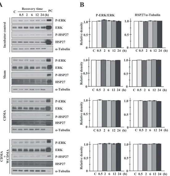
exposed to CDMA or CMDA + WCDMA RF radiation for either a single dose of 4 h (Fig. 3), or for 2 h on 3 consec-utive days (Fig. 4). None of these exposure conditions caused any change in the phosphorylation status of either ERK1/2 or HSP27 (Figs. 3A and 4A). We also observed that ERK1/2 and HSP27 levels remained unaltered. For quanti-tative analysis, the bands in each Western blot were mea-sured by scanning densitometry and normalized relative to internal control proteins (Figs. 3B and 4B). Phosphorylated ERK1/2 was normalized to total ERK1/2, and HSP27 was
Fig. 2. Effects of heat shock and IR exposure on expression of HSPs and phosphorylation of MAPKs in MCF10A cells. A: Western blot analysis of MCF10A cells treated with heat shock for 30 min at 39, 41, or 43°C. C stands for untreated control MCF10A cells and numbers indicate recovery time intervals after each treatment. MCF7 cells were heat shocked at 43°C for 1 h for using a positive control (PC) for Western blot. B: Western blot analysis of MCF10A cells exposed to 0.2 Gy of IR. Data are representative of three independent experiments. C: Statistical analysis of the effect of 0.2 Gy IR on the expression of HSPs and the phosphorylation of HSP27 and MAPKs in MCF10A cells. Bands in each blot were quantified by densitometry. Statistical analysis was performed using the Student’s t-test and is presented as the mean ± standard error of the mean (M ± SEM, n = 3). p < 0.05 (*) was considered to be statistically significant.
normalized to α-tubulin. Phosphorylated HSP27 was too weak to detect reliably by scanning densitometry. No statis-tically significant differences were observed between RF-exposed cells and either sham control or incubator control cells (p > 0.05). These results suggest that exposure to either single or combined RF radiation does not stimulate a stress response in targeted cells under our exposure condition.
DISCUSSION
Recently, as mobile phone users has been increased expo-nentially and mobile electronic devices that employ different frequencies of RF signals were developed, we are exposing to several different RF radiation simultaneously in the living circumstances. However, studies regarding the biological
effects of combined RF radiation exposure have not been reported before we published. To the best of our knowledge, we elucidated firstly no effect of combined RF radiation exposure on the cell cycle and its regulatory protein such as p53, p21, cyclins, and cyclin dependent kinases and on terato-genicity in mice.12,13) Here, we have extended experimental
analysis to assess the effect of simultaneous RF exposure (CDMA plus WCDMA) on changes in HSP expression and MAPK phosphorylation.
In this study, we first examined alterations in HSP27 level and phosphorylation after either CDMA alone or CDMA and WCDMA combined exposure (Figs. 3 and 4). We were unable to detect any statistically significant effect of RF radi-ation exposure on HSP27 level and phosphorylradi-ation under isothermal exposure condition. We have previously
Fig. 3. Effects of 4 h single (CDMA alone) or combined (CDMA + WCDMA) RF radiation exposure on phosphorylation of HSP27 and ERK1/2. A: Western blot analysis of MCF10A cells exposed to a single 4 h dose of RF radiation, either CDMA alone (4.0 W/kg) or CDMA + WCDMA (2.0 W/kg each). Cell lysates were prepared after the indicated recovery periods. Heat-shocked MCF7 cells (43°C, 30 min) without recovery were used as a positive control (PC) for Western blot. B: Quantitative analysis of results in (A). The results shown are the combination of three independent experiments quantified by densitometry. Statistical analysis was performed using the Student’s t-test, and data are presented as M ± SEM (n = 3, p < 0.05 statistically significant).
evidenced no effect of RF radiation on the levels of HSP90, HSP70, and HSP25 in mice, human T-lymphocytes, and rat primary astrocytes.14,15) Other groups also evidenced no
effect of RF radiation on the level of HSPs in a variety of human cell lines, such as neuroblastoma cells, primary monocytes, lymphocytes, glioblastoma, and fibroblasts.16–18)
Furthermore, Dawe et al.19) exposed hsp16-1::lacZ transgenic
C. elegans to 1.8 GHz RF radiation and found no change in reporter gene expression. Cleary et al.20) reported that RF
radiation did not have a detectable effect on the induction of HSPs, and Capri et al.17) found that 1.8 GHz RF did not alter
the level of HSP70. When human peripheral blood was exposed to 0.9 GHz RF, no statistically significant difference in expression of HSP70 and HSP27 was detected.21)
Further-more, exposure of human-derived immune cell lines to 1.9
GHz RF radiation did not induce changes in HSP27 or HSP70 expression,22) and 48 h exposure to GSM-1800 had
no effect on HSP70, HSC70, and HSP27 in either kerati-nocytes or fibroblasts.23) Above-mentioned reports were
con-sistent with our results, demonstrating no effect of RF radiation on HSPs induction.
In contrast, many studies have been reported that RF radi-ation could induce either HSPs expression or HSP27 phosphorylation. Increase of HSP70 protein levels have been reported in in vitro such as human glioma cells,24) human
lens epithelial cells,25) human trophoblast cells,26) in mouse
p53-deficient embryonic stem cells differentiating in vitro,27)
and chick embryos28) due to exposure to RF radiation. In
addition, Weisbrot et al.3) demonstrated that a non-thermal
effect of RF radiation from the GSM mobile phone elevated
Fig. 4. Effects of single or combined RF radiation exposures over time, on phosphorylation of HSP27 and ERK1/2. A: Western blot analysis of MCF10A cells exposed to either CDMA alone (4.0 W/kg) or CDMA + WCDMA (2.0 W/kg each) for 2 h per day for 3 consecutive days. Cell lysates were prepared after the indicated recovery periods. Heat-shocked MCF7 cells (43°C, 30 min) without recovery were used as a positive control (PC) for Western blot. B: Quantitative analysis of results in (A). The results shown are the combination of three independent experiments quantified by densitometry. Statistical analysis was performed using the Student’s t-test and data are presented as M ± SEM (n = 3, p < 0.05 statistically significant).
HSP70 levels in Drosophila melanogaster. Others have dem-onstrated that exposure of MO54 cells to 1.95 GHz RF radi-ation had no effect on cellular proliferradi-ation and expression of HSP70 and HSP27, but may inhibit phosphorylation of HSP27.17) Wang et al.29) reported increased phosphorylation
of HSP27 after 2450 high-frequency electromagnetic fields (HFEMFs) exposure at very high SAR (> 100 W/kg), pos-sibly through a thermal effect. Despite of many studies regarding the effect of RF exposure on HSP induction, positive or negative effect has been obtained and further investigation is still demanding. Such discrepancies could be attributed to thermal effects associated with high levels of SAR and/or exposure condition. To exclude thermal effect in HSP induction, we conducted RF radiation exposure under isothermal condition via circulating cooling water throughout the cavity of exposure chamber for this and previous studies. Experimental and exposure conditions have to be carefully controlled and independent replication studies are necessary for further investigation.
MAPKs are activated by various stressors and can induce intracellular changes to defend against or adapt to extracel-lular stresses.30) It is possible that the non-thermal effects of
RF radiation could affect the levels or activities of key pro-teins that might subsequently alter gene transcription and protein stability. Human epidermoid cancer KB cells exposed to 1.95 GHz RF radiation have increased JNK activ-ity and reduced p38 activactiv-ity, concomitant with increases in HSP70 and HSP27 levels and decreased HSP90 expres-sion.31) Friedman et al.32) suggested a mechanism for the
short-term activation of ERK by RF radiation at frequencies used in mobile phones. They found that RF exposure rapidly generated reactive oxygen species, which in turn caused the release of heparin-binding epidermal growth factor (Hb-EGF) via activation of matrix metalloproteinases. After release, Hb-EGF activated EGF receptors, leading to further activation of ERK but not JNKs or p38. ERK and JNK can be activated by 0.9 GHz RF radiation, which impairs cell cycle progression in human neuroblastoma cells.19) Yu et al.33)
reported that HSP27 and HSP70 expression, as well as ERK and JNK phosphorylation, were observed in human epithelial cells exposed to 1.8 GHz RF. Another report demonstrated that 0.83 GHz RF radiation activated ERK and JNK, but not p38. RF irradiation has also been shown to stimulate neurite out-growth in PC12m3 cells via activation of p38.34) It has been
reported that the class of MAPK that is activated may depend on the SAR of RF exposure.35) Briefly, authors evidenced that
SAR 1.6 W/kg predominantly activated ERK signaling and expression of anti-apoptotic genes and SAR 4.0 W/kg activat-ed JNK signaling and expression of apoptotic genes. Although many studies were evidenced RF radiation could induce MAPKs phosphorylation, we could not detect any statistically significant effect of single and combined RF radiation expo-sure on the phosphorylation of ERK1/2 (Figs. 3 and 4). We also assessed phosphorylation levels of ERK1/2, JNK, and p38
in either mice, human T-lymphocytes and rat primary astrocytes after exposure to RF radiation and failed to find any detectable changes.14,15)
Although a number of studies investigating the effect of RF exposure on HSPs and/or MAPKs have been reported, results are still controversial, partially due to the use of a variety of cell types and RF exposure conditions. Research Agenda for RF Fields (WHO, 2010; www.who.int/peh-emf/ research/agenda/) recommended that further in vitro studies will be performed on a variety of cell types using newer, more sensitive methods. In this study, first of all, we tried to find a sensitive cell line and sensitive markers for the detection of cellular stress response. When we compared stress response between MCF7 and MCF10A cells after heat shock treatment, MCF10A exhibited more sensitive response in inductions of HSP27, HSP70, and HSP90 and more sustainable response in phosphorylations of JNK and ERK1/2 than MCF7 cells did (data not shown). When we assessed the changes in HSP levels and MAPK phosphory-lations in MCF10A after stimuli of heat shock and low dose of IR, we found that phosphorylations of ERK1/2 and HSP27 represented the most sensitive markers for the detection of cellular stress response (Fig. 2). Therefore, we examined phosphorylations of ERK1/2 and HSP27 after RF exposure. Conclusively, we found MCG10A cells which were exposed to combined RF radiation (CDMA plus WCDMA) did not induce levels of expression and phosphorylation of HSP27 and ERK1/2.
Since HSP and MAPK alter evidently their status against thermal change, we suggest that non-thermal effect has to be considered to elucidate effect of RF radiation on cellular stress response. Since RF radiation might not be sufficiently strong enough to cause evident biological alteration, to identify target molecule(s) or mechanism(s) which could be affected by RF radiation exposure has the difficulties. More sensitive and rigorous quantitative methods should be employed and the use of high-throughput techniques would be helpful for further in vitro studies in the research field of RF radiation, which denoting in the research needs of Research Agenda for RF Fields (WHO, 2010).
ACKNOWLEDGEMENT
This work was supported by research fund from the Korea Communications Commission (2010).
REFERENCES
1. McNamee JP and Chauhan V (2009) Radiofrequency radia-tion and gene/protein expression: a review. Radiat Res 172: 265–287.
2. Li Z and Srivastava P (2004) Heat-shock proteins. Curr Protoc Immunol Feb Appendix 1:Appendix 1T pp. 1–6.
3. Weisbrot D, et al (2003) Effects of mobile phone radiation on reproduction and development in Drosophila melanogaster. J
Cell Biochem 89: 48–55.
4. Morimoto RI and Santoro MG (1998) Stress-inducible responses and heat shock proteins: new pharmacologic targets for cytoprotection. Nat Biotechnol 16: 833–838.
5. Lavoie JN, et al (1995) Modulation of cellular thermoresis-tance and actin filament stability accompanies phosphoryla-tion-induced changes in the oligomeric structure of heat shock protein 27. Mol Cell Biol 15: 505–516.
6. Butt E, et al (2001) Heat shock protein 27 is a substrate of cGMP-dependent protein kinase in intact human platelets: phosphorylation-induced actin polymerization caused by HSP27 mutants. J Biol Chem 276: 7108–7113.
7. Minden A and Karin M (1997) Regulation and function of the JNK subgroup of MAP kinases. Biochim Biophys Acta 1333: F85–F104.
8. Weston CR, Lambright DG and Davis RJ (2002) Signal trans-duction. MAP kinase signaling specificity. Science 296: 2345–2347.
9. Rouse J, et al (1994) A novel kinase cascade triggered by stress and heat shock that stimulates MAPKAP kinase-2 and phosphorylation of the small heat shock proteins. Cell 78: 1027–1037.
10. Moros EG, Straube WL and Pickard WF (1999) The radial transmission line as a broad-band shielded exposure system for microwave irradiation of large numbers of culture flasks. Bioelectromagnetics 20: 65–80.
11. Pickard WF, Straube WL and Moros EG (2000) Experimental and numerical determination of SAR distributions within cul-ture flasks in a dielectric loaded radial transmission line. IEEE Trans Biomed Eng 47: 202–208.
12. Lee KY, et al (2011) Effects of combined radiofrequency radiation exposure on the cell cycle and its regulatory pro-teins. Bioelectromagnetics 32: 169–178.
13. Lee HJ, et al (2009) Lack of teratogenicity after combined exposure of pregnant mice to CDMA and WCDMA radiofre-quency electromagnetic fields. Radiat Res 172: 648–652. 14. Lee JS, et al (2006) Radiofrequency radiation does not induce
stress response in human T-lymphocytes and rat primary astrocytes. Bioelectromagnetics 27: 578–588.
15. Lee JS, et al (2005) Subchronic exposure of hsp70.1-deficient mice to radiofrequency radiation. Int J Radiat Biol 81: 781–792. 16. Miyakoshi J, et al (2005) Effects of exposure to a 1950 MHz
radio frequency field on expression of Hsp70 and Hsp27 in human glioma cells. Bioelectromagnetics 26: 251–257. 17. Capri M, et al (2004) 1800 MHz radiofrequency (mobile
phones, different Global System for Mobile communication modulations) does not affect apoptosis and heat shock protein 70 level in peripheral blood mononuclear cells from young and old donors. Int J Radiat Biol 80: 389–397.
18. Buttiglione M, et al (2007) Radiofrequency radiation (900 MHz) induces Egr-1 gene expression and affects cell-cycle control in human neuroblastoma cells. J Cell Physiol 213: 759–767.
19. Dawe AS, et al (2008) Continuous wave and simulated GSM exposure at 1.8 W/kg and 1.8 GHz do not induce hsp16-1 heat-shock gene expression in Caenorhabditis elegans. Bio-electromagnetics 29: 92–99.
20. Cleary SF, et al (1997) Stress proteins are not induced in
mammalian cells exposed to radiofrequency or microwave radiation. Bioelectromagnetics 18: 499–505.
21. Lim HB, et al (2005) Effect of 900 MHz electromagnetic fields on nonthermal induction of heat-shock proteins in human leukocytes. Radiat Res 163: 45–52.
22. Chauhan V, et al (2006) Analysis of proto-oncogene and heat-shock protein gene expression in human derived cell-lines exposed in vitro to an intermittent 1.9 GHz pulse-modulated radiofrequency field. Int J Radiat Biol 82: 347–354.
23. Sanchez S, et al (2007) In vitro study of the stress response of human skin cells to GSM-1800 mobile phone signals compared to UVB radiation and heat shock. Radiat Res 167: 572–580. 24. Tian F, et al (2002) Exposure to 2.45 GHz electromagnetic
fields induces hsp70 at a high SAR of more than 20 W/kg but not at 5 W/kg in human glioma MO54 cells. Int J Radiat Biol
78: 433–440.
25. Lixia S, et al (2006) Effects of 1.8 GHz radiofrequency field on DNA damage and expression of heat shock protein 70 in human lens epithelial cells. Mutat Res 602: 135–142. 26. Valbonesi P, et al (2008) Evaluation of HSP70 expression and
DNA damage in cells of a human trophoblast cell line exposed to 1.8 GHz amplitude-modulated radiofrequency fields. Radiat Res 169: 270–279.
27. Czyz J, et al (2004) High frequency electromagnetic fields (GSM signals) affect gene expression levels in tumor suppres-sor p53-deficient embryonic stem cells. Bioelectromagnetics
25: 296–307.
28. Shallom JM, et al (2002) Microwave exposure induces Hsp70 and confers protection against hypoxia in chick embryos. J Cell Biochem 86: 490–496.
29. Wang J, et al (2006) Effects of a 2450 MHz high-frequency electromagnetic field with a wide range of SARs on the induc-tion of heat-shock proteins in A172 cells. Bioelectromagnetics
27: 479–486.
30. Pearson G, et al (2001) Mitogen-activated protein (MAP) kinase pathways: regulation and physiological functions. Endocr Rev 22: 153–183.
31. Caraglia M, et al (2005) Electromagnetic fields at mobile phone frequency induce apoptosis and inactivation of the multi-chaperone complex in human epidermoid cancer cells. J Cell Physiol 204: 539–548.
32. Friedman J, et al (2007) Mechanism of short-term ERK acti-vation by electromagnetic fields at mobile phone frequencies. Biochem J 405: 559–568.
33. Yu Y, et al (2008) Effects of exposure to 1.8 GHz radiofre-quency field on the expression of Hsps and phosphorylation of MAPKs in human lens epithelial cells. Cell Res 18: 1233–1235. 34. Inoue S, et al (2008) Microwave irradiation induces neurite
outgrowth in PC12m3 cells via the p38 mitogen-activated pro-tein kinase pathway. Neurosci Lett 432: 35–39.
35. Lee KS, et al (2008) Mobile phone electromagnetic radiation activates MAPK signaling and regulates viability in Droso-phila. Bioelectromagnetics 29: 371–379.
Received on March 25, 2011 Revision received on August 19, 2011 Accepted on September 18, 2011