Yonsei Med J http://www.eymj.org Volume 54 Number 4 July 2013
1062
Case Report
http://dx.doi.org/10.3349/ymj.2013.54.4.1062pISSN: 0513-5796, eISSN: 1976-2437 Yonsei Med J 54(4):1062-1065, 2013
Hybrid Method of Transurethral Resection of Ejaculatory
Ducts Using Holmium:Yttriumaluminium Garnet Laser
on Complete Ejaculatory Duct Obstruction
Joo Yong Lee, Richilda Red Diaz, Young Deuk Choi, and Kang Su Cho
Department of Urology, Severance Hospital, Urological Science Institute, Yonsei University College of Medicine, Seoul, Korea. Received: June 19, 2012
Revised: September 10, 2012 Accepted: September 10, 2012 Corresponding author: Dr. Kang Su Cho, Department of Urology, Severance Hospital, Urological Science Institute, Yonsei University College of Medicine, 50 Yonsei-ro,
Seodaemun-gu, Seoul 120-752, Korea. Tel: 82-2-2228-2320, Fax: 82-2-312-2538 E-mail: kscho99@yuhs.ac
∙ The authors have no financial conflicts of interest.
© Copyright:
Yonsei University College of Medicine 2013 This is an Open Access article distributed under the terms of the Creative Commons Attribution Non-Commercial License (http://creativecommons.org/ licenses/by-nc/3.0) which permits unrestricted non-commercial use, distribution, and reproduction in any medium, provided the original work is properly cited.
A 32-year old single man presented with azoospermia and low semen volume which was noted one and half a year ago. Transrectal ultrasonography and seminal vesiculography were performed to evaluate ejaculatory duct obstruction, and trans-urethral resection of the ejaculatory duct was performed using a hybrid technique of holmium:yttriumaluminium garnet laser with monopolar transurethral resection to overcome the narrow prostatic urethra. To our knowledge, this is the first report on the successful outcome of a hybrid technique applied for transurethral resection of the ejaculatory duct.
Key Words: Ejaculatory ducts, semen analysis, infertility, holmium
INTRODUCTION
Infertility, defined as the inability to conceive after 1 year of unprotected intercourse, affects approximately 10% to 15% of reproductive age couples in the United States.1 Ejaculatory duct obstruction (EDO) is a relatively rare cause of infertility as male factor and is surgically correctable following an appropriate treatment. Complete EDO has been reported in 1% of all infertile men. Patients with complete EDO shows absent or low ejaculate, and azoospermia in the presence of a palpable vas deferens.2 EDO may show various symptoms including dysuria, hematospermia, pain during or after ejaculation, and perineal or testicular pain or discomfort.3 EDO may be congenital or acquired. The causes of EDO include congenital atresia, duct cysts, trauma, infection, inflammation and calculus formation.4 The standard method of establishing the diagnosis of EDO is seminal vesiculography. Dilated seminal ves-icles can be fllled with a dye solution under transrectal ultrasonography (TRUS) guidance; if duct obstruction is complete, the efflux of the dye solution cannot be seen during cystoscopy.5 With advances in noninvasive diagnostic methods, such as high-resolution TRUS and magnetic resonance imaging (MRI), the seminal vesicles can be accurately evaluated, thereby facilitating the diagnosis of EDO.
The treatment of choice for distal seminal tract obstruction is transurethral resec-tion of the ejaculatory duct (TUREJD). Approximately half of men undergoing this procedure for EDO show improvement of their semen parameters and half of the
Hybrid Method of TUREJD
Yonsei Med J http://www.eymj.org Volume 54 Number 4 July 2013 1063
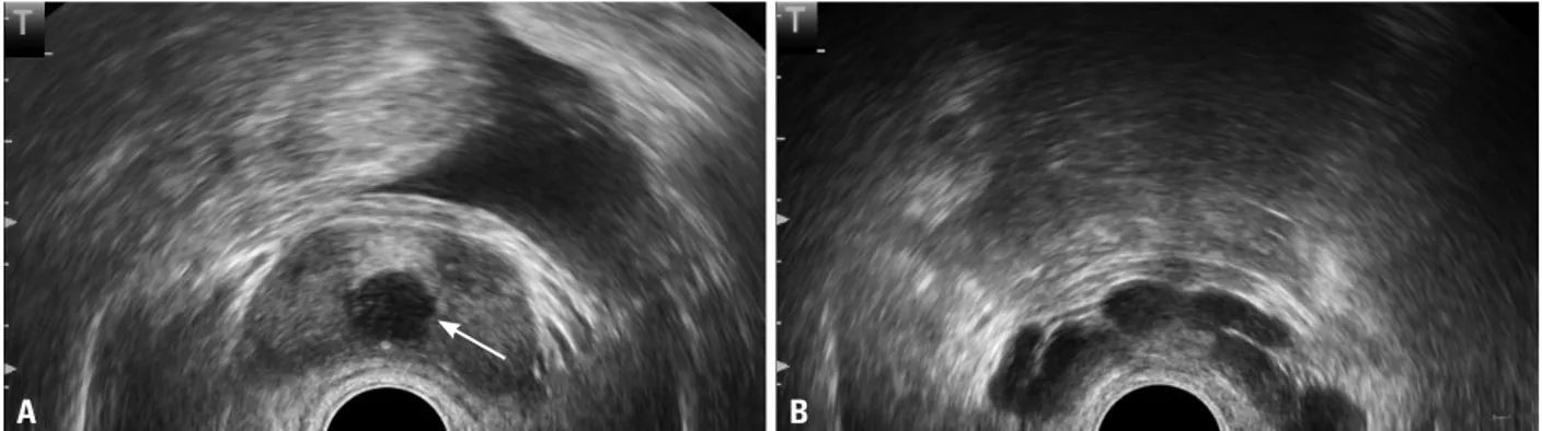
analysis revealed that the total volume was 0.5 mL and had no identifiable spermatozoa. Bilateral vas deferens were palpable on physical examination. TRUS revealed a mid-line prostatic cyst of approximately 1.5 cm in diameter with dilated seminal vesicles (Fig. 1). Pelvic MRI revealed a 2 cm sized prostatic utricular cyst and mild dilatation of the seminal vesicle (Fig. 2A, B, C and D). Seminal vesiculog-raphy was performed to evaluate ejaculatory duct obstruc-tion because of the prostatic ultracular cyst (Fig. 2E). Under seminal vesiculography, the seminal vesicles and midline cyst were found to be filled with contrast media (Fig. 2D). Fluid which was aspirated from the midline cyst contained some spermatozoa on examination under light microscopy. Preoperative cystoscopy revealed a narrowing of the tatic urethra near the verumontanum due to a midline pros-tatic cyst (Fig. 3A). Therefore, a diagnosis of a EDO was men who improve achieve a subsequent pregnancy.6 Here,
we report a single case of EDO with a midline prostatic cyst which resulted in a narrow prostatic urethra. Thus, a hybrid technique using the holmium:yttriumaluminium garnet (Ho:YAG) laser and monopolar transurethral resection (TUR) was performed to overcome the narrow prostatic urethra. To our knowledge, our case is the first report of a hydrid technique used to treat TUREJD.
CASE REPORT
Patient history
A 32-year old, single man presented with azoospermia and low semen volume which was noted one and half a year ago. He had no other known medical problems. Semen
Fig. 1. Transrectal ultrasonography revealed (A) a midline prostatic cyst with an approximate diameter of 1.5 cm (white arrow) with (B) dilated seminal
vesi-cles.
Fig. 2. T2-weighted magnetic resonance image and seminal vesiculography. (A and C) Approximately 2 cm sized cystic lesion (white arrows) was located in
the prostate. (B, C and D) Mild dilatation of the seminal vesicles was observed. (E) The seminal vesicles and midline cyst (white arrow head) were filled with contrast media under seminal vesiculography.
A A C B B D E
Joo Yong Lee, et al.
Yonsei Med J http://www.eymj.org Volume 54 Number 4 July 2013
1064
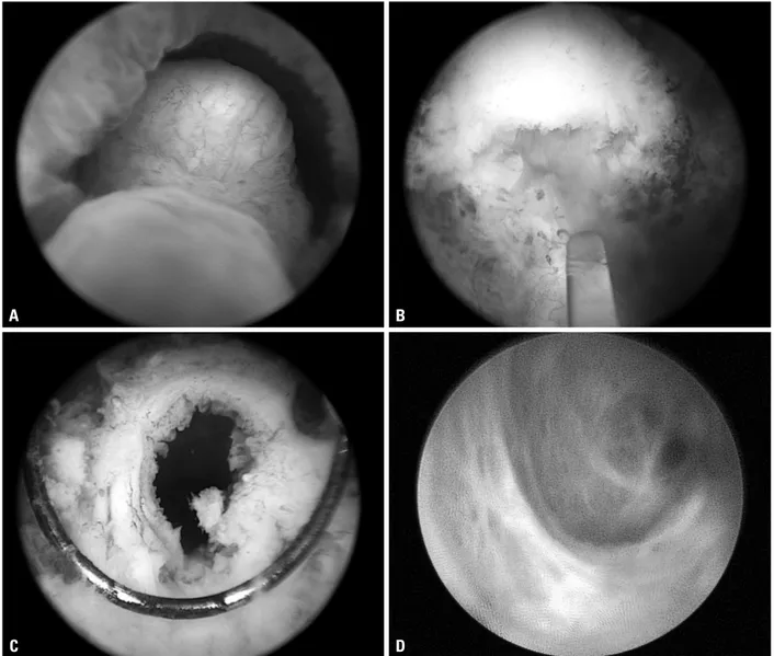
TUR, the cystic wall was completely resected and a guide-wire was inserted through the ejaculatory duct. The ejacula-tory ducts and their wall were visualized using a 6.5 Fr semi-rigid ureteroscope (Richard Wolf GmbH, Knittlingen, Germany) and appeared to be normal (Fig. 3D).
The patient was discharged the following day after the Fol-ey catheter was removed. After a month, the patient did not have any lower urinary tract symptoms. A month later, postop-erative semen analysis showed normal findings with a 3 mL volume. Concentration of spermatozoa was 15.2×10⁶/mL.
DISCUSSION
In patients with suspected EDO, the standard procedure has made and TUREJD was planned.
Surgical procedure
Under spinal anesthesia, the patient was placed in the lithot-omy position. A 10 mL diluted indigo-carmine solution was injected through a midline cyst guided TRUS. Using a 24 Fr resectoscope (Karl Storz, Tuttlingen, Germany) with a laser bridge and a Ho:YAG laser with a 550 µm laser fiber (Lumenis, Yokneam, Israel), unroofing of the midline pros-tate cyst was performed. During the unroofing process, the power was set at 2 J and 10 Hz. After the process, the dilut-ed indigo-carmine solution was extravasatdilut-ed to the sur-rounding area. When sufficient space at the prostatic ure-thra was secured following incision with the Ho:YAG laser, a monopolar TUR was performed (Fig. 3B and C). Using
Fig. 3. Hybrid technique for complete ejaculatory duct obstruction. (A) Small space of the prostatic urethra. (B) Using Ho:YAG laser with a 550 µm laser fiber
(Lumenis, Yokneam, Israel), unroofing of midline prostate cyst was performed. (C) Using TUR, the cystic wall was completely resected and a guidewire was inserted through the ejaculatory duct. (D) The ejaculatory ducts and its wall were visualized using a 6.5 Fr semi-rigid ureteroscope (Richard Wolf GmbH, Knittlingen, Germany). Ho:YAG, holmium:yttriumaluminium garnet; TUR, transurethral resection.
C A
D B
Hybrid Method of TUREJD
Yonsei Med J http://www.eymj.org Volume 54 Number 4 July 2013 1065
using the Ho:YAG laser because of narrow prostatic ure-thra. The advantages of the Ho:YAG laser are as follows: precise unroofing using a small diameter laser fiber and avoidance of unnecessary coagulation, thereby minimizing damage to adjacent structures. In patients with EDO, this hybrid method may be a safe and satisfactory alternative treatment option to conventional TUREJD.
REFERENCES
1. Bang JK, Lim JJ, Choi J, Won HJ, Yoon TK, Hong JY, et al. Re-versible infertility associated with testosterone therapy for symp-tomatic hypogonadism in infertile couple. Yonsei Med J 2013;54: 702-6.
2. Jarow JP. Seminal vesicle aspiration in the management of pa-tients with ejaculatory duct obstruction. J Urol 1994;152:899-901. 3. Ozgök Y, Tan MO, Kilciler M, Tahmaz L, Kibar Y. Diagnosis and
treatment of ejaculatory duct obstruction in male infertility. Eur Urol 2001;39:24-9.
4. Pryor JP, Hendry WF. Ejaculatory duct obstruction in subfertile males: analysis of 87 patients. Fertil Steril 1991;56:725-30. 5. Yurdakul T, Gokce G, Kilic O, Piskin MM. Transurethral
resec-tion of ejaculatory ducts in the treatment of complete ejaculatory duct obstruction. Int Urol Nephrol 2008;40:369-72.
6. Donkol RH. Imaging in male-factor obstructive infertility. World J Radiol 2010;2:172-9.
7. Farley S, Barnes R. Stenosis of ejaculatory ducts treated by endo-scopic resection. J Urol 1973;109:664-6.
8. Park MS, Kim YS, Yoon YR. A case of infertility because of ejac-ulatory duct obstruction. Korean J Urol 1992;33:917-21. 9. Fisch H, Lambert SM, Goluboff ET. Management of ejaculatory
duct obstruction: etiology, diagnosis, and treatment. World J Urol 2006;24:604-10.
10. Schroeder-Printzen I, Ludwig M, Köhn F, Weidner W. Surgical therapy in infertile men with ejaculatory duct obstruction: tech-nique and outcome of a standardized surgical approach. Hum Re-prod 2000;15:1364-8.
11. Johnson CW, Bingham JB, Goluboff ET, Fisch H. Transurethral resection of the ejaculatory ducts for treating ejaculatory symp-toms. BJU Int 2005;95:117-9.
12. Wang H, Ye H, Xu C, Liu Z, Gao X, Hou J, et al. Transurethral seminal vesiculoscopy using a 6F vesiculoscope for ejaculatory duct obstruction: initial experience. J Androl 2012;33:637-43. become TUREJD, which was described by Farley and
Barnes in 1973.7 TUREJD can be performed with the gen-eral equipments for transurethral resection of the prostate.8 After a resectoscope is introduced into the urthera, the proxi-mal verumontanum, which may be enlarged, can be resect-ed by a cutting loop. Classical TUREJD is carriresect-ed out using a pure cutting current without electrocoagulation.9 One to two chips are usually resected, removing the proximal ver-umontanum only. If bleeding is encountered, gentle coagu-lation is recommended to avoid the ejaculatory ducts. A catheter is inserted into the bladder and is left in place for a few hours in the recovery room. Postoperative urinary re-tention can occur after catheter removal, particularly in pa-tients with a prior voiding dysfunction.
Schroeder-Printzen, et al.10 reported their surgical experi-ence in sixteen infertile patients with EDO. They performed a combination of transurethral incision and resection using the Tuner-Warwick hook and TUR roof. With their tech-nique, unroofing of all the central cysts following to posi-tive results of intracystic dye test were carried out. With 15 patients with symptomatic EDO undergoing TUREJD, John-son, et al.11 evaluated the diameter of seminal vesicles and considered dilated when their diameters were 12 mm or longer on TRUS. Each man was treated with TUREJD us-ing a conventional resectus-ing loop in the midline of the proxi-mal verumontanum. They concluded that patients with symptomatic EDO who underwent TUREJD demonstrated improvements in their ejaculation, sensation of orgasm, se-men analysis values and fertility. Recently, Wang, et al.12 re-ported interesting results on transurethral seminal vesicu-loscopy for EDO. Twenty-one patients underwent their procedures, and a stone in the ejaculatory duct was removed in 5 cases, using a Ho:YAG laser. They concluded that their results had fewer complications and more optimal sperm recovery than the conventional method. Previous studies showed relatively good surgical outcomes.