167
Print ISSN 1738-5520 / On-line ISSN 1738-5555 Copyright © 2011 The Korean Society of Cardiology CASE REPORT
DOI 10.4070/kcj.2011.41.3.167
Open Access
Recurrent Infective Endocarditis Associated
With Pyogenic Spondylodiskitis
Jae Hoon Kim, MD, Soon Kil Kim, MD, Dong Chan Kim, MD, Hwan Cheol Park, MD, Sung Il Choi, MD, Jin Ho Shin, MD, Jae Ung Lee, MD, Jeong Hyun Kim, MD, and Heon Kil Lim, MD
Department of Internal Medicine, College of Medicine, Hanyang University, Guri Hospital, Guri, Korea ABSTRACT
Infective endocarditis is a life-threatening condition caused by microbial infection of the heart’s endocardial surface. This condition can also be associated with bacterial infections of other organs. We experienced an unusual case of recurrent infec-tive endocarditis associated with pyogenic spondylodiskitis. A 70-year-old man presented with persistent fever and lower back pain visited our hospital. The patient had a past history of recurrent infective endocarditis. He was diagnosed with infective endocarditis again based on clinical symptoms and echocardiographic findings. Magnetic resonance imaging was used to eval-uate lower back pain, which showed acute spondylodiskitis on L3 and L4 vertebrae. The patient completely recovered follow-ing four weeks of antibiotic therapy. (Korean Circ J 2011;41:167-170)
KEY WORDS: Infective endocarditis; Spondylodiskitis.
Received: June 28, 2010 Revision Received: August 9, 2010 Accepted: August 25, 2010
Correspondence: Soon Kil Kim, MD, Department of Internal Medicine, College of Medicine, Hanyang University, Guri Hospital, 249-1 Gyomun-dong, Guri 471-701, Korea
Tel: 82-31-560-2233, Fax: 82-31-553-7369 E-mail: [email protected]
• The authors have no financial conflicts of interest.
cc This is an Open Access article distributed under the terms of the
Cre-ative Commons Attribution Non-Commercial License (http://creCre-ativecom- (http://creativecom-mons.org/licenses/by-nc/3.0) which permits unrestricted non-commer-cial use, distribution, and reproduction in any medium, provided the origi-nal work is properly cited.
Introduction
Infective endocarditis is a life-threatening condition caus-ed by microbial infection of the heart’s endocardial surface. It most commonly involves the heart valves, but other sites in-cluding septal defects, chordae tendinae, and mural endocar-dium can be involved.
Common symptoms of infective endocarditis include fever, anorexia, weight loss, malaise, night sweats, skin lesions (sp-linter hemorrhage, Osler’s nodes, Janeway’s lesions), conjunc-tival petechiae, and splenomegaly.
Occasionally, serious complications such as septic aneury-sms and septic emboli can occur in major organs (kidney, br-ain, liver, spleen) which may result in death. Septic emboli may
occur in the spine, but the incidence is extremely low. We herein present our experience of a patient with infective endocarditis associated with pyogenic spondylodiskitis who was successfully treated with antimicrobial treatment.
Case
A 70-year-old man presented to our hospital with a two-week history of fever associated with lower back pain. He had a past history of recurrent infective endocarditis, for which he was admitted to our hospital in 2005 and in 2009.
Strep-tococcus bovis was isolated during the first visit in 2005 as
the causative organism of infective endocarditis. In addition, vegetation was found on the posterior leaflet of the mitral val-ve. During the second admission, blood culture showed the growth of a Streptococcus species. However, specific culture results were not reported. Similar to the first admission, veget-ation growth was again found on the posterior leaflet of the mitral valve. On both admissions, the patient successfully re-covered following the appropriate antibiotic treatment with reduction of vegetation.
On admission, the patient’s core body temperature was 39.0°C, blood pressure 130/80 mmHg, and pulse rate was 88 beats/minute. Auscultation revealed a regular heart beat with an early systolic murmur was found at the left lower sternal bor-der (grade III/VI). Neurological examination was
unremark-168 Infective Endocarditis and Pyogenic Spondylodiskitis
able. No peripheral stigmata of infective endocarditis were noted.
Laboratory tests showed absence of leukocytosis (white blood cell 7,400/mm3), but mild anemia (hemoglobin 8.5 mg/
dL), and thrombocytopenia (platelets 120,000/mm3) was
noted. Except for a C-reactive protein (CRP) value of 4.17 mg/ dL (0.1-0.8 mg/dL), no other laboratory tests showed signifi-cant abnormalities (blood urea nitrogen 15 mg/dL, creatinine 0.8 mg/dL, total protein 6.9 g/dL, albumin 3.4 g/dL, aspartate aminotransferase 27 IU/L, alanine aminotransferase 14 IU/L).
Chest radiography showed no pulmonary edema or active lesions in the lungs. In transthoracic echocardiography, a 2.7×
1.4 cm sized vegetation on the posterior leaflet of the mitral valve (Fig. 1) was noted. Furthermore, moderately severe mi-tral regurgitation, trivial aortic regurgitation, and moderate tricuspid regurgitation was also noted. Other findings includ-ed left ventricular (LV) hypertrophy (LV mass 270.5 gm), LV enlargement (LV end diastolic dimension 61 mm, LV end sys-tolic dimension 41 mm), left atrial (LA) enlargement (LA di-mension size 40.1 mm), and mild pulmonary hypertension (pul-monary arterial pressure 37 mmHg).
After diagnosis of infective endocarditis was confirmed, treatment was immediately started with intravenous ceftriax-one and gentamycin, on suspicion of recurrent infective en-docarditis.
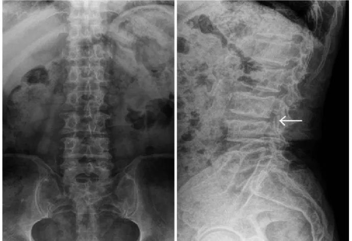
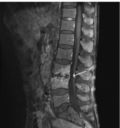
On the second day of antibiotic therapy, fever and other signs of infection resolved. We continued antibiotic treatment for four weeks. However, blood culture demonstrated no gr-owth of bacteria and fungi. Although his clinical signs and symptoms related to infective endocarditis improved, and CRP level decreased, his back pain did not completely resolve. Con-sequently, we decided to order a plain lumbar spine radio-graph to evaluate the lower back pain, which showed disc sp-ace narrowing and endplate erosion of the L3 and L4 vertebral bodies (Fig. 2). Magnetic resonance imaging of the lumbar spine was performed to further evaluate the erosive lesions, which confirmed acute spondylodiskitis of L3, L4 vertebrae and L3-4 disc space (Fig. 3). Pyogenic spondylodiskitis was highly suspicious to be associated with infective endocarditis according to the clinical course and patient’s history. The
pa-Fig. 1. A 2.7×1.4 cm sized vegetation on the posterior leaflet of
mitral valve.
Fig. 2. Plain radiograph shows narrowing of the disc space at L3-L4 level. Destruction of the superior endplate of the L4 vertebra, and the
Jae Hoon Kim, et al. 169
tient was discharged after four weeks of intravenous antibi-otic treatment without complication. We decided to conduct follow-up evaluation of the patient’s spinal lesions at the or-thopedic outpatient clinic.
During follow-up at the outpatient clinic, the size of mitral valve vegetation was shown to be reduced on echocardiogra-phy (1.35×0.67 cm). There were no symptoms of infective en-docarditis or pyogenic spondylodiskitis.
Discussion
Infectious diskitis (spondylodiskitis, spondylodiscitis, infec-tious spondylitis) is an inflammatory process that involves one or more extradural components of the spine.1) Although it
affects a small proportion (2-7%) of all patients with osteo-myelitis, it has clinical importance due to potential morbidi-ty and mortalimorbidi-ty. Spondylodiskitis commonly involves the lum-bar (45%), thoracic (35%), and cervical (10-20%) spines.2)
Se-condary epidural abscess formation occurs most frequently in the cervical spine, followed in frequency by the thoracic and lumbar spine.2)
Although the clinical presentation of patients with spondy-lodiskitis varies, it generally commences with insidious de-velopment of localized back pain combined with non-specif-ic symptoms, such as malaise, fever, and weight loss. Our pa-tient also experienced these symptoms. As fever and leuko-cytosis are often absent, symptoms of spondylodiskitis may be present for months before the diagnosis is confirmed. This may result in progression of the local disease process. Pa-tients may present with hip contracture or paralysis
second-ary to abscess formation in the paraspinal or epidural spaces. Pyogenic spinal infections are most commonly caused by
Staphylococcus aureus (in 60% of all patients) and Enterobac-ter species (in 30% of all patients).1)3)4) Pseudomonas
aerugi-nosa, Serratia species, and Candida species most often affect
patients with a history of intravenous drug abuse.
Mycobac-terium tuberculosis causes most non-pyogenic spinal
infec-tions. However, fungi (e.g., Cryptococcus species, Aspergillus species, coccidioidomycosis) also may cause infections.5-8) In
spondylodiskitis, the three main routes of infection are hema-togenous spread, direct inoculation, and contiguous spread. In adults, most cases result from direct inoculation after spi-nal instrumentation procedures, including surgery, discogra-phy, and epidural injections. Spontaneous infections result from a hematogenous source (e.g., bacteremia, intravenous drug ab-use) usually beginning at a lumbar or thoracic vertebral body subjacent to the vertebral endplate. Loss of disk height may oc-cur as pyogenic organisms release enzymes that dissolve the nu-cleus pulposus. Non-pyogenic organisms, such as tuberculosis do not produce proteolytic enzymes. Therefore, they tend to spare the disk from destruction. Spread from an adjacent so-urce, such as a psoas abscess, is an uncommon mechanism. CSF and lymphatic spread are also uncommon routes of infection.9)
In our patient, pyogenic spondylodiskitis was associated with recurrent infective endocarditis. Several cases of infec-tive endocarditis associated with spondylodiskitis have been reported, however to the best of our knowledge, there was no reported case in Korea.10)11) Pigrau et al.12) investigated the
incidence and risk factors of infective endocarditis in patients with pyogenic vertebral osteomyelitis. A retrospective record review was conducted on all cases of vertebral osteomyelitis from January 1986 to June 2002 that occurred at a tertiary re-ferral hospital. Among 606 patients with infective endocar-ditis, 28 cases (4.6%) had pyogenic vertebral osteomyelitis. Among 91 cases of pyogenic vertebral osteomyelitis, 28 cases (30.8%) were associated with infective endocarditis. In 6 pa-tients, there were no clinical signs of infective endocarditis, and the diagnosis was established only by routine echocardiog-raphy. When specifically investigated, the incidence of infec-tive endocarditis is high in patients with pyogenic vertebral osteomyelitis. Therefore, in pyogenic vertebral osteomyelitis patients, routine echocardiography is valuable for early detec-tion of infective endocarditis.
REFERENCES
1) Hopkinson N, Stevenson J, Benjamin S. A case ascertainment study
of septic discitis: clinical, microbiological and radiological features. QJM 2001;94:465-70.
2) Cottle L, Riordan T. Infectious spondylodiscitis. J Infect 2008;56:
401-12.
3) Ponte CD, McDonald M. Septic discitis resulting from Escherichia coli
urosepsis. J Fam Pract 1992;34:767-71.
4) Park CB, Kim JJ, Song JK, et al. Right-Sided Infective Endocarditis in
Fig. 3. Spine MRI shows abnormal high signal density in the disc
within the adjacent vertebral bodies at L3-L4 level (arrow). Bony destruction of vertebral bodies is seen in L3 and L4.
170 Infective Endocarditis and Pyogenic Spondylodiskitis
Korea. Korean Circ J 2005;35:633-8.
5) Moreillon P, Que YA. Infective endocarditis. Lancet 2004;363:139-49. 6) Duval X, Leport C. Prophylaxis of infective endocarditis: current
te-ndencies, continuing controversies. Lancet Infect Dis 2008;8:225-32.
7) Kim MK, Song JK, Kang DH, et al. Recent trends and clinical outcomes
of infective endocarditis. Korean J Med 2000;58:28-38.
8) Wilson W, Taubert KA, Gewitz M, et al. Prevention of infective
endo-carditis: guidelines from the American Heart Association: a guideline from the American Heart Association Rheumatic Fever, Endocarditis, and Kawasaki Disease Committee, Council on Cardiovascular Dis-ease in the Young, and the Council on Clinical Cardiology, Council on Cardiovascular Surgery and Anesthesia, and the Quality of Care and Outcomes Research Interdisciplinary Working Group. Circulation
2007;116:1736-54.
9) Yi MZ, Lee SH, Park CB, et al. Clinical characteristics of
nosocomi-al infective endocarditis in a tertiary referrnosocomi-al hospitnosocomi-al. Korean Circ J 2006;36:236-41.
10) Talsania N, Ogundipe O. Infective discitis mimicking infective
endo-carditis and osteoarthritic back pain. Internet J Rheumatol 2005;2:3.
11) Udayaraj UP, Gendi NS, Osman EM. Septic discitis as a complication
of infective endocarditis caused by Streptococcus oralis. J Rheumatol 2003;30:632-3.
12) Pigrau C, Almirante B, Flores X, et al. Spontaneous pyogenic
verte-bral osteomyelitis and endocarditis: incidence, risk factors, and out-come. Am J Med 2005;118:1287.