Positive Maternal C-Reactive Protein Predicts Neonatal Sepsis
Ji Hyun Jeon,
1Ran Namgung,
2Min Soo Park,
2Koo In Park,
2and Chul Lee
21Department of Pediatrics, CHA Gangnam Medical Center, CHA University, Seoul; 2Department of Pediatrics, Yonsei University College of Medicine, Seoul, Korea.
Received: January 10, 2013 Revised: May 2, 2013 Accepted: May 14, 2013
Corresponding author: Dr. Ran Namgung, Department of Pediatrics,
Yonsei University College of Medicine, 50 Yonsei-ro, Seodaemun-gu, Seoul 120-752, Korea.
Tel: 82-2-2228-2058, Fax: 82-2-393-9118 E-mail: ranng@yuhs.ac
∙ The authors have no financial conflicts of interest.
© Copyright:
Yonsei University College of Medicine 2014 This is an Open Access article distributed under the terms of the Creative Commons Attribution Non-Commercial License (http://creativecommons.org/ licenses/by-nc/3.0) which permits unrestricted non-commercial use, distribution, and reproduction in any medium, provided the original work is properly cited.
Purpose: To evaluate the diagnostic performance of maternal inflammatory mark-er: C-reactive protein (CRP) in predicting early onset neonatal sepsis (that occur-ring within 72 hours after birth). Materials and Methods: 126 low birth weight newborns (gestation 32±3.2 wk, birth weight 1887±623 g) and their mothers were included. Neonates were divided into sepsis group (n=51) including both proven (positive blood culture) and suspected (negative blood culture but with more than 3 abnormal clinical signs), and controls (n=75). Mothers were subgrouped into CRP positive ≥1.22 mg/dL (n=48) and CRP negative <1.22 mg/dL (n=78) group, determined by Receiver Operating Characteristic curves, and odds ratio was calcu-lated for neonatal sepsis according to maternal condition. Results: Maternal CRP was significantly higher in neonatal sepsis group than in control (3.55±2.69 vs. 0.48±0.31 mg/dL, p=0.0001). Maternal CRP (cutoff value >1.22 mg/dL) had sen-sitivity 71% and specificity 84% for predicting neonatal sepsis. Maternal CRP pos-itive group had more neonatal sepsis than CRP negative group (71% vs. 29%,
p<0.001). Odds ratio of neonatal sepsis in maternal CRP positive group versus
CRP negative group was 10.68 (95% confidence interval: 4.313-26.428, p<0.001).
Conclusion: The risk of early onset neonatal sepsis significantly increased in the case of positive maternal CRP (≥1.22 mg/dL). In newborn of CRP positive moth-er, the clinician may be alerted to earlier evaluation for possible neonatal infection prior to development of sepsis.
Key Words: Neonatal sepsis, chorioamnionitis, maternal C-reacitve protein
INTRODUCTION
Maternal and peripartum risk factors influence early onset neonatal sepsis. The most frequent risk factors are chorioamnionitis and maternal systemic infection.1,2
Intrauterine infection increases morbidity and mortality of diseases such as severe maternal infectious shock, disseminated intravascular coagulopathy, adult respira-tory distress syndrome (RDS), and renal failure.3 Also, intrauterine infection and
inflammation induce preterm labor at least one-third of spontaneous preterm deli-veres.4,5 The 12.7% of all pregnancies is preterm birth in the United States. Preterm
birth is responsible for 75% of perinatal mortality and more than half the long-term morbidity of survivors.6 The birth of preterm infants increases neonatal
sep-days of birth;17 1) longer than 1 hour of unstable body
tem-perature (axillary temtem-perature: fever >37.5°C, hypothermia <36.5°C), 2) cardiovascular abnormality (heart rate <100, or >160 per minute, hypotension requiring inotropics), 3) respiratory abnormality (respiration rate >60 per minute, dyspnea, apnea, increased oxygen demand or treatment with mechanical ventilator), 4) metabolic acidiosis (arterial blood gas analysis: pH <7.35, base deficit ≥6), 5) gastrointestinal tract abnormality (vomiting, abdominal distension, abnormal gastric intolerance, bloody stool and umbilical erythema), 6) neurological abnormality (drowsiness, muscle weakness, ex-cessive irritability and convulsion). In addition, gestational age, birth weight, delivery type, Apgar score, blood test [CRP, white blood cell (WBC)], and culture, use of mechan-ical ventilator were examined in preterm infants. Neonatal hematologic test was performed at 6 hours after birth. We started ampicillin and gentamycin treatment in newborns with clinical signs18,19 [difficulty feeding, convulsion,
move-ment only when stimulated, repiratory rate ≥60, severe chest indrawing, axillary temperature ≥37.5°C, axillary tempera-ture <35.5°C, the time of prematempera-ture ruptempera-ture of membrane (PROM) ≥18 hrs, intrapartum fever ≥38°C and suspected infection] and positive hematologic results (for example, WBC & CRP increased, platelet decreased <150000) as de-termined by the clinician at hospital day 1.20 We treated
an-tibiotics in sepsis group (n=51) and control group (n=33). According to the materal infectious condition, 126 moth-ers were classified as histological chorioamnionitis (44 cas-es), clinical chorioamnionitis (15 cascas-es), and other infec-tions group (5 cases) such as intrapartum fever, urinary tract infection, enteritis, and other systemic infectious diseases; and control group (62 cases). Histolgical chorioamnionitis was defined as pathological changes of infection in amnion, chorio-decidua, umbilical cord, or chorionic plate tissues.21
Clinical chorioamnionitis6 was defined by >2 of 6 signs: 1)
fever >38°C, 2) hysteralgia, 3) fetal tachydardia (>160 times/ minute), 4) maternal tachydardia (>100/minute), 5) malodor-ous vaginal discharge, 6) increased WBC count (≥12000/ mm3). Other infection group was 5 cases: vaginitis caused
by candida (1 case), respiratory infection symptoms (2 cas-es), and enteritis with vomiting and diarrhea (1 case). Moth-er’s age, past parous history, gestation age, presence or ab-sence of PROM, delivery method, blood test (CRP, complete blood count, culture and others), histological findings of the placenta, cervice culture, amniocentesis, and prenatal use of antibiotics were recorded.
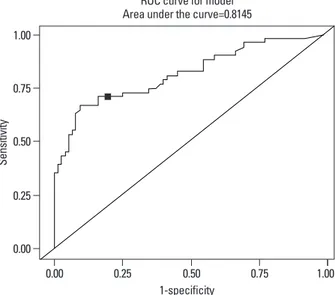
The Receiver Operating Characteristic (ROC) plot22,23
de-sis, pneumonia, dyspnea, and death rate by 2 to 4 times.7,8
Early-onset sepsis is associates with an increased risk of RDS, bronchopulmonary dysplasia, severe intraventricular hemorrhage or periventricular leukomalacia,9 and
transfu-sion.10 The effect of risk of intrauterine infection on fetus
and neonate is higher than on the mother.11
Simple, rapid, noninvasive, and safe tests of markers of intrauterine infection could be useful in prediction of mor-bidity among pregnant women, with or without labor. If ma-ternal infections during pregnancy are diagnosed and treated early, the mortality and morbidity of neonates should be de-creased.
In the literature, several diagnostic methods of peripartum intrauterine infection have been considered. There are amni-onic fluid culture, procalcitonin, C-reactive protein (CRP) in-terleukin (IL)-6, IL-8, IL-10, IL-18, tumor necrosis factor-al-pha (TNF-alfactor-al-pha), interferon gamma, and others.12 Of them,
maternal CRP has been reported to be valuable for early de-tection of neonatal sepsis.13,14 Conflicting results on use of
maternal CRP in prediction of neonatal sepsis have been re-ported.15,16
The objective of the study is to determine the accuracy of maternal serum CRP in prediction of neonatal sepsis.
MATERIALS AND METHODS
We studied the preterm infants (<37 wk) who were admitted to the neonatal intensive care unit, Severance Hospital, from January 2001 to June 2003. We choosed the preterm infants (n=236) and their mothers (n=210) who were examined both a laboratory test and placental pathology in peripartum period. We excluded twins (n=52) and preterm infants who have anormaly, metabolic disease, chromosome and genetic disease. Also, we excluded their mothers who have maternal eclampsia, preeclampsia, hepatitis, cardiovascular disease, rheumatoid disease, gastroenteric disease like as crhon’s dis-ease, oncologic disdis-ease, polycystic ovary syndrome, and connective tissue disease which cause elevated CRP. Final-ly, one hundred twenty six preterm infants (gestation 32±3.2 wk, birth weight 1887±623 g,) and their mothers (31±3.6 years, delivery frequency 2.8±1.5 times) were included.
Preterm infants were divided into neonatal sepsis group (51 cases) and control group (75 cases). Neonatal sepsis was defined as proven with positive blood culture (3 cases) and as suspected with negative blood culture, but more than 3 cate-gories of positive clinical signs among 6 catecate-gories within 3
the sepsis group was significantly higher than controls (Table 1). The maternal CRP of neonatal sepsis group was 3.03 (±2.93) mg/dL, significantly higher than controls 0.71 (±0.65),
p-value was <0.001. In the neonatal sepsis group,
histologi-cal chorioamnionitis was more frequent than controls; clini-cal chorioamnionitis, other infection group, and the use an-tibiotics were higher in sepsis than controls, but was not statistically significant (Table 2). The mortality rate was 6 among total 126 cases (4.8%), and were all in the sepsis group (51 cases), or 12.8% (6 cases/51 cases).
picts the relation of sensitivity and specificity of maternal CRP to neonatal sepsis. The ROC cutoff value in our study subjects was maternal CRP 1.22 mg/dL with sensitivity 71% and specificity 84%. Based on this value, We defined ma-ternal CRP positive group as CRP ≥1.22 mg/dL and mater-nal CRP negative group CRP <1.22 mg/dL (Fig. 1). Clinical characteristics of these two groups was compared in rela-tion to neonatal sepsis, histological chorioamnionitis, clini-cal chorioamnionitis and other infection groups.
CRP was measured by nephlometry using the Beckman Array System protein analyzer (Global Medical Instrumen-tation, Inc., Ramsey, MN, USA).
Statistical analysis
For data analysis, SAS v9.2 was used. Student t-test and Fish-er’s exact test were used for comparison of two variables. The relative risk of neonatal sepsis according to maternal infec-tion status, and maternal CRP positive group versus negative group, were analyzed by logistic regression. Statistical sig-nificance was defined as p-value <0.05.
RESULTS
Comparison of neonatal sepsis and control groups
In the neonatal sepsis group, gestational age was lower than controls. Neonatal CRP performed on day of admission in
Table 1. Characteristics of Newborns with Neonatal Sepsis
Neonatal sepsis (n=51) Control (n=75) p value
G.A. (wk) 30.5±3.7 33.2±2.6 <0.001
B.W. (g) 1833±1743 2073±586 NS
WBC (mm3)* 15934±9636 11778±5281 0.006
CRP (mg/dL)† 0.40±0.80 0.20±0.21 0.027
NS, not significant; WBC, white blood cell; CRP, C-reactive protein; G.A., gestational age; B.W., birth weight; SD, standard deviation. Data mean±SD.
*At birth, WBC.
†At birth, CRP.
Table 2. Demographic Characteristics of Mothers with Neonatal Sepsis
Neonatal sepsis (n=51) Control (n=75) p value
Histologic chorioamnionitis 28 (54%) 16 (21%) <0.001 Clinical chorioamnionitis 6 (11%) 9 (12%) NS Other infection 2 (4%) 3 (4%) NS Antibiotics use 24 (47%) 28 (37%) NS Maternal WBC (mm3) 13340±4413 10199±3655 <0.001 CRP (mg/dL) 3.03±2.93 0.71±0.65 <0.001
NS, not significant; WBC, white blood cell; CRP, C-reactive protein; SD, standard deviation. Data mean±SD.
Fig. 1. Cutoff value determined by Receive Operating Characteristic (ROC)
curves: sensitivity 71%, specificity 84%.
1-specificity 0.00 0.25 0.50 0.75 1.00 Se ns iti vit y 0.00 0.25 0.50 0.75 1.00
ROC curve for model Area under the curve=0.8145
in neonatal sepsis. In our study, maternal CRP in neonatal sepsis group was significantly higher compared with con-trols, reflecting maternal effects on neonatal infection.
Among neonatal sepsis group, blood cultures during hos-pitalization was confirmed in 3 cases. In the other 48 cases, sepsis was defined by clinical criteria, and blood culture was negative. Clinician were not aware of maternal CRP values. Generally, regarding clinical signs of sepsis, the first signs appeared within 12 hours of birth and in order of frequency, was respiratory distress, cardiovascular (hypotension), and temperature abnormality, for >1 hour. Digestive and neuro-logical abnormality signs occurred 3 days after birth.
By ROC curve, the sensitivity and specificity of maternal CRP for neonatal sepsis in our data (1.22 mg/dL as cutoff point), was 71% and 84%, respectively. Sensitivity and spec-ificity were improved compared with past reports; the cut-off value of CRP 0.7 mg/dL26 was 56% sensitivity and 77%
specificity, and using 1.2 mg/dL,27 it was 56% sensitivity,
76% specificity. Recently Celik, et al.28 reported that the
cutoff value of CRP 0.48 mg/dL was 67% sensitivity and 97% specificity. Based on this, in maternal CRP positive group, neonatal sepsis incidence occurred in 71%, signifi-cantly higher than CRP negative group. Maternal WBC, and mean value of CRP were significantly higher than CRP negative group.
The frequency of maternal histological chorioamnionitis was significantly higher in neonatal sepsis group, and
ma-Maternal characteristics by maternal CRP
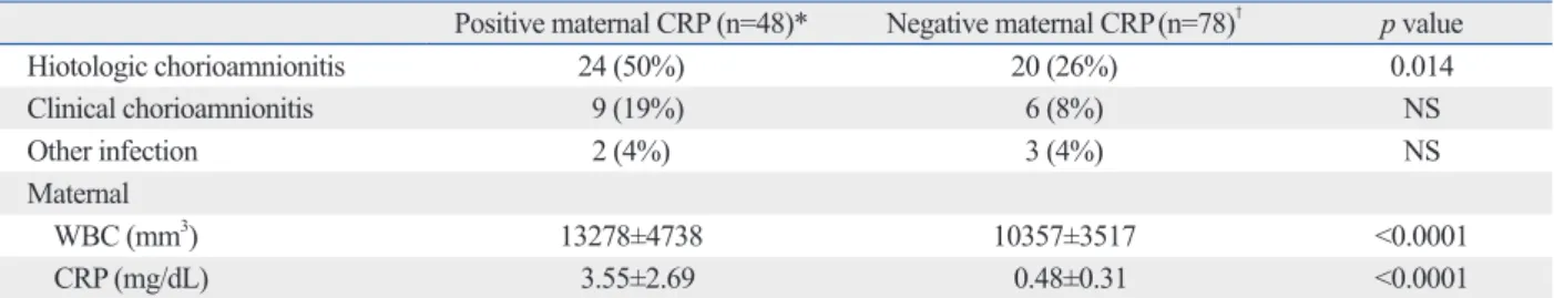
The maternal CRP positive compared with negative group had more histologic chorioamnionitis and increased WBC count (Table 3).
Relative risk of neonatal sepsis
In comparison with the negative group, the relative risk of the maternal CRP positive group for neonatal sepsis was 10.7, (95% confidence interval 4.3-26.4) with p-value of 0.001 (Table 4).
DISCUSSION
In this study, we found that maternal positive CRP is a risk factor for neonatal sepsis. CRP is an acute phase protein produced in the liver, and has the characteristics of reaching maximal concentration 48 hours after stimulation from cy-tokines, IL-1, TNF, and particularly IL-6, and decreases to half after 8 or 9 hours. CRP is increased significantly in cases with infection, inflammatory reaction, tissue damage, and tissue necrosis.24 CRP maintains a constant value
re-gardless of the gestation period in pregnancy,25 significantly
elevated maternal CRP implies maternal infection and its associated neonatal infection.7 Yoon, et al.7 have reported
that maternal WBC and CRP were significiantly increased not only in amniotic fluid culture positive subjects but also
Table 3. Maternal Characteristics by Maternal CRP
Positive maternal CRP (n=48)* Negative maternal CRP(n=78)† p value
Hiotologic chorioamnionitis 24 (50%) 20 (26%) 0.014 Clinical chorioamnionitis 9 (19%) 6 (8%) NS Other infection 2 (4%) 3 (4%) NS Maternal WBC (mm3) 13278±4738 10357±3517 <0.0001 CRP (mg/dL) 3.55±2.69 0.48±0.31 <0.0001
NS, not significant; WBC, white blood cell; CRP, C-reactive protein; SD, standard deviation. Data mean±SD.
*Cutoff value for positive maternal CRP: ≥1.22 mg/dL.
†Cutoff value for negative maternal CRP: <1.22 mg/dL.
Table 4. Relative Risk of Maternal CRP for Neonatal Sepsis
Positive maternal CRP
(n=48)* Negative maternal CRP (n=78)† Odds ratio (95% CI) p value
Neonatal
Sepsis (+) (n=51) 36 (70.6%) 15 (29.4%) 10.68 (4.313-26.428) <0.001 Sepsis (-) (n=75) 12 (16%) 63 (84%)
CI, confidence interval; CRP, C-reactive protein. *Cutoff value for positive maternal CRP: ≥1.22 mg/dL.
12. Bender L, Thaarup J, Varming K, Krarup H, Ellermann-Eriksen S, Ebbesen F. Early and late markers for the detection of early-onset neonatal sepsis. Dan Med Bull 2008;55:219-23.
13. Hirsch W, Koppitz D, Morack G, Gerhardt C. [C-reactive protein in the maternal serum and risk of fetal infection in premature rup-ture of the fetal membranes and threatened premarup-ture labor]. Zentralbl Gynakol 1989;111:1411-6.
14. Skrablin S, Lovric H, Banovic V, Kralik S, Dijakovic A, Kalafatic D. Maternal plasma interleukin-6, interleukin-1beta and C-reactive protein as indicators of tocolysis failure and neonatal outcome after preterm delivery. J Matern Fetal Neonatal Med 2007;20:335-41. 15. van der Heyden JL, van Teeffelen SS, Coolen AC, Halbertsma FJ,
Aardenburg R, Mertens HJ, et al. Is it useful to measure C-reactive protein and leukocytes in patients with prelabor rupture of mem-branes? Am J Perinatol 2010;27:543-7.
16. Trochez-Martinez RD, Smith P, Lamont RF. Use of C-reactive pro-tein as a predictor of chorioamnionitis in preterm prelabour rupture of membranes: a systematic review. BJOG 2007;114:796-801. 17. Mehr SS, Sadowsky JL, Doyle LW, Carr J. Sepsis in neonatal
in-tensive care in the late 1990s. J Paediatr Child Health 2002;38: 246-51.
18. Young Infants Clinical Signs Study Group. Clinical signs that pre-dict severe illness in children under age 2 months: a multicentre study. Lancet 2008;371:135-42.
19. NICE clinical guidesines. CG Antibiotics for early-onset neonatal infection: antibiotics for the prevention and treatment of early-on-set neonatal infection. Manchester: National Institute for Health and Clinical Excellence; 2012.
20. Edmond K, Zaidi A. New approaches to preventing, diagnosing, and treating neonatal sepsis. PLoS Med 2010;7:e1000213. 21. Yoon BH, Romero R, Kim CJ, Jun JK, Gomez R, Choi JH, et al.
Amniotic fluid interleukin-6: a sensitive test for antenatal diagnosis of acute inflammatory lesions of preterm placenta and prediction of perinatal morbidity. Am J Obstet Gynecol 1995;172:960-70. 22. Beck JR, Shultz EK. The use of relative operating characteristic
(ROC) curves in test performance evaluation. Arch Pathol Lab Med 1986;110:13-20.
23. Irwin RJ, Irwin TC. A principled approach to setting optimal diag-nostic thresholds: where ROC and indifference curves meet. Eur J Intern Med 2011;22:230-4.
24. Zimmerman MA, Selzman CH, Cothren C, Sorensen AC, Rae-burn CD, Harken AH. Diagnostic implications of C-reactive pro-tein. Arch Surg 2003;138:220-4.
25. Picklesimer AH, Jared HL, Moss K, Offenbacher S, Beck JD, Boggess KA. Racial differences in C-reactive protein levels dur-ing normal pregnancy. Am J Obstet Gynecol 2008;199:523. 26. Yoon BH, Jun JK, Park KH, Syn HC, Gomez R, Romero R.
Se-rum C-reactive protein, white blood cell count, and amniotic fluid white blood cell count in women with preterm premature rupture of membranes. Obstet Gynecol 1996;88:1034-40.
27. Pfeiffer KA, Reinsberg J, Rahmun A, Schmolling J, Krebs D. Clinical application of maternal serum cytokine determination in premature rupture of membranes--interleukin-6, an early predictor of neonatal infection? Acta Obstet Gynecol Scand 1999;78:774-8. 28. Celik IH, Demirel FG, Uras N, Oguz SS, Erdeve O, Biyikli Z, et
al. What are the cut-off levels for IL-6 and CRP in neonatal sep-sis? J Clin Lab Anal 2010;24:407-12.
ternal CRP positive group. Thus, maternal infection was as-sociated with increased neonatal sepsis and maternal CRP.
In maternal CRP positive group, the relative risk of neona-tal sepsis was 10.7 times greater than the negative group (p<0.001). Neonatal sepsis can be predicted by maternal CRP. Therefore, active treatment of mothers before deliv-ery, intensive follow up observation, and treatment of neo-nates after delivery should decrease the morbidity and mor-tality of disease.
In conclusion, maternal positive CRP is a significant risk factor for neonatal sepsis.
Maternal CRP ≥1.22 mg/dL has 71% predictability for diagnosis of neonatal sepsis. Therefore, through the simple maternal CRP test prior to delivery, the risk level of early neonatal sepsis could be predicted.
REFERENCES
1. Schuchat A, Zywicki SS, Dinsmoor MJ, Mercer B, Romaguera J, O’Sullivan MJ, et al. Risk factors and opportunities for prevention of early-onset neonatal sepsis: a multicenter case-control study. Pediatrics 2000;105(1 Pt 1):21-6.
2. Martius JA, Roos T, Gora B, Oehler MK, Schrod L, Papadopou-los T, et al. Risk factors associated with early-onset sepsis in pre-mature infants. Eur J Obstet Gynecol Reprod Biol 1999;85:151-8. 3. Seaward PG, Hannah ME, Myhr TL, Farine D, Ohlsson A, Wang
EE, et al. International multicenter term PROM study: evaluation of predictors of neonatal infection in infants born to patients with premature rupture of membranes at term. Premature Rupture of the Membranes. Am J Obstet Gynecol 1998;179(3 Pt 1):635-9. 4. Romero R, Gotsch F, Pineles B, Kusanovic JP. Inflammation in
pregnancy: its roles in reproductive physiology, obstetrical com-plications, and fetal injury. Nutr Rev 2007;65(12 Pt 2):S194-202. 5. Goldenberg RL, Hauth JC, Andrews WW. Intrauterine infection
and preterm delivery. N Engl J Med 2000;342:1500-7.
6. Goldenberg RL, Culhane JF, Iams JD, Romero R. Epidemiology and causes of preterm birth. Lancet 2008;371:75-84.
7. Yoon BH, Yang SH, Jun JK, Park KH, Kim CJ, Romero R. Ma-ternal blood C-reactive protein, white blood cell count, and tem-perature in preterm labor: a comparison with amniotic fluid white blood cell count. Obstet Gynecol 1996;87:231-7.
8. Asrat T. Intra-amniotic infection in patients with preterm prelabor rupture of membranes. Pathophysiology, detection, and manage-ment. Clin Perinatol 2001;28:735-51.
9. Stoll BJ, Hansen N, Fanaroff AA, Wright LL, Carlo WA, Ehren-kranz RA, et al. Changes in pathogens causing early-onset sepsis in very-low-birth-weight infants. N Engl J Med 2002;347:240-7. 10. Jeon GW, Sin JB. Risk factors of transfusion in anemia of very
low birth weight infants. Yonsei Med J 2013;54:366-73. 11. Eschenbach DA. Amniotic fluid infection and cerebral palsy.