1)
서 론
심실중격결손이 없는 폐동맥 폐쇄(pulmonary atresia with intact ventricular septum, PA/IVS)는 폐동맥판과 우심실의 다 양한 형태학적 이상을 특징으로 하며 관상동맥 이상을 동반하는 비교적 희귀한 선천성 심장질환으로 다양한 형태학적 특징을 갖 접수 : 2002년 9월 24일, 승인 : 2002년 10월 28일 책임저자 : 최재영, 연세대학교 의과대학 심장혈관병원 소아심장과 Tel : 02)361-7085 Fax : 02)312-9538 E-mail : cjy0122@yumc.yonsei.ac.kr 는 만큼 다양한 치료방법이 적용되고 있지만 예후가 비교적 불 량한 질환이다1-4). 이상적인 치료의 목표는 가능한 한 체순환과 폐순환이 독립된 정상적인 순환의 달성, 즉 양심실 재건에 있다 고 할 수 있겠는데 이를 위해서는 초기에 우심실의 감압 및 혈 류 확보를 통하여 우심실이 충분히 자랄 수 있는 여건을 제공하 여 폐순환을 우심실이 담당할 수 있도록 하는 것이 필수적이라 하겠다4, 6, 18). 이를 위하여 과거에는 수술적 우심실 유출로 재건
술(surgical right ventricle outlet tract repair, surgical RVOT repair)을 시행하여 왔으나 수술 사망률이 보고에 따라
서는 43%로 매우 높고6) 또 본 질환의 경우 주 폐동맥의 크기는
심실중격 결손이 없는 폐동맥 폐쇄의 내과-외과적 협동치료
연세대학교 의과대학 심장혈관병원 심혈관연구소, 소아심장과, 심장혈관외과*
김경식·권병철·이종균·최재영·설준희·이승규·박영환*·조범구*
Medico-Surgical Cooperative Treatment of
Pulmonary Atresia with Intact Ventricular Septum
Kyeong Sik Kim, M.D., Byeong Chul Kweon, M.D., Jong Kyun Lee, M.D. Jae Young Choi, M.D., Jun Hee Sul, M.D., Sung Kyu Lee, M.D.
Young Whan Park, M.D.* and Bum Koo Cho, M.D.*
Division of Pediatric Cardiology, Cardiovascular Surgery*, Yonsei Cardiovascular Center, Cardiovascular Research Institute, Yonsei University College of Medicine, Seoul, Korea
Purpose : The actual clinical examples of co-appliance of catheter intervention with surgical proce-dures in the treatment of pulmonary atresia with an intact ventricular septum(PA/IVS) which we have experienced in our institution are here shown, and the anatomical and hemodynamical profiles between each method is compared.
Methods : Medical records of 33 patients with PA/IVS who underwent various treatment from Jan-uary, 1995 to December, 2000 were reviewed for a retrograde study.
Results : In three out of 10 patients who underwent percutaneous balloon pulmonary valvotomy (PPV), residual pulmonary stenosis were observed in their out patient department(OPD) follow-ups, eventually necessitatig balloon pulmonary valvuloplasty(BPV). One out of three patients exhibited deterioration of tricuspid regurgitation after BPV, requiring surgical tricuspid annuloplasty(TAP). Two out of the seven patients who received primarily surgical right ventricle outlet tract(RVOT) repair without any systemic-pulmonary shunt or intervention needed additional intervention employ-ing cardiac catheterization after operation. Two patients received interventional catheterization before surgical RVOT repair. In five out of 11 cases of Fontan type operation, coil embolization of collateral circulation was done before total cavo-pulmonary connection(TCPC), and in three cases, interven-tional catheterization was needed after TCPC.
Conclusion : Both medical and surgical treatment modalities are widely used in management of PA/IVS patients, and recent results prove that medico-surgical cooperative treatment is essential. (J Korean Pediatr Soc 2003;46:250-258)
Key Words : Percutaneous pulmonary balloon valvotomy(PPV), Surgical RVOT repair, Total cavo-pulmonary connection(TCPC), Right ventricle dependent coronary circulation(RVDCC), Balloon pulmonary valvuloplasty(BPV)
대부분 비교적 잘 발달되어 있으며 폐동맥 판막의 막성 폐쇄만 이 동반된 경우가 흔하기 때문에 기존의 수술적 우심실 유출로 재건술 대신에 풍선도자를 이용한 경피적 폐동맥 판막 절개술 (percutaneous pulmonary balloon valvotomy, PPV)이 도입되
어 시행되고 있다8-19). 또한 수술적 우심실 유출로 재건술 또는
체폐 단락술(systemic-pulmonary shunt, SP shunt)을 단계적 으로 시행함에 있어서도 수술 전후에 다양한 형태의 비수술적 치료를 필요로 하는 경우가 많은데 이에 저자들은 심실중격 결 손이 없는 폐동맥 폐쇄 치료의 실제에 있어 수술-비수술적 협동 치료의 효용성을 확인하고 각각의 치료방법에 대한 치료 성적과 치료전후의 해부학적 혈역학적 지표들을 비교하기 위하여 본 연 구를 시행하였다. 대상 및 방법 1. 대 상 1995년 1월부터 2000년 12월까지 본원에서 심실중격 결손이 없는 폐동맥 폐쇄로 진단되어 치료받은 환자들 중 현재 추적관 찰이 가능한 총 33명의 환자를 대상으로 하였다. 2. 방 법 환아들의 의무 기록지, 심도자 검사 및 심혈관조영술 결과를 후향적으로 분석하였다. 심도자 검사는 Optimus 200 양면 X-선 발생기(Philips Medical System, Netherlands)를 사용하여 시행 하였으며, 심혈관 조영술시 사용한 조영제로는 Optiray (Mallin-crodt Medical Inc., Quebec, Canada)로서 자동 주입기를 이용 하여 체중 1 kg당 1-2 mL를 1-2초에 걸쳐서 주입하였다.
초기 치료방침에 따라 양심실 교정이 가능했던 환자들 가운데 풍선도자를 이용한 경피적 폐동맥 판막 절개술(percutaneous pulmonary balloon valvotomy, PPV)을 시행 받은 환아군, 수 술적 우심실 유출로 재건술(surgical right ventricle outlet tract repair, surgical RVOT repair)을 시행 받은 환아군, 양심
실 교정이 불가능하여 단계적인 체폐 단락술(systemic-pul-monary shunt, SP shunt)을 거쳐 폰탄 타입 수술(Fontan type operation)을 시행 받은 환아군 등 3개 군으로 나누어 치료성적 과 치료 전후의 해부학적, 혈역학적 지표들을 비교하였으며 현재 까지 추적 관찰하는 동안 필요에 의해 시행되어져 왔던 다양한 형태의 수술, 비수술적 치료를 도식화하였다. 결과는 평균±표준편차 및 범위로 표시하였으며, 자료의 통계 처리는 Student t-test를 이용하여 양측검정을 하였으며, 신뢰구 간을 95%로 하여 유의성을 분석하였다. 결 과 33명의 환아 중 경피적 폐동맥 판막 절개술(PPV)을 시행 받 은 환아는 10명이었고 수술적 우심실 유출로 재건술(surgical RVOT repair)을 시행 받은 환아는 12명이었으며 폰탄 타입 수 술을 시행 받은 환아는 모두 11명이었다(Table 1).
Table 1. Patient Population & Anatomical Feature of Pulmonary Atresia with Intact Ventricular Septum(PA/IVS) Patients from 1995 to 2000
Parameter PPV Surgical RVOT repair Fontan type operation
Number of patients Male : Female
Age at Op. or intervention(day) TV diameter(Z-value) PV diameter(Z-value) RVDCC(n) 10 5 : 5 13±12(3-72) -0.5±2.0(-4.5-2.0) -3.9±1.7(-6.0- -1.0) 0 12 5 : 7 25±19(3-132) -0.6±1.6☨ (-2.5-3.0) -3.3±1.7(-5.0-0) 0 11 7 : 4 23±32*(3-82) 850±300†(565-1602) -3.5±1.3§(-5.0-2.0) -2.8±2.7(-6.0-2.5) 4
Abbreviations : PPV, percutaneous pulmonary balloon valvotomy; RVOT, right ventricle outlet tract; Op, operation; TV, trocuspid valve; PV, pulmonary valve; RVDCC, right ventricle dependent coronary circulation
*
age at 1st palliation(modified Blalock-Taussig shunt or Glenn shunt)
†age at Fontan type operation ☨P>0.05 vs PPV group §
P<0.0001 vs biventricular repair group(PPV group plus Surgical RVOT repair group)
Fig. 1. Flow chart showing catheter or surgical intervention in the treatment of pulmonary atresia with intact ventricular septum(PA/IVS) patients who were underwent percutaneous pulmonary balloon valvotomy as initial management. Abbrevia-tions : PPV, percutaneous pulmonary balloon valvotomy; BVP, balloon valvuloplasty; TAP, tricuspid annulo-plasty; SP, sys-temic-pulmonary *due to ARF(1 case) & poor RV compliance (1 case). Re-intervention(4) PPV(10) BVP→TAP(1)BVP(2) SP shunt(1) Died(1) Alive(3) Died(1) (n=8) (n=2)* Alive(5)
심도자를 이용한 경피적 폐동맥 판막 성형술(n=10)은 평균 생후 13±12(3-72)일에 시행 받았으며 삼첨판 Z값은 평균 -0.5±2.0(-4.5-2.0)이었고 우심실 의존성 관상동맥 순환(right ventricle dependent coronary circulation, RVDCC)을 가진 경 우는 없었다(Table 1).
경피적 폐동맥 판막 성형술(PPV)을 시행 받은 10명의 환아 중 외래추적 과정에서 지속되는 폐동맥 판막 협착으로 3례에서 풍선 판막 성형술(balloon pulmonary valvuloplasty, BPV)의 재시행이 필요하였으며 그 중 1례에서는 풍선 판막 성형술에도 불구하고 심한 삼첨판 폐쇄부전이 동반되어 임상증상의 호전이
뚜렷하지 않아 삼첨판륜 성형술(tricuspid annulo-plasty, TAP) 을 시행한 후 호전되었다(Fig. 1).
수술적 우심실 유출로 재건술을(n=12) 시행 받은 환아의 수 술시 연령은 평균 생후 25±19(3-132)일이었고 삼첨판 Z값은 평균 -0.6±1.6(-2.5-3.0)이었으며 우심실 의존성 관상동맥 순환 (RVDCC)을 가진 경우는 없었다(Table 1).
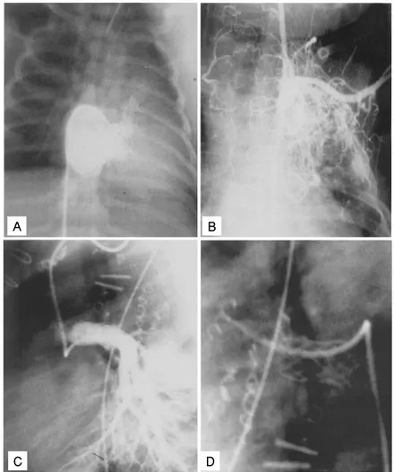
체폐 단락술(SP shunt)이나 심도자 중재술(catheter inter-vention) 없이 일차적으로 우심실 유출로 재건술을 시행 받은 7 례 중 2례에서 시술 후 추가적인 심도자 중재술을 필요로 하였 으며 그 중 1례는 지속적인 폐동맥 판막 협착에 대한 풍선 판막
Fig. 2. Flow chart showing catheter or surgical intervention in the treatment of pulmonary atresia with intact ventricular septum(PA/IVS) patients who were underwent surgical RVOT repair as initial manage-ment. Abbreviations : RVOT, right ventricle outlet tract; MBTS, modified Blalock-Taussig shunt; Gl, Glenn shunt; CE, coil embolization; PDA, patent ductus arteriosus; BAS, balloon atrial septostomy.
*
due to right side heart failure(2 cases) and sepsis/multi-organ failure(1 case).
Post Op intervention(2) Pre Op intervention(2) 1stage Surgical RVOT repair(n=7) Alive(3) PPV(1) PDA CE(1) Died(2) Alive(2) (n=9) (n=3)* SP shunt(4) Central shunt(1) MBTS(2) MBTS→Gl(1) CE(1) BAS(1) Surgical RVOT repair(n=5) Died(1) Alive(4)
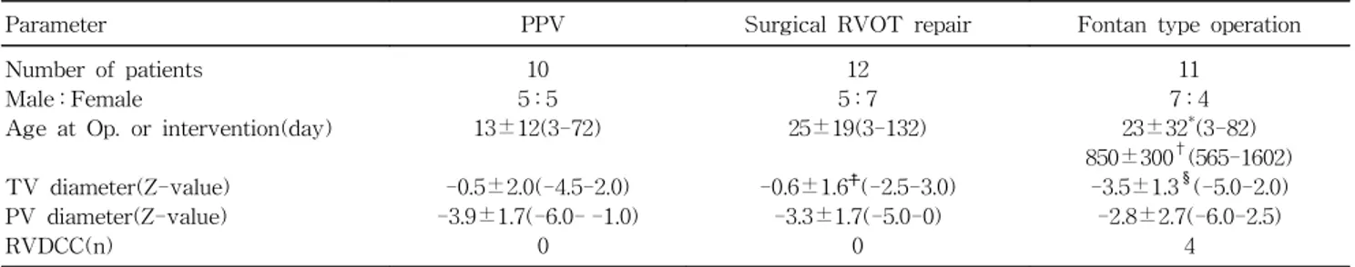
Fig. 3. Flow chart showing catheter or surgical intervention in the treatment of pulmonary atresia with intact ventricular septum(PA/IVS) patients who were underwent Fontan type operation. Abbreviations : PTA, percutaneous transluminal angioplasty; TCPC, Total cavo-pulmonary connection. *due to Fontan circulation failure. BAS(1) MBTS → Gl(5) MBTS → Gl(4) Gl(1) 1 stage Fontan Op.(1) CE(4) CE→PTA→CE(1) Fontan Op.(11) (TCPC=10) Alive(8) CE →PTA(1) stent(2) alive (2) Died* (1) (n=10) Pre intervention(5) Intraatrial baffle(5) Extracardiac conduit(4) Lateral tunnel(1)
성형술(BPV)이었고 1례는 잔류 동맥관개존에 대한 코일 색전술 이었다(Fig. 2, 4).
또한 총 2례에서 수술적 우심실 유출로 재건술 이전에 심도 자 중재술을 시행 받았는데 그 중 1례는 생후 14일에 시행한 풍 선 심방중격 절개술(balloon atrial septostomy, BAS)이었고 1 례는 생후 1개월에 시행 받은 중심성 체폐 단락술(central shunt) 후의 측부순환에 대한 코일 색전술(coil embolization)이 었다(Fig. 2).
모두 11례에서 폰탄 타입 수술을 시행 받았으며 그중 10명의 환아가 총 폐정맥-폐동맥 문합술(total cavo-pulmonary con-nection, TCPC)로 전환하였고 총 폐정맥-폐동맥 문합술(TCPC) 이전 체폐 단락술은 평균 생후 23일에 시행 받았으며 폰탄 타입 수술은 평균 생후 2년4개월에 시행 받았다(Table 1). 폰탄 타입 수술을 시행한 11명 환아의 삼첨판 Z값은 평균 -3.5±1.3(-5.0-2.0)이었고 우심실 의존성 관상동맥 순환(RVDCC) 을 가진 경우는 4례에서 있었다(Table 1). 총 5례에서 총 폐정맥-폐동맥 문합술(TCPC) 시행 전 심도자 중재술을 필요로 하였는데 이중 3례에서는 각각 한차례씩 측부 순환에 대한 코일 색전술을 시행하였고 다른 1례에서는 양 방향 성 상대정맥-폐동맥 문합술(bidirectional cavopulmonary shunt, BCPC) 시행 후 측부순환에 대한 2차례의 코일 색전술을 시행 하였고 이후 좌폐동맥 저형성증으로 인한 풍선 혈관 확장술(per-cutaneous transluminal angioplasty, PTA)을 시행한 후 다시 측부순환에 대한 3차 코일 색전술을 추가로 시행한 후 TCPC를 시행하였다. 또 다른 1례에서는 체폐 단락술(modified Blalock-Taussig shunt) 시행 전 생후 5일 풍선 심방중격 절개술(BAS) 을 시행 받았다(Fig. 3).
총 3례에서 총 폐정맥-폐동맥 문합술(TCPC) 시행 후에 심도
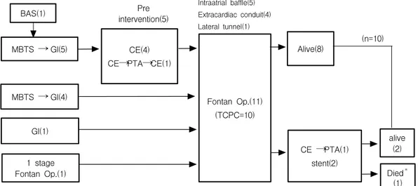
Fig. 4. Selected cineangiogram in a patient with PA and IVS who re-ceived surgical RVOT repair at 24 days of age and coil embolization at 5 years of age. (A) The right ventriculography performed at 5 years of age demonstrates severe tricuspid regurgitation. (B) The right ventriculography performed at 5 years of age demonstrates adequate blood supply through pulmonary artery and adequate growth of right ventricle. (C, D) Selected frames from aortogram showing residual PDA and PDA coil embolization performed at 5 years of age.
A B
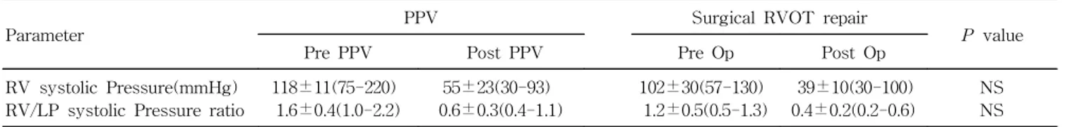
자 중재술을 필요로 하였는데 1례에서는 측부순환에 대한 코일 색전술 후 우폐동맥의 풍선 혈관 성형술(PTA)을 시행하였고 2 례에서는 폐동맥지 협착에 대한 경피적 스텐트 삽입술을 시행하 였다(Fig. 3, 5). 심도자를 이용한 폐동맥 판막 절개술(PPV) 시술 전 우심실/ 좌심실 수축기 압력비는 1.6±0.4(1.0-2.2)에서 시술 후 0.6± 0.3(0.4-1.1)으로 감소하였으며 수술적 우심실 유출로 재건술의 경우 시술 전 1.2±0.5(0.5-1.3)에서 0.4±0.2(0.2-0.6)로 감소하 였으나 두 치료 방법 사이에 통계적으로 유의한 차이는 없었 다(Table 2). 총 공정맥-폐동맥 문합술(TCPC) 시행 전, 후에 좌, 우 폐동 맥압을 포함한 폰탄 순환로(Fontan circuit) 각 부위의 압력은 수술 전후에 유의한 차이가 없었다. 또한 수술 후 총 폐동맥 단 면적 지표(cross sectional area index; Nakata index)는 수술
전 278±67 mm2/m2 BSA(body surface area)에서 수술 후 288±129 mm2/m2 BSA로 유의한 차이가 없어 수술 후에도 폐 동맥의 퇴행 없이 형태학적으로 잘 유지되는 양상을 보였다. 경피적 폐동맥 판막 절개술(PPV)을 시행 받은 환아(n=10) 중 1명의 환아에서 좌측 중뇌동맥의 혈전에 의한 손상이 유발되었 으나 이외에 중대한 합병증은 없었다. 시술 후 2명의 환아가 사 망하였으며, 이 중 1례는 시술 후 동반된 요로기 기형으로 인한 급성신부전으로, 나머지 1례는 시술 후 우심실 유출로의 협착이 있어 우심실 구출이 제한을 받았으며, 시술 후에 PGE1을 지속적 으로 정주 하였으나 산소 포화도가 유지되지 않아 응급으로 체 폐 단락술을 시행하였음에도 불구하고 수술 후 1일 사망하였다.
수술적 우심실 유출로 재건술(surgical RVOT reconstruc-tion)을 시행 받은 환아(n=12) 중 3명의 환아가 사망하였으며 이중 2례는 수술 후 우심실 기능 부전으로 인해 나머지 1례는
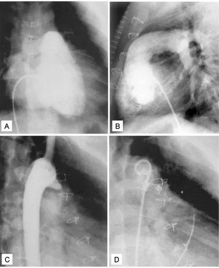
Fig. 5. Selected cineangiogram in a patient with PA and IVS who received coil embolization before and immediately after TCPC, and later received stent implantation 12 months after the procedure. (A) The right ventriculo-graphy performed at 12 days of age demonstrates severely hypoplastic right ventricle. (B) The patient received coil embolization due to formation of col-laterals after TCPC at 20 months of age. (C, D) The patient received stent implantation due to LPA stenosis at 12 months after TCPC.
A B
패혈증과 다기관 기능 부전으로 사망하였다(Fig. 2). 폰탄 타입 수술을 시행 받은 환아(n=11) 중 1명의 환아에서 저산소성 뇌손상의 합병증이 있었으며 총 폐정맥-폐동맥 문합술 시행 전 단백 소실성 장병증이 발병하였던 3명 중 2례에서 수술 후에도 저 알부민 혈증이 지속되어 알부민 및 헤파린 투여로 호 전되었다. 시술 후 1명의 환아가 폰탄 기능 부전으로 사망하였 다(Fig. 3). 고 찰 신생아기에 시행되는 심실중격 결손이 없는 폐동맥 폐쇄(PA/ IVS)의 일차적 치료 목표는 적절한 폐혈류 확보로 저산소증에 의한 위험 및 합병증을 최소화하고, 가급적 우심실을 통한 혈류 를 확보하여 양심실 교정 혹은 1과 1/2심실로의(one and half ventricle) 교정 기회를 증가시키며, 만일 기능적으로 우심실을 폐심실로 사용할 수 없다면 적절한 폐혈관의 발생과 성장을 유 도하여 궁극적으로 성공적인 단심실 교정이 가능하도록 유도하 는데 있다고 하겠다. 이러한 세 가지 치료목표를 달성하기 위한 방법으로 과거에는 일반적으로 우심실 유출로 성형술, 우심실 유 출로 성형술과 체폐 동맥 단락술, 체폐 동맥 단락술만 시행하는 세 가지의 치료방법이 이용되어져 왔다7). PA/IVS의 수술적 치료 방침은 삼첨판의 크기20), 우심실의 형 태2, 22, 23), 우심실 누두부의 발달상태24) 등에 따라 다양하게 제시 되어 왔는데 Hanley 등25)은 우심실 의존성 관상 동맥 순환의 존재 여부와 삼첨판 Z값에 따른 수술 치료 방침을 권유하였다. 이에 따르면 우심실 의존성 관상 동맥 순환을 갖고 있지 않고 삼첨판 Z값이 0에서 -2인 경우 우심실 유출로 재건술만을 권유 하였고, 우심실 의존성 관상 동맥 순환이 없으며 삼첨판 Z값이 -2에서 -3인 경우 양심실 교정이 가능할 수 있기 때문에 우심 실 유출로 재건술과 함께 체폐동맥 단락술을 권유한다고 하였다 25) . 현재까지 양심실 교정이 가능한 우심실 발육부전의 정도가 정해진 바는 없으나 삼첨판 Z값이 -3 이하이면 양심실 교정이 가능한 경우가 많지 않은 것으로 알려져 있는데25), 본 연구 대상 중 경피적 폐동맥 판막 절개술을 시행한 10명의 환아의 경우 삼 첨판 Z값은 평균 -0.5±2.0(-4.5-2.0)이었고 우심실 의존성 관상 동맥 순환(RVDCC)을 가진 경우는 없었다. 이중 삼첨판 Z값이 Table 2. Hemodynamic Profile of Patients Before or After Biventricular Repair
Parameter PPV Surgical RVOT repair P value
Pre PPV Post PPV Pre Op Post Op
RV systolic Pressure(mmHg) RV/LP systolic Pressure ratio
118±11(75-220) 1.6±0.4(1.0-2.2) 55±23(30-93) 0.6±0.3(0.4-1.1) 102±30(57-130) 1.2±0.5(0.5-1.3) 39±10(30-100) 0.4±0.2(0.2-0.6) NS NS Abbreviations : PPV, percutaneous pulmonary balloon valvotomy; RVOT, right ventricle outlet tract; Op, operation; NS, not sig-nificant
Table 3. Summary of Pulmonary Balloon Valvotomy for Treatment of Pulmonary Valve Atresia from Published Data & This Study
Reference No. of patients Procedures Success complications/deathSevere procedural Requiring MBTS
* or intervention Latson30) Parsons, et al.12) Qureshi, et al.8) Rosenthal, et al.10) Rosenthal, et al.9) Gournay, et al.13) Fedderly, et al.19) Schneider, et al.14) Justo, et al.17) Ruiz, et al.26) Total Lee, et al. 1 1 5 10 4 15 2 1 8 4 51 10 Wire puncture+balloon Laser+balloon Laser+balloon Laser+balloon Radiofrequency+balloon 14 wire Puncture+balloon 1 radiofrequency+balloon Wire puncture+balloon Radiofrequency+balloon+stent 6 Wire puncture+balloon 2 radiofrequency+balloon Wire puncture+balloon Wire puncture+balloon 1 1 4 8 4 9 2 1 8 3 41(80%) 8(80%) 0/0 0/0 1/0 2/1 1/1 5/1 0/0 0/0 0/0 0/0 9(18%)/3(6%) 1/2(10%/20%) 0 0 3 4(before valvotomy) 1 5 1 1(before valvotomy) 7 1 23/41(56%)† or 18/36(50%)☨ 3/8(38%)§ *
MBTS : modified Blalock-Taussig shunt
†
The percentage of total patients requiring MBTS was calculated based on 41 patients with successful ballon valvotomy
☨
The percentage of patients requiring MBTS post-ballon valvotomy was calculated based on 36 patients, excluding 5 patients who had MBTS prior to valvotomy
-4.5인 환아는 동맥관 개존, 심방중격 결손, 삼첨판 역류, 삼첨판 협착 등의 동반 기형을 갖고 있었으며 심한 우심실 형성부전과 폐동맥 판륜의 Z값이 -4이었던 환아로 생후 3일에 경피적 폐동 맥 판막 절개술 및 풍선 확장술을 시행하였다. 시술 전 우심실/ 좌심실 수축기 압력 비는 1.71 에서 시술 후 0.93으로 감소하였 고 별다른 합병증은 없었으나 1년 3개월의 외래 추적 관찰 후 시행한 심초음파 상 우심실 형성 부전 정도는 아직 심하여 삼첨 판 Z값은 -4이고 폐동맥 판륜의 Z값은 -2이었다. 수술적 우심실 유출로 재건술을 시행 받은 환아의 삼첨판 Z 값은 평균 -0.6±1.6(-2.5-3.0)이었으며 역시 우심실 의존성 관 상동맥 순환을 가진 예는 없었고 경피적 폐동맥 판막 절개술을 시행 받은 환아 군의 삼첨판 Z값과 수술적 우심실 유출로 재건 술을 시행 받은 환아 군의 삼첨판 Z값 사이에 통계적으로 유의 한 차이는 없었다(Table 1). 심도자를 이용한 폐동맥 판막 절개술 시술 전 우심실/좌심실 수축기 압력 비는 1.6±0.4(1.0-2.2)에서 시술 후 0.6±0.3(0.4-1.1)로 감소하였으며 수술적 우심실 유출로 재건술의 경우 시술 전 1.2±0.5(0.5-1.3)에서 0.4±0.2(0.2-0.6)로 감소하였으나 결과 에 있어 두 치료 방법 사이에 통계적으로 유의한 차이는 없었다 (Table 2). 결국 우심실 유출로 재건술로 경피적 폐동맥 판막 성형술을 시행 받은 대상은 수술적 우심실 유출로 재건술을 시행 받은 대 상과 유사한 형태학적 특성을 가지고 있었으며 시술 전후의 혈 역학적 지표의 개선도에 있어서도 차이가 없음을 확인할 수 있 었다. 양심실 재건이 가능하다고 판단되는 환아에서 경피적 폐동맥 판막 절개술을 시행할 것인가 수술적 우심실 유출로 재건술을 시행할 것인가의 선택에 대해서는 아직까지 명확한 기준이 제시 되어 있지 않지만 Fedderly 등19)은 삼첨판륜의 크기가 11 mm (또는 Z값이 -0.5) 이상이며 폐동맥 판륜의 크기가 7 mm(또는 Z값이 -1.5) 이상이고 이완기말 우심실 용적이 30 mL/m2 이상 인 막성 PA/IVS의 경우 경피적 폐동맥 판막 절개술이 효과적 이라 하였으며 Cheatham27)은 위의 조건과 함께 누두부가 잘 발 달된 tripartite RV인 경우 낮은 합병증 가능성과 추가적 재수술 의 필요성 없이 성공적인 시술을 기대할 수 있다고 하였다. 본 연구대상 중 성공적으로 경피적 폐동맥 판막 절개술을 시 행한 환아(n=8)에서 풍선을 이용한 폐동맥 판막 성형술이나 수 술적 삼첨판륜 성형술까지 포함한 추가적 재시술이 필요했던 예 는 모두 3례이었으며 추가적인 재시술이 필요치 않았던 5명의 환아들의 삼첨판 Z값의 크기는 평균 -0.5±1.5(-2.0-1.5)이었으 며 폐동맥 판륜 Z값의 크기는 평균 -1.5±1.5(-3.0-1.0)로서 상 기 기준에 부합되었다. 1997년 Ruiz 등은 1991년부터 1997년까지의 다기관 조사에서 심도자를 이용한 경피적 폐동맥 판막 성형술의 치료 성적을 요 약하였는데8-14, 17-19) 모두 51례 중 시술과 관련된 중대한 합병증 이나 사망 없이 시술에 성공한 예는 모두 41례(80%)였으며 합 병증은 9례(18%), 사망한 예는 모두 3례(6%) 이었다. 시술에 성 공한 41례 중 5례에서는 시술 전에, 18례에서는 시술 후에 체폐 단락술을 필요로 하여 시술 전후를 합친다면 모두 23례(23/41; 56%)에서 체폐 단락술을 필요로 하였다(Table 3). 본원에서 경피적 폐동맥 판막 절개술을 시행한 10명의 환아 에서 시술에 성공한 예는 8례(80%)이었으며 시술 전 체폐 단락 술을 시행한 환아는 없었으나 시술 후 체폐 단락술을 시행한 환 아는 1례(central shunt, 사망)에서 있었고 시술에 성공한 8례 중 풍선을 이용한 반복적 폐동맥 판막 성형술(BVP) 등 심도자 를 이용한 추가적 재시술이 필요한 경우는 3례(3/8; 38%)에서 있었다(Fig. 1). 수술적 우심실 유출로 재건술을 시행한 12례의 경우 3명(25 %)의 환아가 사망하였으며, 수술 성공률은 9/12(75%)로서 경피 적 폐동맥 판막 절개술(PPV)을 시행한 경우와 치료 결과상의 차이는 없다고 볼 수 있겠다. 또한 수술적 우심실 유출로 재건술에 성공한 9례 중 수술 전 후에 심도자를 이용한 중재술이 필요했던 경우는 모두 4명의 환 아(BAS 1례, CE 2례, PPV 1례, 4/9; 44%)에서 있었고 폰탄 타입 수술(n=10) 전후에도 9명의 환아에서 모두 11번의 심도자 를 이용한 중재술이 시행되어(CE 7례, PTA 2례, stent im-plantation 2례) PA/IVS의 수술적 치료 전후에 심도자를 이용 한 다양한 형태의 비수술적 치료방법이 시행되고 있음을 확인할 수 있었다(Fig. 2, 3). 폰탄 술식의 적응은 1) 우심실 의존성 관상동맥 순환, 2) 우 심실 형성부전 및 삼첨판 Z값이 -4 미만, 3) 크기가 큰 우심실-관상동맥루 형성, 4) 심한 Ebstein 양 변화로 우심실의 기능을 기대하기 어려운 경우 등으로 볼 수 있는데 28, 29) 본원에서 폰탄 타입 수술을 시행한 11명의 환아 중 우심실 의존성 관상동맥 순환을 가진 예는 4례에서 있었으며 삼첨판 Z 값은 평균-3.5±1.3(-5.0-2.0)으로 경피적 폐동맥 판막 절개술과 수술적 우심실 유출로 재건술을 포함하여 본원에서 양심실 교정 이 가능하였던 환아[0.2±1.9(-4.5-3.0)]와 비교하였을 때 통계적 으로 유의한 차이를 보였다(P<0.0001). 이상과 같이 심도자를 이용한 경피적 폐동맥 판막 절개술과 수술적 우심실 유출로 재건술의 시술 전후 해부학적 지표와 혈 역학적 개선도 등을 비교하였을 때 통계적으로 유의한 차이가 없어 경피적 폐동맥 판막 절개술이 수술적 우심실 유출로 재건 술을 대치할 수 있는 치료법이며 이미 제시된 기준에 부합되는 대상들에서는 추가적인 재치료가 필요 없는 성공적인 치료 결과 를 기대할 수 있음을 확인하였다. 또한 초기 치료 방침이 어떻 게 결정되어지든지 환아의 추적 관찰 기간동안 심도자를 이용한 추가적인 중재술과 추가적인 재수술이 환아의 형태학적 혈역학 적 필요성에 의해 행해지고 있음을 확인하여 심실중격 결손이 없는 폐동맥 폐쇄(PA/IVS)의 치료에 있어 효과적인 궁극적 치 료목표의 달성을 위해 효율적인 내과 외과적 협동치료가 절실히 요구됨을 확인하였다.
요 약 목 적 : 심실중격결손이 없는 폐동맥 폐쇄의 치료에 있어 수 술적 우심실 유출로 재건술 대신 심도자를 이용한 경피적 폐동 맥 판막 절개술이 도입되었고 고식적으로 폰탄 술식을 단계적으 로 시행함에 있어서도 다양한 형태의 비수술적 치료 방법이 시 행되고 있어 수술-비수술적 협동 치료의 중요성이 강조되고 있 는 가운데 실제 심실중격결손이 없는 폐동맥 폐쇄의 치료에 있 어 수술-비수술적 협동 치료가 행해지는 실례를 확인하고 각 치 료 방법들 사이의 치료 성적과 해부학적 혈역학적 지표를 비교 하기 위하여 본 연구를 시행하였다. 방 법 : 1995년 1월부터 2000년 12월까지 본원에서 심실중격 결손이 없는 폐동맥 폐쇄로 치료받은 33명의 환아(경피적 폐동 맥 판막 절개술 10례, 수술적 우심실 유출로 재건술 12례, 폰탄 형 술식 11례, 남 : 녀=17 : 16)를 대상으로 환자 기록지를 후향적 으로 검토하였다. 결 과 : 경피적 폐동맥 판막 성형술을 시행 받은 10명의 환아 중 외래추적 과정에서 지속되는 폐동맥판막 협착으로 3례에서 풍선을 이용한 폐동맥판막 성형술을 필요로 하였으며 1례에서는 풍선을 이용한 폐동맥 판막 성형술에도 불구하고 삼첨판륜 성형 술로 호전 되었다. 체폐 단락술이나 심도자 중재술 없이 일차적 으로 우심실 유출로 재건술을 시행 받은 7례 중 2례에서 시술 후 추가적인 심도자 중재술을 필요로 하였으며 2례 에서 수술적 우심실 유출로 재건술 이전에 심도자 중재술을 시행 받았다. 폰 탄 타입 수술을 시행한 11례 중 모두 5례에서 총 폐정맥-폐동맥 문합술 전에 측부순환에 대한 코일 색전술을 시행 받았고 모두 3례에서 총 폐정맥-폐동맥 문합술 후에 심도자 중재술을 필요로 하였다. 결 론 : 심실중격결손이 없는 폐동맥 폐쇄 환아의 치료에 있 어 수술적 치료와 비수술적 치료가 병행되고 있으며 수술-비수 술적 협동치료가 필수적으로 요구된다. 참 고 문 헌
1) Cobanoglu A, Metzdorff MT, Pinson CW, Grunkemeir GL, Sunderland LO, Starr A. Valvotomy for pulmonary atresia with intact ventricular septum. J Thoracic Cardiovasc Surg 1985;89:482-90.
2) de Leval M, Bull C, Hopkin R, Rees P, Deanfield J, Taylor JF, et al. Decision making in the definitive repair of the heart with a small right ventricle. Circulation 1985;72(2 Suppl):52S-62S.
3) Coles JG, Freedom RM, Lightfoot NE, Dasmahapatra HK, William WG, Trusler GA, et al. Long term results in neo-nates with pulmonary atresia and intact ventricular septum. Ann Thorac Surg 1989;47:213-7.
4) Rao PS. Comprehensive management of pulmonary atresia with intact ventricular septum. Ann Thorac Surg 1985;40:
409-13.
5) Rao PS, Liebman J, Borkat G. Right ventricular growth in a case of pulmonic stenosis with intact ventricular septum and hypoplastic right ventricle. Circulation 1976;53:389-94. 6) 이종균. Transcatheter valvotomy of pulmonary atresia with
intact ventricular septum. 순환기 2001;31(4 Suppl):59S-60S. 7) 성시찬. 온전한 심실중격의 폐동맥 폐쇄증에 대한 수술. 순환기
2001;31(4 Suppl):61S-66S.
8) Qureshi SA, Rosenthal E, Tynan M, Anjos R, Baker EJ. Transcatheter laser-assisted balloon pulmonary valve dila-tion in pulmonic valve atresia. Am J Cardiol 1991;67:428-31.
9) Rosenthal E, Qureshi SA, Chan KC, Martin RP, Skehan DJ, Jordan SC, et al. Radiofrequency-assisted balloon dila-tation in patients with pulmonary valve atresia and an in-tact ventricular septum. Br Heart J 1993;69:347-51.
10) Rosenthal E, Qureshi SA, Kakadekal AP, Anjors R, Baker EJ, Tynan M. Technique of percutaneous laser assisted valve dilatation for valvular atresia in congenital heart dis-ease. Br Heart J 1993;69:556-62.
11) Redington AN, Cullens S, Rigby MC. Laser or radiofre-qency pulmonary valvotomy in neonates with pulmonary valve atresia and intact ventricular septum : Description of a new method avoiding arterial catheterization. Cardiol Young 1992;2:387-90.
12) Parsons JM, Rees MR, Gibbs JL. Percutaneous laser valvo-tomy with balloon dilatation of the pulmonary valve as primary treatment for pulmonary atresia. Br Heart J 1991; 66:36-8.
13) Gournay V, Piechand J, Delogu A, Sidi D, Kachancer J. Baloon valvotomy for critical stenosis or atresia of pulmo-nary valve in newborns. J Am Coll Cardiol 1995;26:1725-31.
14) Schneider M, Schranz D, Michel-Behnke I, Oelert H. Transcatheter radiofrequency perforation and stent implan-tation for palliation of pulmonary atresia in a 3,060 gm infant. Cathet Cardiovasc Diagn 1995;34:42-5.
15) Wright SB, Radtke WA, Gillette PC. Percutaneous radio-frequency valvotomy using a standard 5-Fr electrode cath-eter for pulmonary atresia in neonates. Am J Cardiol 1996; 73:1370-2.
16) Akagi T, Hashino K, Maeno Y, Ishii M, Sugimura T, Kawano T, et al. Ballon dilatation of the pulmonary valve in a patient with pulmonary atresia with intact ventricular septum using a commercially available radiofrequency cath-eter. Pediatr Cardiol 1997;18:61-3.
17) Justo RN, Nykanen DG, Williams WG, Freedom RM, Ben-son LN. Transcatheter perforation of the right ventricular outflow tract as intial therapy for pulmonary valve and in-tact ventricular septum in the newborn. Cathet Cardiovasc Diagn 1997;40:408-13.
18) Siblini G, Rao PS, Singh GK, Tinker K, Balfour IC. Trans-catheter management of neonates with pulmonary atresia and intact ventricular septum. Cathet Cardiovasc Diagn 1997;42:395-402.
19) Fedderly RT, Lloyd TR, Mendelsohn AM, Beekman RH. Determinants of successful balloon valvotomy in infants with critical pulmonary stenosis or membranous pulmonary atresia with intact ventricular septum. J Am Coll Cardiol
1995;25:460-5.
20) Hanley FL, Sade RM, Blackstone EH, Kirklin JW, Freedom RM, Nanda NC. Outcome in neonatal pulmonary atresia with intact ventricular septum. A multiinstitutional study. J Thorac Cardiovasc Surg 1993;105:406-27.
21) Rychik J, Levy H, Gaynor JW, DeCampli WM, Spray TL. Outcome after operations for pulmonary atresia with intact ventricular septum. J Thorac Cardiovasc Surg 1998;116: 924-31.
22) Billingsley AM, Laks H, Boyce SW, George B, Santulli T, Williams RG. Definitive repair in patients with pulmonary atresia and intact ventricular septum. J Thorac Cardiovasc Surg 1989;97:746-54.
23) de Leval MR, Bull C, Stark J, Anderson RH, Taylor JF, Macartney FJ. Pulmonary atresia and intact ventricular septum : Surgical management based on a revised classifi-cation. Circulation 1982;66:272-80.
24) Pawade A, Capuani A, Penny DJ, Karl TR, Mee RB. Pul-monary atresia with intact ventricular septum : surgical management based on right ventricular infundibulum. J Card Surg 1993;8:371-83.
25) Reddy VM, Hanley FM. Pulmonary atresia with intact ventricular septum. Early palliation, subsquent management,
and possible role of fetal surgical intervention. In : Baue AE, Geha AS, Hammond GL, Laks H, Naunheim KS, edi-tors. Glenn's Thoracic and cardiovascular surgery. 6th ed. London : Prentice-Hall international Inc, 1996;1315-32. 26) Ruiz CE, Zhang HP. Is balloon a challenge to scalpel in
membranous pulmonary valve atresia or just a partner? Cathet Cardiovasc Diagn 1997;40:414-5.
27) Cheatham JP. To perforate or not to perforate - That's the question. or is it? Just ask Richard. Cathet Cardiovasc Diagn 1997;42:403-4.
28) Najm HK, Williams WG, Coles JG, Rebeyka IM. Freedom RM. Pulmonary atresia with intact ventricular septum : Re-sults of the Fontan procedure. Ann Thorac Surg 1997;63: 669-75.
29) Laks H, Plunkett MD. Pulmonary atresia and pulmonary atresia with intact septum. In : Kaiser L, Klon IL, Spray TL, editors. Mastery of cardiothoracic surgery. New York : Lippincott-Raven Co, 1998:805-18.
30) Latson LA. Nonsurgical treatment of a neonate with pul-monary atresia and intact ventricular septum by transcath-eter puncture and balloon dilatation of the atretic valve membrane. Am J Cardiol 1991;68:277-9.