MicroRNAs are Potential Biomarkers and Therapeutic Targets of Atopic Dermatitis
Won Suck Yoon1,2, Young Yoo2,3, Tae Ho Lee4, Sung Ha Park5, Byeong Mo Kim*51Department of Biotechnology, School of Life Sciences and Biotechnology, Korea University, Republic of Korea 2Institute of Allergy Immunology, Korea University, Seoul, Republic of Korea
3Deparment of Pediatrics, College of Medicine, Korea University, Republic of Korea
4Division of Gerontology, Department of Medicine, Beth Israel Deaconess Medical Center, Harvard Medical School, USA
5Severance Integrative Research Institute for Cerebral & Cardiovascular Diseases (SIRIC), Yonsei University College of Medicine, Republic of Korea
*Corresponding author:
Dr. Byeong Mo Kim, Severance Integrative Research Institute for Cerebral & Cardiovascular Diseases
(SIRIC), Yonsei University College of Medicine, 50 Yonsei-ro, Seodaemun-gu, Seoul 120-752, Republic of Korea, Tel:
82-2-2228-0789; Fax: 82-2-2227-7906; E-mail: bkim2@yuhs.ac
Received:
01-05-201
5Accepted:
01-21-201
5Published: 02-05-2015
Copyright:
© 2015 Byeong Mo
Review Article AbstractmicroRNAs (miRNAs) are a class of small, non-coding RNAs that regulate the expression of a diverse array of genes and path-ways, with important roles in disease pathogenesis. Many of these miRNAs are currently under investigation as biomarkers or therapeutic options in a range of diseases. Here, we discuss the role of miRNAs in atopic dermatitis, a relapsing chronic pruritic inflammatory skin disease of unknown etiology, as they have been strongly implicated in the pathogenesis of skin inflammation. We outline the history and application of miRNAs for the detection and treatment of atopic dermatitis in comparison with other diseases, such as cancer and cardiovascular disease, which may assist the development of diagnostic and therapeutic strategies.
Keywords: MiRNA; Atopic Dermatitis; Biomarker; Therapeutic Application
Abbreviations
miRNA : microRNA; AD : Atopic Dermatitis; CVD : Cardiovascular Diseases; Th cells : T Helper Cells;
MDC : Macrophage-Derived Chemokine;
ST-miRCCL22 : Salmonella typhimurium Expressing CCL22 miRNA
Introduction
microRNAs (miRNA) are a class of small, non-coding RNAs ~21-25 nucleotides in length. Over 1000 human miRNAs have been identified to date, primarily located in the intronic re-gions of other genes. These structures represent an important form of post-transcriptional gene regulation by directly inhib-iting gene translation through their binding of the 3′ untrans-lated regions of their target messenger RNA (mRNA), resulting in lower mRNA stability, or translation inhibition [1,2]. The effects of these miRNAs are far reaching, with more than 30% of the entire genome affected, including critical processes, such as development, differentiation, cell growth, and apop-tosis. This central role of miRNAs in overall cell function has drawn considerable interest from cancer researchers, as can-cers ultimately arise as a result of aberrant gene expression. As miRNAs are readily detected in accessible body fluids, such as saliva, blood, urine, and even hair follicles, this suggests the possibility of miRNAs as potential biomarkers of human dis-eases. This review summarizes the molecular mechanisms of miRNA activity, highlights recent studies demonstrating their application as both biomarkers and therapy targets, and ex-plores their therapeutic potential for the treatment of atopic dermatitis (AD).
miRNAs as biomarkers and therapeutic targets in cancer and cardiovascular disease
miRNAs were first identified in the mid-1990s with the discov-ery of Let-7 and Lin-4 in the model organism Caenorhabditis
elegans [3,4]. Homologs have since been identified in nearly
all eukaryotic organisms, with significant conservation among species. Given the strong conservation of these structures, and their central role in processes such as development, differenti-ation, cell growth, and apoptosis, it was not surprising reports of aberrant miRNAs expressed in cancers and other diseases emerged soon thereafter.
Ongoing clinical trials are currently assessing the correlation between miRNA expression and disease prognosis in cancer. As in vitro expression profiles of many tumor-derived miRNAs have shown promise for the diagnosis of patients, miRNA ex-pression profiles might be used to precisely classify various cancer types, and might be superior to gene expression pro-files in classification of tumors. More widespread screening before the onset of disease is also possible; stable miRNAs de-tected in easily accessible fluids, such as serum, plasma, and urine, as well as hair, have shown distinctive patterns of miR-NA expression among patients and controls highlighting their possible use as diagnostic markers [5-7].
Interestingly, a single miRNA can simultaneously regulate both tumor suppressive and oncogenic target genes within a single cancer [8-11]. For example, miR-196b can target not only the
HOXA9/MEIS1 oncogenes, but also FAS tumor suppressor gene
in mixed lineage leukemia-rearranged leukemia [8,9]. This implies that tumor initiation and development, as influenced by miRNAs, might be more complex than previously thought, which has important implications for using miRNAs as thera-peutic agents. To date, several drugs have been shown to alter miRNA expression, including the bioactive agent docosahex-aenoic acid, which inhibits the expression of miR-21, a protu-morigenic miRNA [12]. Researchers are currently designing inhibitors for oncogenic miRNAs, along with mimics for tu-mor-suppressor miRNAs, which can act alone or synergistical-ly with currentsynergistical-ly approved treatments [12,13].
In addition to cancer, many miRNAs have been identified as novel biomarkers and potential therapeutic targets for car-diovascular diseases (CVD). miRNAs play a crucial role in the biogenesis and function of the cardiovascular gene regulato-ry system, and have been implicated as dynamic regulators of cardiac and vascular signaling and arterial remodeling [14-16]. For example, in atherosclerosis, miRNA expression is directly regulated by blood flow, with endothelial cell miRNA expres-sion increased in response to laminar or high flow, and other decreased in cases of low or disturbed flow [15,16]. Together, this regulatory dynamic suggests a possible therapeutic use of miRNAs as inhibitors of atherosclerosis development. The en-dothelial cell-derived mechano-sensitive miR-143/145 can be delivered to smooth muscle cells via endothelial extracellular vesicles, exerting atheroprotective effects in smooth muscle cells [16]. Recent research has also suggested the use of sta-ble miRNAs circulating in body fluids as potential biomarkers for cardiovascular diseases. The muscle-enriched 1, miR-133 and miR-499-5p, as well as the cardiomyocyte-specific miR-208, have been extensively investigated for their diagnos-tic ability in plasma of patients with coronary artery disease [17,18]. These observations strongly suggest a role for circu-lating miRNAs as a blood-based biomarker for cardiovascular diseases.
Several studies have demonstrated the potential therapeutic use of miRNAs to modulate disease processes by antagoniz-ing miRNA expression or increasantagoniz-ing their inhibitor functions. For example, targeted expression of miR-590 and miR-199a in heart cells using an adeno-associated viral vector demonstrat-ed a re-entry of cardiomyocytes into the cell cycle, resulting in reduced infarct size and improved cardiac function [18].
miRNAs and allergic diseases
Atopic dermatitis (AD) is a chronically relapsing inflamma-tory skin disease that results from a combination of genetic susceptibility and heightened immunologic responses to envi-ronmental allergens. Recently, the role of miRNAs in disease pathogenesis has begun expanding outside of the realms of cancer and CVD to allergic diseases, such as AD, with
signif-alleviation of airway inflammation in a murine model of asth-ma [27]. Similarly, the miR-200 family of miRNAs regulates ex-pression of E-cadherin, with supex-pression of E-cadherin is as-sociated with increases in CCL17, a Th2 cell chemoattractant [28]. Other miRNAs, such as miR-1 and miR-155, also play a role in allergic inflammation, as suppression of these miRNAs by VEGFA contributes to Th2 inflammation in the endothelium and promotes recruitment of activated T cells and subsequent eosinophilic inflammation, together with Th2 cytokine pro-duction [29,30].
Recently, our group demonstrated that miRNA targeting CCL22 suppressed inflammatory responses in macrophages and in an animal model of AD [31,32]. This MDC/CCL22 axis was direct-ly implicated in Th2-associated skin inflammatory reactions with significant increases in serum concentrations strongly correlated with disease severity in AD.
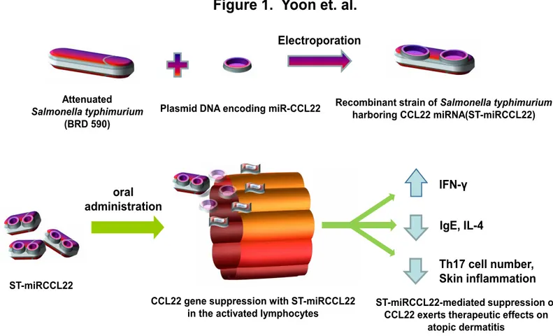
In those studies, a recombinant strain of Salmonella
typhimuri-um expressing CCL22 miRNA (ST-miRCCL22) was used for the in vivo knockdown of CCL22 as a treatment for AD.
ST-miRC-CL22 was shown to significantly downregulate CST-miRC-CL22 expres-sion in activated lymphocytes in vitro. Subsequent in vivo anal-yses in a mouse model of AD revealed decreases in both IL-4 and IgE expression, alongside increases in IFNγ; Th17 cells were also suppressed in AD mice treated with ST-miRCCL22. Together, these data suggest that targeted miRNA delivery may be an effective method for the treatment of AD (Figure 1). icant attention paid to both genetic and environmental
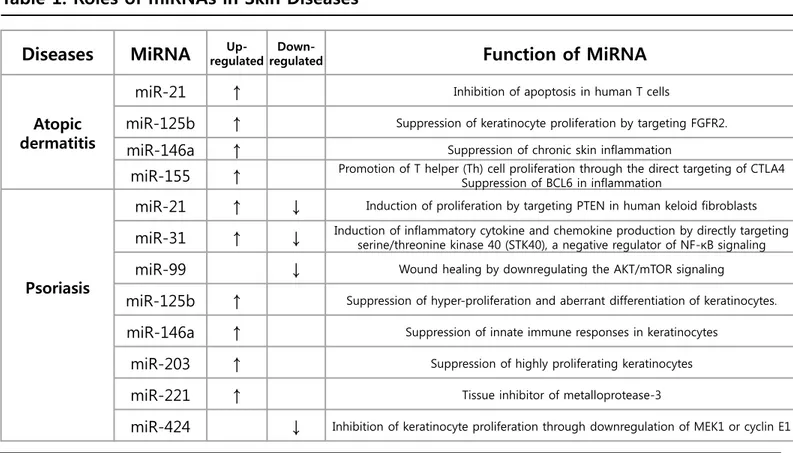
fac-tors. AD is an inflammatory skin disease that results from a combination of genetic predisposition, imbalanced immune responses, epidermal barrier abnormalities, and severe pru-ritus. A Th1/Th2 imbalance is a key factor in the pathogenesis of allergic diseases such as AD, with some reports implicating miRNA regulation of innate and adaptive immune responses in Th2 polarization [19-23]. Immune cells, including monocytes/ macrophages, and dendritic cells, as well as T and B lympho-cytes, play a key role in AD [23]. miRNAs have been shown to regulate an array of immune cell functions, including macro-phage-derived cytokines (MDC) and the expression of inflam-matory mediators in the context of AD [23,24]. miRNA expres-sion profiling of human skin with psoriasis and atopic eczema revealed differential miRNA expression compared to healthy subjects, with multiple miRNAs differentially expressed in le-sions skin relative to that of healthy controls [25]. Depending on the miRNA involved, these structures can function as sup-pressors or activators of various skin diseases (Table 1). The Th1/Th2 imbalance plays a central role in the clinical expression of allergy and asthma, with Th2 cytokines acting as a driving factor in the pathophysiology of allergic diseases [19,22,23]. This balance of immune responses is heavily influ-enced by miRNAs, with serum levels providing insights into the pathology of allergic diseases [26]. The let-7 family of miR-NAs has been shown to regulate IL-13 production by human T cells, resulting in reduced IL-13 production in the lungs, and
Table 1. Roles of miRNAs in Skin Diseases
Diseases
MiRNA
regulated Up- regulated Down-Function of MiRNA
Atopic dermatitis
miR-21
↑
Inhibition of apoptosis in human T cellsmiR-125b
↑
Suppression of keratinocyte proliferation by targeting FGFR2.miR-146a
↑
Suppression of chronic skin inflammationmiR-155
↑
Promotion of T helper (Th) cell proliferation through the direct targeting of CTLA4 Suppression of BCL6 in inflammationPsoriasis
miR-21
↑
↓
Induction of proliferation by targeting PTEN in human keloid fibroblastsmiR-31
↑
↓
Induction of inflammatory cytokine and chemokine production by directly targeting serine/threonine kinase 40 (STK40), a negative regulator of NF-κB signalingmiR-99
↓
Wound healing by downregulating the AKT/mTOR signalingmiR-125b
↑
Suppression of hyper-proliferation and aberrant differentiation of keratinocytes.miR-146a
↑
Suppression of innate immune responses in keratinocytesmiR-203
↑
Suppression of highly proliferating keratinocytesmiR-221
↑
Tissue inhibitor of metalloprotease-3Conclusions
miRNAs have been suggested as potential biomarkers and therapeutic agents for the treatment of human diseases, such as cancer and cardiovascular disease; similar strategies may also be applicable for the treatment of allergic diseases, such as AD, with multiple products currently in development. Al-though the current state of knowledge regarding the expres-sion, regulation, pharmacokinetics, and safety of miRNAs as a therapeutic strategy remain limited, these compounds hold tremendous promise for the treatment of all stages of AD.
Acknowledgement
This work was supported by a grant of the Korea University
and by NRF Fund grant number NRF-2012R1A1A2038549. This study was also supported by a grant of the Korea Health-care technology R&D Project, Ministry for Health & Welfare Af-fairs, Republic of Korea (HI08C2149).
References
1. Bartel DP. MicroRNAs: genomics, biogenesis, mechanism, and function. Cell. 2004, 116(2):281-297.
2. Bartel DP. MicroRNAs: target recognition and regulatory functions. Cell. 2009, 136(2):215-233.
3. Lee RC, Feinbaum RL, Ambros V. The C. elegans heterochron-ic gene lin-4 encodes small RNAs with antisense
complemen-Attenuated
Salmonella typhimurium
(BRD 590)
Plasmid DNA encoding miR-CCL22 Recombinant strain of Salmonella typhimurium harboring CCL22 miRNA(ST-miRCCL22)
Electroporation
oral
administration
ST-miRCCL22-mediated suppression of CCL22 exerts therapeutic effects on
atopic dermatitis CCL22 gene suppression with ST-miRCCL22
in the activated lymphocytes
IFN-γ
IgE, IL-4
Th17 cell number,
Skin inflammation
Figure 1. Oral administration of miRNA-CCL22 using Salmonella typhimurium. Upper panel, The recombinant plasmid pcDNATM6.2-GW/EmGFP-miR were electroporated into attenuated S. typhimurium. Lower panel, After oral
administration of ST-miRCCL22, the therapeutic effects of ST-miRCCL22 on atopic dermatitis-like skin in mice were assessed. Changes in IL-4, IFN-γ, and IgE levels in serum were measured by ELISA; Th17 cells were enumerated by FACS analysis. Samples were collected 7 days after oral inoculation.
ST-miRCCL22
tarity to lin-14. Cell. 1993, 75(5):843-854.
4. Lagos-Quintana M, Rauhut R, Lendeckel W, Tuschl T. Identi-fication of novel genes coding for small expressed RNAs. Sci-ence. 2001, 294 (5543): 853–858.
5. Tay HL, Plank M, Collison A, Mattes J, Kumar RK et al. Mi-croRNA: Potential biomarkers and therapeutic targets for al-lergic asthma? Ann Med. 2014, 46(8):633-639.
6. Rychahou PG, Jackson LN, Farrow BJ, Evers BM. RNA inter-ference: mechanisms of action and therapeutic consideration. Surgery. 2006, 140(5):719-725.
7. Zen K, Zhang CY. Circulating microRNAs: a novel class of biomarkers to diagnose and monitor human cancers. Med Res Rev. 2012, 32(2):326-348.
8. Li Z, Huang H, Chen P, He M, Li Y et al. miR-196b directly tar-gets both HOXA9/MEIS1 oncogenes and FAS tumour suppres-sor in MLL-rearranged leukaemia. Nat commun. 2012, 3:688. 9. Watashi K, Yeung ML, Starost MF, Hosmane RS, Jeang KT. Identification of small molecules that suppress microRNA function and reverse tumorigenesis. The J Biol Chem. 2010, 285(32):24707-24716.
10. Jiang H, Wang P, Wang Q, Wang B, Mu J et al. Quantitative-ly controlling expression of miR-17~92 determines colon tu-mor progression in a mouse tutu-mor model. Am J Pathol. 2014, 184(5):1355-1368.
11. Jiang H, Wang P, Li X, Wang Q, Deng ZB et al. Restoration of miR17/20a in solid tumor cells enhances the natural killer cell antitumor activity by targeting Mekk2. Cancer Immunol Res. 2014, 2(8):789-799.
12. Mandal CC, Ghosh-Choudhury T, Dey N, Choudhury GG, Ghosh-Choudhury N. miR-21 is targeted by omega-3 polyun-saturated fatty acid to regulate breast tumor CSF-1 expression. Carcinogenesis. 2012, 33(10):1897-1908.
13. Lennox KA, Behlke MA. Chemical modification and design of anti-miRNA oligonucleotides. Gene Ther. 2011, 18(12):1111-1120.
14. Weber C, Schober A, Zernecke A. MicroRNAs in arterial re-modelling, inflammation and atherosclerosis. Curr Drug Tar-gets. 2010, 11(8):950-956.
15. Arunachalam G, Upadhyay R, Ding H, Triggle CR. MicroRNA signature and cardiovascular dysfunction. J Cardiovasc Phar-macol. 2014.
16. Hergenreider E, Heydt S, Treguer K, Boettger T, Horrevoets AJ et al. Atheroprotective communication between endothelial cells and smooth muscle cells through miRNAs. Nat Cell Biol. 2012, 14(3):249-256.
17. Tijsen AJ, Pinto YM, Creemers EE. Circulating microRNAs as diagnostic biomarkers for cardiovascular diseases. Am J Physi-ol Heart Circ PhysiPhysi-ol. 2012, 303(9):H1085-1095.
18. Eulalio A, Mano M, Dal Ferro M, Zentilin L, Sinagra G et al. Functional screening identifies miRNAs inducing cardiac re-generation. Nature. 2012, 492(7429): 376-381.
19. Mamessier E, Botturi K, Vervloet D, Magnan A. T regula-tory lymphocytes, atopy and asthma: a new concept in three dimensions. Rev Mal Respir. 2005, 22(2 Pt 1):305-311. 20. Globinska A, Pawelczyk M, Kowalski ML. MicroRNAs and the immune response to respiratory virus infections. Expert Rev Clin Immunol. 2014, 10(7): 963-971.
21. Rebane A, Akdis CA. MicroRNAs in allergy and asthma. Curr Allergy Asthma Rep. 2014, 14(4): 424.
22. Ait-Oufella H, Sage AP, Mallat Z, Tedgui A. Adaptive (T and B cells) immunity and control by dendritic cells in atherosclero-sis. Circ Res. 2014, 114(10):1640-1660.
23. Mamessier E, Botturi K, Vervloet D, Magnan A. T regula-tory lymphocytes, atopy and asthma: a new concept in three dimensions. Rev Mal Respir. 2005, 22(2 Pt 1): 305-311. 24. Hashimoto S, Nakamura K, Oyama N, Kaneko F, Tsunemi Y et al. Macrophage-derived chemokine (MDC)/CCL22 produced by monocyte derived dendritic cells reflects the disease ac-tivity in patients with atopic dermatitis. J Dermatol Sci. 2006, 44(2):93-99.
25. Sonkoly E, Wei T, Janson PC, Saaf A, Lundeberg L et al. Mi-croRNAs: novel regulators involved in the pathogenesis of pso-riasis? PloS One. 2007, 2(7):e610.
26. Takyar S, Vasavada H, Zhang JG, Ahangari F, Niu N et al. VEGF controls lung Th2 inflammation via the miR-1-Mpl (my-eloproliferative leukemia virus oncogene)-P-selectin axis. J Exp Med. 2013, 210(10):1993-2010.
27. Kumar M, Ahmad T, Sharma A, Mabalirajan U, Kulshreshtha A et al. Let-7 microRNA-mediated regulation of IL-13 and al-lergic airway inflammation. J Allergy Clin Immunol. 2011, 128(5):1077-1085 e1071-1010.
28. Gregory PA, Bert AG, Paterson EL, Barry SC, Tsykin A et al. The miR-200 family and miR-205 regulate epithelial to
mes-enchymal transition by targeting ZEB1 and SIP1. Nat cell Biol. 2008, 10(5):593-601.
29. Heijink IH, Kies PM, Kauffman HF, Postma DS, van Ooster-hout AJ et al. Down-regulation of E-cadherin in human bron-chial epithelial cells leads to epidermal growth factor recep-tor-dependent Th2 cell-promoting activity. J Immunol. 2007, 178(12):7678-7685.
30. Takyar S, Vasavada H, Zhang JG, Ahangari F, Niu N et al. VEGF controls lung Th2 inflammation via the miR-1-Mpl (my-eloproliferative leukemia virus oncogene)-P-selectin axis. J Exp Med. 2013, 210(10):1993-2010.
31. Yoon WS, Lee SS, Chae YS, Park YK. Therapeutic effects of recombinant Salmonella typhimurium harboring CCL22 miR-NA on atopic dermatitis-like skin in mice. Exp Mol Med. 2011, 43(2):63-70.
32. Yoon WS, Ryu SR, Lee SS, Chae YS, Kim EJ et al. Suppression of inflammation by recombinant Salmonella typhimurium har-boring CCL22 microRNA. DNA Cell Biol. 2012, 31(3):290-297.