J. Korean Soc. Appl. Biol. Chem. 51(4), 338-341 (2008)
Short Communication
338
A Simple and Rapid Method for
Screening of
Nocardioides simplex
Mutant with High Cholesterol
Oxidase Activity
Young-Sun Jang1 and Jong-Moon Jeong2,* 1Ben’s Lab Co., Ltd. and 2Department of Life Science, University of Suwon, Hwaseong-si, 445-743, Republic of Korea
Key words: cholesterol oxidase, ethyl methanesulfonate, Nocardioides simplex, mutagenesis, screening method
Received August 11, 2008; Accepted September 23, 2008
CHO (EC 1.1.3.6) is a widely used enzyme that has industrial and clinical uses related to hypertension and obesity. This enzyme is used for the clinical determination of total or free serum cholesterol by coupling with the related enzymes [Allain et al., 1974]. CHO catalyzes the first step of the cholesterol degradation, oxidizing the cholesterol to 4-cholestene-3-one through the reduction of the flavin adenine dinucleotide cofactor [Uwajima et al., 1974]. The reduced cofactor is recycled to the oxidized form by the reduction of oxygen to hydrogen peroxide (Fig. 1A). The hydrogen peroxide is then coupled to the horseradish peroxidase as a substrate, and oxidizes a color reagent into a colored product [Murooka and Yamashita, 2001; Xiang and Sampson, 2004]. This colored product has been used as the basis for the measurement of CHO activity.
The CHOs have been isolated from various micro-organisms, including Arthrobacter [Liu et al., 1988],
Brevibacterium [Uwajima et al., 1974], Corynebacterium
[Shirokane et al., 1977], Nocardia [Cheethan et al., 1982], Pseudomonas [Lee et al., 1989; Doukyu and Aono, 2001], Rhodococcus [Johnson and Somkuti,
1991], Schizophyllum [Fukuyama and Miyake, 1979],
Streptomyces [Tomioka et al., 1976], Streptoverticillium
[Inouye et al., 1982], and Burkholderia [Doukyu and Aono, 1998], and their properties have been reported.
To increase the CHO production from Nocardioides simplex, we performed mutagenesis by treatment with EMS. However, to obtain mutants with increased CHO activity, the EMS-treated cells needed to be screened on a large scale. Therefore, the establishment of a simple and rapid screening method was important. In this study, we describe a simple, rapid method for screening mutant strains with increased CHO activity.
N. simplex (ATCC6946), obtained from the Korean culture center of microorganisms (Seoul, Korea), was grown in a YM medium containing 0.3% (w/v) yeast extract, 0.3% (w/v) malt extract, 0.5% (w/v) peptone, and 1% (w/v) D-glucose and incubated at 30°C on a rotary shaker (200 rpm). The ODA stock solution (3%, w/v) was prepared by dissolving 150 mg ODA in 5 mL deionized water. The cholesterol stock solution (1%, w/v) was prepared as follows; 1 mL Triton X-100 was added to a 50-mL tube and stirred constantly in a water bath (about 80oC). After the Triton X-100 was warmed up, 100
mg cholesterol was added and stirred thoroughly until all cholesterol was dissolved. Subsequently, 9 mL deionized water was slowly added to the tube with stirring for complete mixing. When the solution became opaque, it was warmed again in the same water bath for about 1 min.
For EMS mutagenesis, the cells were grown in 1 mL YM broth for 15 to 18 h until the optical density reached 0.7 at 600 nm. The cells that had been incubated in 1 mL broth were pelleted by centrifugation at 10,770×g for 2
min, and were resuspended in 1 mL of 0.1 M sodium phosphate buffer (pH 7.0). EMS at the final concentration of 0.08% (v/v) to 0.64% (v/v) was added to each cell suspension, which was then mixed by gentle vortexing. The mixture was allowed to stand for 1, 10 or 30 min at room temperature, and was washed four times with 0.1 M sodium phosphate buffer. After the EMS treatment, the cells were plated on the YM agar medium and incubated at 30oC for about 2 days. Each colony was transferred
onto the master YM agar plate using sterilized toothpicks, and the plate was then incubated at 30oC for 24 h. After
the colony formation, paper discs (6 mm in diameter, Advantec, Toyo, Japan) were placed on each colony, and 25µL reaction cocktail (0.5% ODA, 0.2% cholesterol,
and 0.75 unit peroxidase in 0.1 M sodium phosphate buffer) was dropped per paper disc. The plates with the paper disc-loaded colonies were incubated for additional 3 h at 30oC. As the CHO of each colony reacted with the
*Corresponding author
Phone: +82-31-222-6514; Fax: +82-31-222-6552 E-mail: jmjeong@suwon.ac.kr
Abbreviations: 4-AAP, 4-aminoantipyrine; CHO, cholesterol
oxidase; EMS, ethyl methanesulfonate; ODA, o-Dianisidine; YM, yeast and malt extract medium.
Screening method of N. simplex with high CHO 339
cholesterol in the paper disc, a bright reddish brown color developed.
Each bacterial colony obtained after the CHO paper disc screening was cultivated in 10 mL YM broth at 30oC
for 5 days with constant shaking (200 rpm). Following incubation, the supernatant was prepared by centrifugation at 10,770×g for 2 min. CHO activity of the supernatant
was measured according to the method of Masurekar and Goodhue [1978] in a reaction solution containing 0.01% (w/v) ODA, 0.015% (w/v) cholesterol, and 0.75 unit peroxidase in 0.05 M potassium phosphate buffer (pH 7.0). The enzymatic reaction was initiated by the addition of the cell supernatant at 37oC for 100 min, and the
production of hydrogen peroxide was monitored by measuring the absorbance of ODA (quinoneimine form) at 500 nm. One unit of the cholesterol oxidase activity was defined as the formation of 1µmole of hydrogen
peroxide per min at 37oC.
When various concentrations of EMS were applied at
different exposure times to induce mutagenesis, the number of colonies decreased as the EMS concentration and exposure time increased (Fig. 2). At the EMS concentration of 0.64%, the survival rate of bacteria after treatment for 30 min was found to be slightly lower than 5%. Therefore, this condition was used for the mutagenesis of N. simplex.
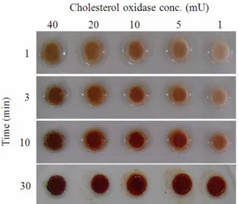
To estimate the eligibility of the paper disc colorimetric method, the correlation between the concentration of CHO and the development of color was evaluated. CHO (Sigma, St. Louis, MO) was diluted to 1, 5, 10, 20, and 40 milliunits per paper disc with the buffer (0.1 M sodium phosphate buffer, pH 7.0) and dropped on to the paper disc. After the addition of the reaction cocktail, the color of the paper discs intensified as the incubation time increased from 1 to 10 min (Fig. 3). However, after 30 min, it was difficult to differentiate the color intensity developed by the CHO activity. This result indicates that optimum incubation time for easy comparison of the color intensity is between 1-10 min.
This method was then tested for the measurement of the bacterial CHO activity on the agar plate (Fig. 4). The reaction cocktail in the paper disc on each colony began to develop a reddish brown color as the incubation continued (Fig. 4M). As a negative control, paper discs without bacterial colonies did not show any color development (Fig. 4C).
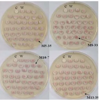
To evaluate the applicability of this method for the screening of colonies with high CHO productivity, we applied the paper disc colorimetric method on the master plate. The results obtained through this method are presented in Fig. 5. The paper disc with the reaction
Fig. 1. (A): Enzymatic reaction catalyzed by CHO with cholesterol. (B): The principle of enzymatic assay of the CHO.
Fig. 2. Survival rate of N. simplex upon exposure to EMS at various concentrations. Symbols: ▲, 0.08%
EMS; △, 0.16% EMS; ■, 0.32% EMS; □, 0.64% EMS.
Values indicate means±S.E. (n=3).
Fig. 3. Different color intensities depending on the amount of CHO and the reaction time.
340 Young-Sun Jang and Jong-Moon Jeong
cocktail only, used as a negative control, did not develop any color (Fig. 5C). Some mutant colonies revealed distinct coloration intensities. The color intensities of some of the paper discs (indicated with arrows in Fig. 5) were stronger than those of the wild type colony (Fig. 5W). We expected that these selected colonies would produce higher CHO levels than the wild type. To confirm our expectation, the selected mutant colonies, referred to as M5-35, M8-33, M10-7, and M13-39, were measured for CHO activity using the conventional quantitative analysis (Fig. 6). The actual activities of the CHOs from the selected colonies were higher than that of the wild type. In the case of M5-35, the color intensity of
the paper disc was the highest among all of the colonies, and showed the highest CHO activity at the same time, indicating that the expression levels of these mutant colonies are increased by the EMS gene mutation.
Conventional cholesterol oxidase activity assay methods include one developed by Masurekar and Goodhue [1978] and the other described by Allain et al. [1974].
Each method uses a different color reagent for the detection of CHO activity. The color reagent described by Masurekar and Goodhue [1978] was prepared by dissolving 0.01% (w/v) ODA in 50 mM potassium phosphate buffer (pH 7.5). In the case of Allain et al., the color reagent was
prepared by mixing 0.032% (w/v) 4-AAP and 0.2% (v/v) phenol in 0.1 M potassium phosphate buffer (pH 7.0). The amount of hydrogen peroxide produced by CHO during the cholesterol oxidation was measured indirectly as the amount of quinoneimine formed by the oxidation of ODA or 4-AAP in the presence of hydrogen peroxide and peroxidase (Fig. 1B). For these methods, each colony must be inoculated into the liquid broth and incubated for more than 5 days. After the colonies produce sufficient levels of CHO in the broth, the supernatant is separated from the broth by centrifugation at 10,770×g for 2 min.
This supernatant is then measured by mixing with chemicals, followed by incubation for 100 min at 37oC.
Therefore, application of these methods is difficult for a large scale screening due to the multiple procedures and considerable time required for each sample.
As a simple screening method for obtaining a high-activity strain, Xiang and Sampson [2004] used cholesterol agar plates. This method utilized the different optical properties of cholesterol/Triton X-100 micelles versus 4-cholestene-3-one/Triton X-100 micelles. Because the
Fig. 4. Color development after addition of colony into reaction cocktail. (C) incubated with reaction cocktail only. (M) incubated with reaction cocktail plus bacterial colony.
Fig. 5. Screening of high CHO-producing strain by paper disc colorimetric method. (c) only reaction cocktail in paper disc without bacterial colony, (w) wild type N. simplex as a control. The arrows indicate high CHO-producing colony types when compared with the wild type.
Fig. 6. Determination of the CHO activity of selected mutant strains. The cells were grown in 10 mL YM broth at 30oC for 5 days. One milliliter of culture broth was
centrifuged, and its supernatant was assayed for the CHO activity. (w) wild type N. simplex as a control. Values indicate means±S.E. (n=3).
Screening method of N. simplex with high CHO 341
agar plates containing cholesterol were opaque, bacterial colonies producing catalytically active CHO developed halos surrounding the colonies as a result of the oxidation of the cholesterol in the agar. However, in the present study, when this method was used for colony screening, discerning each colony-surrounding halo was difficult, probably due to the insufficient amount of the CHO secreted from each colony to create clear halos on the cholesterol-containing opaque agar plates.
In contrast, Nishiya et al. [1997] used indicator agar
plates to screen for CHO in the bacterial cells. The indicator agar plates were prepared by adding 0.1% (w/v) cholesterol, 0.1% (w/v) Triton X-100, 0.01% (w/v) ODA, and 25 units of peroxidase to the agar medium. Results could be obtained after incubation of the indicator agar plates for 15 to 24 h. However, the ODA sometimes oxidized spontaneously to form quinoneimine during incubation, and at the same time changes the agar medium color to dark brown; it was thus difficult to determine whether the color was developed through spontaneous oxidation or due to the presence of CHO. Based on these reasons, we determined that a simple, easy, and rapid method for screening a large number of colonies on agar plates is needed.
In conclusion, the present report describes a paper disc colorimetric method for the measurement of CHO activity. This method is applicable to the screening of mutant colonies with high CHO productivity on agar plates. The major advantages of this method include its reliability and simple, rapid screening.
Using this screening method, several high CHO-producing mutant strains were obtained from a random EMS mutagenesis, and quantitative assays were then performed for the actual measurement of their activities. These results revealed that CHO activity of the selected mutants was higher than that of the wild type; thus, the paper disc colorimetric method for the screening of CHO can be useful for a simple, rapid screening of mutant colonies on agar plates.
References
Allain CC, Poon LS, Chan CSG, Richmond W, and Fu PC (1974) Enzymatic determination of total serum choles-terol. Clin Chem20, 470-475.
Arlma K, Nagasawa M, Bae M, and Tamura G (1969) Microbial transformation of steroids. Part I: Decomposi-tion of cholesterol by microorganisms. Agric Biol Chem
33, 1636-1643.
Cheethan PSJ, Dunnill P, and Lilly MD (1982) The charac-terization and interconversion of three forms of choles-terol oxidase extracted from Nocardia rhodochrous.
Biochem J201, 515-521.
Doukyu N and Aono R (1998) Purification of extracellular cholesterol oxidase with high activity in the presence of organic solvents from Pseudomonas sp. strain ST-200.
Appl Environ Microbiol64, 1929-1932.
Doukyu N and Aono R (2001) Cloning, sequence analysis and expression of a gene encoding an organic solvent-and detergent-tolerant cholesterol oxidase of Burkhold-era cepacia strain ST-200. Appl Microbiol Biotechnol
57, 146-152.
Fukuyama M and Miyake Y (1979) Purification and some properties of cholesterol oxidase from Schizophyllum commune with covalently bound flavin. J Biochem 85,
1183-1193.
Inouye Y, Taguchi K, Fuhii A, Ishimaru K, Nakamura S, and Nomi R (1982) Purification and characterization of extracellular 3â-hydroxysteroid oxidase produced by
Streptoverticillium cholesterolicum. Chem Pharm Bull
30, 951-958.
Johnson TL and Somkuti GA (1991) Isolation of choles-terol oxidase from Rhodococcus equi ATCC 33706. Bio-technol Appl Biochem13, 196-204.
Lee SY, Rhee HI, Tae WC, Shin JC, and Park BK (1989) Purification and characterization of cholesterol oxidase from Pseudomonas sp. and taxonomic study of the strain. Appl Microbiol Biotechnol31, 542-546.
Liu WH, Meng MH, and Chen KS (1988) Purification and some properties of cholesterol oxidases produced by an inducible and a constitutive mutant of Arthrobacter sim-plex. Agric Biol Chem52, 413-418.
Masurekar PS and Goodhue CT (1978) United States Patent No. 4,093,517. Eastman Kodak Company. Rochester. NY
Murooka Y and Yamashita M (2001) Genetic and protein engineering of diagnostic enzymes, cholesterol oxidase and xylitol oxidase. J Biosci Bioeng91, 433-441.
Nishiya Y, Harada N, Teshima SI, Yamashita M, Fujii I, Hirayama N, and Murooka Y (1997) Improvement of thermal stability of Streptomyces cholesterol oxidase by random mutagenesis and a structural interpretation. Pro-tein Eng10, 231-235.
Shirokane Y, Nakamura K, and Mizusawa K (1977) Purifi-cation and some properties of an extracellular 3β
-hydroxysteroid oxidase produced by Corynebacterium cholesterolicum. J Ferment Technol55, 337-346.
H, Kag H, Kagawa M, and Nakamura S (1976) Some enzymatic properties of 3β-hydroxysteroid oxidase
pro-duced by Streptomyces violascens. J Biochem 79,
903-915.
Uwajima T, Yagi H, and Terada O (1974) Properties of crystalline 3β-hydroxysteroid oxidase of Brevibacterium
sterolicum. Agric Biol Chem38, 1149-1156.
Xiang J and Sampson NS (2004) Library screening studies to investigate substrate specificity in the reaction cata-lyzed by cholesterol oxidase. Protein Eng Des Sel 17,