Alteration of MicroRNAs Targeted Integrins by PD-MSCs Transplantation Is Involved in Hepatic Regeneration in a Rat Model with BDL
Sohae Park*
Department of Biomedical Science, CHA University, Seongnam 13488, Korea Received May 13, 2021 /Revised June 18, 2021 /Accepted June 29, 2021
Placenta-derived mesenchymal stem cells (PD-MSCs) are promising candidates for cell-based therapy in regenerative medicine. The migration and homing potential of PD-MSCs to injured sites is a critical property of MSC engraftment. MicroRNAs (miRNAs) have recently been shown to regulate the critical functions of MSCs, such as proliferation, survival, and migration. The objective of the present study was to identify the miRNA and target genes involved in PD-MSCs homing in a bile duct ligation (BDL) rat model. We selected candidate miRNAs targeting genes for PD-MSCs homing based on mi- croarray analysis. PD-MSC engraftment in BDL-injured rat liver was identified by immunofluo- rescence assay and human-specific Alu gene expression by quantitative real-time polymerase chain re- action (qRT-PCR) one week after transplantation. Compared with migrated naïve PD-MSCs under hy- poxic and normoxic conditions (Hyp/Nor), the transplanted group with PD-MSCs (Tx) showed dis- tinct differences in miRNA expressions in BDL-injured rat liver. We also validated the miRNAs and their target genes for PD-MSCs homing. The expressions of integrin α4 (ITGA4) and integrin α5 (ITGA5) target genes for miR-199a-5p and miR-148a-3p were significantly upregulated in the Tx group (p<0.05).
In addition, integrin β1 (ITGB1) and integrin β8 (ITGB8) were upregulated by suppressing miR-183-5p and miR-145-5p, respectively. These results demonstrated that PD-MSCs regulate miRNA expression related to the integrin family for their homing effects on the BDL-injured rat liver. The findings fur- ther suggest that miRNA-mediated regulation of the integrin family contributes to the therapeutic effi- cacy of PD-MSCs in the rat hepatic fibrosis model by BDL.
Key words : Integrins, liver failure, microRNA, Placenta-derived mesenchymal stem cells (PD-MSCs), stem cell migration
*Corresponding author
*Tel : +82-31-881-7145, Fax : +82-31-881-7249
*E-mail : [email protected]
This is an Open-Access article distributed under the terms of the Creative Commons Attribution Non-Commercial License (http://creativecommons.org/licenses/by-nc/3.0) which permits unrestricted non-commercial use, distribution, and reproduction in any medium, provided the original work is properly cited.
Introduction
Hepatic failure induced by chronic liver injury is a com- mon response to liver diseases that are difficult to cure with significant morbidity and mortality worldwide [30]. Despite the powerful regenerative ability of the liver, repetitive and continuous hepatic damages lead to progressive liver fib- rosis and ultimately end-stage liver disease [40]. Although a number of specific therapies for liver fibrosis have been developed, liver transplantation is the most effective treat- ment for patients with chronic liver disease. However, this treatment has many limitations such as organ shortages, im- munological rejection and high medical costs [4, 18].
Recently, multipotent mesenchymal stem cells (MSCs)
have been increasingly applied in clinical trials for the treat- ment of various diseases, especially liver disorders based on their unique therapeutic properties in preclinical and clin- ical studies [www.clinicaltrials.gov, 7, 35]. MSCs are able to self-renewal and differentiation into multiple cell lineages, including hepatocytes, which play key roles in tissue healing and regenerative medicine [1, 27]. MSCs are found in almost tissues, including bone marrow, adipose tissue, placenta and cord blood as well as other tissues [3]. Among them, pla- centa-derived MSCs (PD-MSCs) obtained from fetal tissue origin have advantages for strong immunomodulatory ca- pacity, anti-inflammatory effects and easily accessible to ob- tain abundant cells in vitro with free from ethical concern [20]. These properties are considered to maximize the ther- apeutic efficacy of PD-MSCs-based therapy in regenerative medicine. Furthermore, PD-MSCs can migrate toward in- jured areas and secrete various molecules to create a micro- environment in response to cellular damage signals which induce homing of MSCs [31]. The homing capacity of MSCs is also affected by the dynamic expression of cell-adhesion molecules including integrins during their engraftment.
Previous studies reported that PD-MSCs enhanced endothe- lial cells migration in a hypoxic condition via integrin alpha 4 (ITGA4) and Ras homolog (RHO) signaling, and integrin- dependent signaling improved MSC migration into injured target tissues [8].
MicroRNAs (miRNA) are small, non-coding RNAs (~22 nucleotides) that can regulate protein-coding genes by bind- ing to their target mRNAs through various post-transcrip- tional mechanisms [25]. MiRNA have recently been shown to play critical functions in MSCs to regulate the cellular properties, proliferation, migration and paracrine activity [24]. MiRNAs are also involved in regulating multiple fac- tors for liver regeneration [6, 19]. Recent reports suggest that miRNAs can control the expression levels of many im- portant adhesion molecules [33]. The integrin family of cell matrix adhesion receptors is a key regulator of cell‐ex- tracellular matrix interactions and certain miRNA involved in regulating the specific integrin. It has been reported that miR-31 and miR-92a suppressed the expression of mRNA encoding integrin α5, and miR-30 and let-7a regulated in- tegrin β1 expression [2, 5, 32]. Furthermore, upregulation of miR-10b promoted the migration of bone marrow MSCs in vitro by targeting E-cadherin [39]. However, the profiles and functions of miRNAs induced by PD-MSCs trans- plantation on the MSC homing process for liver regeneration in the BDL-injured rat model remains poorly understood.
Therefore, the objective of this study was to identify the ex- pression of miRNAs and their target genes associated with cell-adhesion molecules for PD-MSCs homing and to con- firm the correlation between miRNAs and their predicted target genes in a BDL-rat model transplanted with PD-MSCs.
Materials and Methods
Cell culture
The collection of placental samples and their use for re- search purposes were approved by the Institutional Review Board (IRB) of CHA Gangnam Medical Center, Seoul, Korea (IRB07-18). PD-MSCs were harvested as described previously [22] and cultured in α-modified minimal essential medium (HyClone, Logan, UT, USA) supplemented with 10% fetal bovine serum (Gibco, Grand Island, NY, USA), 1% pen- icillin/streptomycin (Gibco), 1 μg/ml heparin (Sigma-Al- drich, St. Louis, MO, USA) and 25 ng/ml human fibroblast growth factor-4 (PeproTech, Rocky Hill, NJ, USA). Cells were maintained below 5% CO2 at 37℃. To induce hypoxia
condition, the cells were placed in a hypoxia chamber (C- chamber, BioSpherix, Ltd., Lacona, NY, USA) and main- tained at 1% O2 and 37℃ in a 5% CO2 humidified condition.
Animal models and transplantation of PD-MSCs Seven-week-old male Sprague-Dawley rats (Orient Bio Inc., Seongnam, Korea) were housed under specific patho- gen-free conditions and generated chronic liver cirrhosis us- ing common BDL as previously described [19]. After one week post-surgery, PD-MSCs (2×10⁶ cells, 9-10 passages) were stained with the PKH67 Fluorescent Cell Linker Kit (Sigma-Aldrich) and transplanted through the tail vein in the transplanted group (Tx ; n=20) . Non-transplanted BDL group (NTx; n=20) were maintained as the normal group (Nor; n=5). The liver tissue samples were harvested at 1, 2, 3, and 5 weeks from rats in all groups. All animal experi- mental protocols were approved by the Institutional Animal Care and Use Committee of CHA University, Bundang, Korea (IACUC-190048)
Quantitative real-time polymerase chain reaction (qRT-PCR) analysis
Total RNA was extracted from PD-MSCs and rat liver tis- sues using TRIzol (Invitrogen, Carlsbad, CA, USA). Comple- mentary DNA (cDNA) was synthesized from 500 ng total RNA using SuperScript III reverse transcriptase (Invitrogen), according to the manufacturer’s instructions. cDNA syn- thesis for miRNAs was performed using miR-X miRNA First-Strand Synthesis kit (Takara Bio, Kusatsu, Shiga, Japan).
qRT-PCR was performed using SYBR Master Mix (Roche, Basel, Switzerland) in a CFX Connect™ Real-Time System (Bio-Rad,Hercules, CA, USA). Target gene and miRNA ex- pression were normalized to glyceraldehyde 3-phosphate dehydrogenase (GAPDH) and U6 small RNA, respectively.
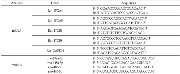
The sequences of the primers for target genes and miRNAs are shown in Table 1. All reactions were performed at least in triplicate.
Transwell migration assay
The migration of PD-MSCs was performed using a Trans- well assay under normoxic and hypoxic (1% O2) conditions.
PD-MSCs (2×10⁴ cells/well) were added into Transwell membrane inserts (8 μm pore size; Corning, NY, USA) and incubate for 24 hr at 37℃. The migrated cells from each con- dition were analyzed for miRNA expression.
Table 1. Primer sequences used in the present study for qRT-PCR analysis
Analysis Genes Sequences
mRNA
Rat ITGA4 F: 5'-GGAAGCCCCAGTGGAGAAC-3'
R: 5'-ATTGTCACTCCCAGCCACTGA-3'
Rat ITGA5 F: 5'-AGCCCCAGGGACTTACAACT-3'
R: 5'-CTTCATAGGGCCCATCTTCA-3'
Rat ITGB1 F: 5'-AACAGTGAAGACATGGATGC-3'
R: 5'-CTCTCTCTTCCTGCACACAC-3'
Rat ITGB8 F: 5'-AGTGCCCTCCAAGCTTAACCAC-3'
R: 5'-CGTCCCACCTCTCTCTCGAA-3'
Rat GAPDH F: 5'-TCCCTCAAGATTGTCAGCAA-3'
R: 5'-AGATCCACAACGGATACATT-3'
miRNA
rno-199a-5p rno-148a-3p rno-183-5p rno-145-5p
F: 5'-CCCAGUGUUCAGACUACCUGUUC-3' F: 5'-UCAGUGCACUACAGAACUUUG-3' F: 5'-UAUGGCACUGGUAGAAUUCACU-3' F: 5'-GUCCAGUUUUCCCAGGAAUCCCU-3'
Analysis of microRNA profiling
To analyze microRNAs profiling, miRNAs sequencing ex- periments was performed using the Illumina platform (Illu- mina Inc., San Diego, CA, USA) according to the manu- facturer’s instructions. The construction of small RNA li- braries and sequences were performed by LAS Inc. (Gimpo, Republic of Korea). The miRNA expression levels were esti- mated by transcripts per 10 million reads (TPTM) according to normalizing the miRNA counts with the total number of clean reads in the small RNA libraries.
Statistical analysis
The experimental data are expressed as the means± stand- ard deviation. Statistical analysis was performed using Student’s t-test, and p-value < 0.05 was considered statisti- cally significant. Each experiment was performed at least in triplicate.
Results
MicroRNA profiling of migrated PD-MSCs under hy- poxic condition and in BDL-injured rat liver
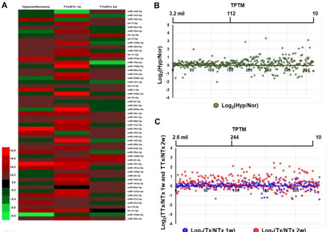
To determine miRNA-mediated gene expression related to the PD-MSC migration and homing toward injured sites, we analyzed miRNA profiles in BDL-induced cirrhotic liver tissues at 1 week and 2 weeks after PD-MSCs transplantation and migrated PD-MSCs under hypoxic conditions using miRNA microarray (Fig. 1A). In the heatmap analysis, 54 miRNAs showing differential expression between groups were selected. In particular, the expression of miRNA in the
transplanted /non-transplanted group at 1 week (Tx/NTx 1w) was significantly different compared to that in the trans- planted /non-transplanted group at 2 weeks (Tx/NTx 2w).
Comparative expression analysis of scatter plot between hy- poxic and normoxic conditions revealed significant change in miRNA expression. Moreover, in BDL-rat liver, remark- ably different event of miRNA expression between trans- planted /non-transplanted group at 1 week (Tx/NTx 1w) and transplanted /non-transplanted group at 2 weeks (Tx/
NTx 2w) was identified (Fig. 1B, Fig. 1C). These data suggest that variable miRNA expression regulates PD-MSCs migra- tion under hypoxic condition and in the BDL-injured rat liv- er transplanted with PD-MSCs.
Engraftment of PD-MSCs in BDL-injured rat liver.
The homing of transplanted PD-MSCs to injured sites is a critical property of engraftment of MSCs [12]. After tail- vein transplantation of PD-MSCs into BDL-injured rat, PD-MSC engraftment in the rat liver was identified by im- munofluorescence assay using PKH67(+) signals, and ge- nomic human Alu expression by qRT-PCR. As shown in Fig.
2A, PKH67-positive green signals in the frozen liver sections of BDL-injured rat were found at 1 week and 2 weeks after transplantation (Fig. 2A). Also genomic human Alu ex- pression was increased at 1 week and gradually decreased after 2 weeks (Fig. 2B). These data suggest that PD-MSCs successfully migrated to the damaged liver tissue and en- grafted following their administration as a live condition for a short term, which in turn led to providing the signals through paracrine effects on damaged hepatocytes.
A B
C
Fig. 1. MiroRNA expression profiling of migrated PD-MSCs under hypoxic condition and in BDL-injured rat liver. (A) Heatmap showing the different expression of miRNA in migrated PD-MSCs under hypoxic versus normoxic conditions, and trans- planted (Tx) versus non-transplanted (NTx) groups at one week (Tx/NTx 1w) and two weeks (Tx/NTx 2w). (B) Scatterplot of miRNA in migrated PD-MSCs under hypoxic and normoxic conditions (C) Scatterplot of miRNA in Tx/NTx 1w compared to Tx/NTx 2w groups.
A B
Fig. 2. PD-MSC engraftment in BDL-injured rat liver. (A) Representiative images of PKH67 (green)-labeled PD-MSCs in BDL-injured rat liver by immunofluo- rescence microscopy at 1, 2 and 5 weeks after PD-MSCs injection. (B) Human-specific genomic Alu express- ion in BDL-injured rat liver by qRT- PCR * NTx vs. Tx groups.
Decreased miRNA-199a-5p and miRNA-148a-3p by PD-MSCs regulate the expression of targeting in- tegrin α4 and α5 in BDL-injured rat liver
One of the key functions of PD-MSCs for cell-based thera-
pies is the ability to home to the sites of damaged tissues.
Integrins are known to play an important role in cell migra- tion. Validated expression of miRNAs targeted to integrins revealed distinct differences in the expression of integrin.
A
B
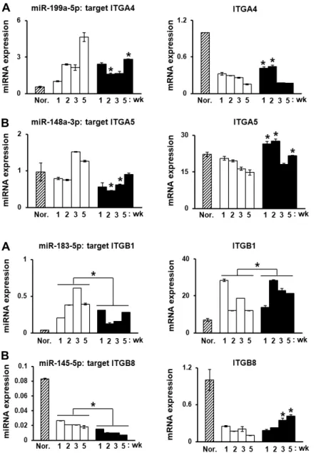
Fig. 3. MiRNAs induced by PD-MSCs regulate in- tegrin α subunits in BDL-injured rat liver.
(A) ITGA4-targeted rno-miR199a-5p ex- pression and (B) ITGA5-targeted rno-148a- 3p expression in BDL-injured rat liver at 1, 2, 3 and 5 weeks after PD-MSCs injection as determined by qRT-PCR. The experi- ments were performed at least in triplicate.
Data from each group are indicated as the mean ± SD, determined by Student’s t-test.
* p<0.05 versus NTx group.
A
B
Fig. 4. MiRNAs induced by PD-MSCs regulate in- tegrin β subunits in BDL-injured rat liver.
(A) ITGB1-targeted rno-miR183-5p express- ion and (B) ITGB8-targeted rno-miR-145-5p expression in BDL-injured rat liver at 1, 2, 3 and 5 weeks after PD-MSCs injection as determined by qRT-PCR. The experiments were performed at least in triplicate. Data from each group are indicated as the mean
± SD, determined by Student’s t-test.
* p<0.05 versus NTx group.
From miRNAs database, we analyzed that miRNA-199a-5p and miRNA-148a-3p are predicted to ITGA4 and ITGA5, respectively. To determine the correlation between miR- 199a-5p and ITGA4 in the BDL rat liver after PD-MSCs trans- plantation, qRT-PCR was applied to evaluate the expression levels of miR-199a-5p and ITGA4A. As shown in Fig. 1A, miR-199a-5p expression was upregulated in non-trans- planted (NTx) BLD rat liver compared with normal liver tis- sues, and the mRNA level of predicted target gene, ITGA4, was decreased. Whereas ITGA4 expression was significantly increased in the transplanted group (Tx) compared with the NTx group at 1 week and 2 weeks (Fig. 3A, *p<0.05).
Generally, it is well known that ITGA5 expression is one of major factors on adhesion molecules involved in migra- tion of cell [17]. Moreover, the ITGA5 expression of the Tx
group was remarkably upregulated compared with the NTx group by suppressing rno-miR-148a-3p (Fig. 4B, *p<0.05).
Especially, their expression levels were higher (max. 50- folds) than those of ITGA4. These results suggest that de- creased miR- 199a-5p and miR-148a-3p regulates the integrin family for homing and engraftment of PD-MSCs in a BDL-injured rat model as well as ITGA4 and ITGA5 might be important factors in PD-MSC homing in damaged liver tissues.
Down-regulated miRNA-183-5p and miRNA-145- 5p by PD-MSCs regulate the expression of targeting integrin β1 and β8 in BDL-injured rat liver
Basically, integrin consists of two components such as in- tegrin alpha and integrin beta subunits among several in-
tegrin subunits [16]. The major integrin-to-integrin complex is ITGA5 and ITGB1 in cellular events including migration and homing [34]. To validate the expression of the miRNAs, miR-183-5p and miR-145-5p, and the predicted targets of each miRNA in the BDL rat liver after PD-MSCs trans- plantation, the expression levels of these miRNAs and mRNAs were examined using qRT-PCR analysis. As shown in Fig. 4A, the expression of miR-183-5p was significantly downregulated and the target gene ITGB1 expression was correlatively increased in the Tx group compared to those in the NTx group (Fig. 4A, *p<0.05). Additionally, the ex- pression of miR-145-5p and target gene ITGB8 revealed sig- nificant correlation in the Tx group compared to the NTx group. Comparative expression levels of target genes, ITGB1 and ITGB8, between 1 week and 5 weeks revealed significant increases by suppressing miRNAs, miR-183-5p and 145-5p, respectively. Especially, miRNA-183-5p and their target IGTB1 levels were higher than those of miRNA-145-5p and ITGB8. These results suggest that alterative miRNA ex- pression regulates the integrin family for homing and en- graftment of PD-MSCs in a BDL-injured rat model.
Discussion
Placenta derived-MSCs (PD-MSCs) are promising candi- dates for stem cell therapy in the treatment of various degen- erative diseases. The efficacy of PD-MSCs in cell-based thera- pies depends on their homing ability and engraftment into the injury site [14]. Recent studies have shown that modu- lation of miRNA expression by MSCs has been shown to change their biological properties that drive cellular adhe- sion between MSCs and injured cells [9, 10]. Moreover, the potential of PD-MSCs engraftment has also been associated with regulation of miRNAs expression in a liver-injured rat model [19]. In the present study, different profiles of miRNA were confirmed in migrated PD-MSCs under hypoxic con- ditions and in the PD-MSCs transplanted BDL-induced cir- rhotic liver tissues at 1 week and 2 weeks. In addition, the relevance of miRNAs in the PD-MSCs transplanted BDL-in- duced cirrhotic liver tissues (Tx group) was identified in the experiments showing that different expression profiles re- lated to the integrin targeting miRNAs (e.g., miR-199a-5p, miR-148a-3p, miR-183-5p, and miR-145-5p).
Several cytokines secreted from injured tissues have been demonstrated to stimulate the migration of MSCs by inter- acting with their receptors and activating migration-related
signaling cascades. MiRNA also found to affect cell migra- tion at downstream of cytokines and receptors [15]. Especially, miR-27b and miR-27a have been reported to affect the migra- tion of MSCs through targeting stromal cell-derived factor (SDF)-1 and its chemokine receptor type 4(CXCR4) [26].
Also, phosphatase of regulating liver-1(PRL-1)-overexpressed PD-MSCs enhanced miRNA-mediated MSC migration through integrin-dependent signaling with improvement of liver re- generation [19]. Based on these results, PD-MSCs may have multiple effects on their migration and tissue regeneration through altered expression of cell adhesion molecules affect- ing by miRNAs in the injured tissues.
Among the differently expressed miRNAs in the BDL-in- duced liver tissues (NTx), miR-199a-5p and miR-148a-3p were found to be capable of regulating the expression levels of integrin α4 (ITGA4) and integrin α5 (ITGA5), respectively.
Downregulation of miR-199a-5p increased the expression of ITGA4 in the Tx group compared to those in the NTx group.
MiR-199a-5p has been demonstrated to participate in the regulation of multiple biological processes, including cell proliferation, migration and invasion [36, 38]. It was shown that miR-199a-5p protected hepatocyte damage induced by bile acid [11] and functioned as a tumor suppressor by downregulating ITGA3 [29]. Also, ITGA4 involved in in- crease MSC homing to the bone marrow [21]. Comparison with these data, our results indicate that PD-MSCs play an important role in promoting their migration by regulating miR-199a-5p expression for ITGA4 in BDL-induced rat liver tissues. The expression levels of integrin α5 (ITGA5) were also significantly increased by downregulation of miR-148a- 3p in the Tx group. Especially, their expression levels were higher than those of ITGA4. ITGA5 was found to be regu- lated by several miRNAs (e.g., miR-31, miR-17-92 cluster, and miR-148b) [5]. Wu et al [37] reported that overex- pression of miR-143a-3p reduced laryngeal squamous cell carcinoma cell migration and proliferation.
Integrins are αβ heterodimeric cell-surface receptors that facilitate cell adhesion and migration [16]. Among the in- tegrin superfamily, α5β1 is one of the best characterized in- tegrins for cell migration [34]. Like most integrins, integrin β1 (ITGB1) is also regulated by multiple miRNAs. The miRNA reported to directly regulate ITGB1 expression in- clude miR-29b and miR-124a [13, 28]. Chen et al [5] reported that ITGB1 is a critical factor for cell adhesion and invasive- ness affected by miR-183, which led to negative regulation on the invasive activity of endometrial stromal cells. It has
been reported that integrin α5β1 mediates MSC migration at sites of vascular remodeling by activation of focal adhe- sion kinase and platelet-derived growth factor receptor-β phosphorylation [34]. Our results showed that PD-MSCs in- duced significantly downregulated miR-183-5p expression resulting in upregulation of ITGB1 target gene expression in the BDL-injured rat liver. These data suggest that PD- MSCs in vivo contribute to the enhanced cell migration regu- lated by miR-183-5p and target gene ITGB1 expression. In addition, the expression of miR-145-5p by PD-MSCs was markedly downregulated during the experimental period, and its target gene, ITGB8, was gradually increased in the BDL-injured rat liver. It has been reported that miR-145 was identified as a tumor suppressor in a variety of cancers, and downregulated the expression of ITGB8 in human corneal epithelial cells [23].
The therapeutic effect of MSCs depends on their ability to home the injured site, which is affected by several factors derived from MSCs as their paracrine actions. Many studies have demonstrated that miRNAs may be involved in the paracrine action of MSCs [14, 15]. We confirmed that PD- MSCs successfully migrated to the damaged rat liver tissue and engrafted for 2 weeks after intravenous injection. During the 5-week experimental periods, PD-MSCs-transplantation in a liver failure rat model induced alterative expression of miRNA targeting integrin genes, which lead to better migra- tion and engraftment of PD-MSCs into injured liver tissues, and enhancement of liver regeneration. Among the various mechanisms regulating integrin expression, miRNAs regu- lated by PD-MSCs have an important target in liver regen- eration. Further study is ongoing to miRNA target binding validation by luciferase reporter assay and requires the eluci- dation of detailed mechanism..
In conclusion, the present study demonstrates that PD- MSCs regulate alterative miRNA expression related to in- tegrin family for their homing effects on BDL-injured rat liver. These findings provide a fundamental mechanism for miRNA-mediated effects of PD-MSCs transplantation in hepatic regeneration and support the development of stem cell-based therapy.
The Conflict of Interest Statement
The authors declare that they have no conflicts of interest with the contents of this article.
References
1. Ballini, A., Cantore, S., Scacco, S., Coletti, D. and Tatullo, M. 2018. Mesenchymal stem cells as promoters, enhancers, and playmakers of the translational regenerative medicine.
Stem Cells Int. 18, 6927401.
2. Bonauer, A., Carmona, G., Iwasaki, M., Mione, M., Koyanagi, M., Fischer, A., Burchfield, J., Fox, H., Doebele, C. and Ohtani, K. 2009. MicroRNA-92a controls angiogenesis and functional recovery of ischemic tissues in mice. Science 324, 1710-1713.
3. Campagnoli, C., Roberts, I. A. G., Kumar, S., Bennett, P.
R., Bellantuono, I. and Fisk, N. M. 2001. Identification of mesenchymal stem/progenitor cells in human first-trimes- ter fetal blood, liver, and bone marrow. Blood 98, 2396-2402.
4. Cao, Y., Ji, C. and Lu, L. 2020. Mesenchymal stem cell ther- apy for liver fibrosis/cirrhosis. Ann. Transl. Med. 8, 562.
5. Chen, W., Harbeck, M. C., Zhang, W. and Jacobson, J. R.
2013. MicroRNA regulation of integrins. Transl. Res. 162, 133-143.
6. Chen, X., Zhao, Y., Wang, F., Bei, Y., Xiao, J. and Yang, C. 2015. MicroRNAs in liver regeneration. Cell Physiol.
Biochem. 37, 615-628.
7. Chen, Y., Shao, J. Z., Xiang, L. X., Dong, X. J. and Zhang, G. R. 2008. Mesenchymal stem cells: A promising candidate in regenerative medicine. Int. J. Biochem. Cell Biol. 40, 815-820.
8. Choi, J. H., Lim, S. M., Yoo, Y. I., Jung, J., Park, J. W. and Kim, G. J. 2016. Microenvironmental interaction between hypoxia and endothelial cells controls the migration ability of placenta-derived mesenchymal stem cells via alpha4 in- tegrin and Rho signaling. J. Cell Biochem. 117, 1145-1157.
9. Clark, E. A., Kalomoiris, S., Nolta, J. A. and Fierro, F. A.
2014. Concise review: MicroRNA function in multipotent mesenchymal stromal cells. Stem Cells 32, 1074-1082.
10. Collinl, F., Bruno, S., Lindoso, R. S. and Camussi, G. 2014.
miRNA expression in mesenchymal stem cells. Curr. Patho- biol. Rep. 2, 101-107.
11. Dai, B. H., Geng, L., Wang, Y., Sui, C. J., Xie, F., Shen, R.
X., Shen, W. F. and Yang, J. M. 2013. microRNA-199a-5p protects hepatocytes from bile acid-induced sustained endo- plasmic reticulum stress. Cell Death Dis. 4, e604.
12. De Becker, A. and Riet, I. V. 2016. Homing and migration of mesenchymal stromal cells: How to improve the efficacy of cell therapy? World J. Stem Cells 8, 73-87.
13. Fowler, A., Thomson, D., Giles, K., Maleki, S., Mreich, E., Wheeler, H., Leedman, P., Biggs, M., Cook, R., Little, N., Robinson, B. and McDonald, K. 2011. miR-124a is frequently down-regulated in glioblastoma and is involved in migra- tion and invasion. Eur. J. Cancer 47, 953-963.
14. Fu, X., Liu, G., Halim, A., Ju, Y., Luo, Q. and Song, A. G.
2019. Mesenchymal stem cell migration and tissue repair.
Cells 8, 784.
15. He, L. and Zhang, H. 2019. MicroRNAs in the migration of mesenchymal stem cells. Stem Cell Rev. Rep. 1, 3-12.
16. Hynes, R. O. 2002. Integrins: bidirectional, allosteric signal- ing machines. Cell 110, 673-687.
17. Kawamura, M., Yamamoto, T., Yamashiro, K., Kochi, S., Yoshihara-Hirata, C., Ideguchi, H., Aoyagi, H., Omori, K.
and Takashiba, S. 2019. Induction of migration of perio- dontal ligament cells by selective regulation of integrin subunits. J. Cell Mol. Med. 23, 1211-1223.
18. Kim, G., Eom, Y. W., Baik, S. K., Shin, Y., Lim, Y. L., Kim, M. Y., Kwon, S. O. and Chang, S. J. 2015. Therapeutic effects of mesenchymal stem cells for patients with chronic liver diseases: Systematic review and meta-analysis. J. Kor. Med.
Sci. 30, 1405-1415.
19. Kim, J. Y., Jun, J. H., Park, S. Y., Yang, S. W., Bae, S. H.
and Kim, G. J. 2019. Dynamic regulation of miRNA ex- pression by functionally enhanced placental mesenchymal stem cells promotes hepatic regeneration in a rat model with bile duct ligation. Int. J. Mol. Sci. 20, 5299.
20. Kim, M. J., Shin, K. S., Jeon, J. H., Lee, D. R., Shim, S. H., Kim, J. K., Cha, D. H., Yoon, T. K. and Kim, G. J. 2011.
Human chorionic-plate-derived mesenchymal stem cells and Wharton's jelly-derived mesenchymal stem cells: a com- parative analysis of their potential as placenta-derived stem cells. Cell Tissue Res. 346, 53-64.
21. Kumar, S. and Ponnazhagan, S. 2007. Bone homing of mes- enchymal stem cells by ectopic alpha 4 integrin expression.
FASEB J. 21, 3917-3927.
22. Lee, M. J., Jung, J., Na, K. H., Moon, J. S., Lee, H. J., Kim, J. H., Kim, G. I., Kwon, S. W., Hwang, S. G. and Kim, G.
J. 2010. Anti-fibrotic effect of chorionic plate-derived mesen- chymal stem cells isolated from human placenta in a rat model of CCl(4)-injured liver: potential application to the treatment of hepatic diseases. J. Cell Biochem. 111, 1453-1463.
23. Lee, S. K., Teng, Y., Wong, H. K., Ng, T. K., Huang, L., Lei, P., Choy, K. W., Liu, Y., Zhang, M., Lam, D. S., Yam, G. H. and Pang, C. P. 2011. MicroRNA-145 regulates human corneal epithelial differentiation. PLoS One 6, e21249.
24. Li, N., Long, B., Han, W., Yuan, S. and Wang, K. 2017.
microRNAs: Important regulators of stem cells. Stem Cell Res. Ther. 8, 110.
25. Li, Z. and Rana, T. M. Therapeutic targeting of microRNAs:
Current status and future challenges. Nat. Rev. Drug Discov.
13, 622-638.
26. Lü, M. H., Hu, C. J., Chen, L., Peng, X., Chen, J., Hu, J.
Y., Teng, M. and Liang, G. P. 2013. miR-27b represses migra- tion of mouse MSCs to burned margins and prolongs wound repair through silencing SDF-1a. PLoS One 8, e68972.
27. Pittenger, M. F., Discher, D. E., Péault, B. M., Phinney, D.
G., Hare, J. M. and Caplan, A. I. 2019. Mesenchymal stem cell perspective: cell biology to clinical progress. NPJ Regen.
Med. 4, 22.
28. Sekiya, Y., Ogawa, T., Yoshizato, K., Ikeda, K. and Kawada, N. 2011. Suppression of hepatic stellate cell activation by microRNA-29b. Biochem. Biophys. Res. Commun. 412, 74-79.
29. Tian, L., Chen, M., He, Q., Yan, Q. and Zhai, C. 2020. Micro RNA-199a-5p suppresses cell proliferation, migration and invasion by targeting ITGA3 in colorectal cancer. Mol. Med.
Rep. 3, 2307-2317.
30. Trefts, E., Gannon, M. and Wasserman, D. H. 2017. The liver. Curr. Biol. 27, R1147-R1151.
31. Ullah, M., Liu, D. D. and Thakor, A. S. 2019. Mesenchymal stromal cell Homing: mechanisms and strategies for im- provement. iScience 15, 421-438.
32. Valastyan, S., Reinhardt, F., Benaich, N., Calogrias, D., Szász, A. M., Wang, Z. C., Brock, J. E., Richardson, A. L. and Wein- berg, R. A. 2009. A pleiotropically acting microRNA, miR- 31, inhibits breast cancer metastasis. Cell 137, 1032-1046.
33. Valastyan, S. and Weinberg, R. A. 2011. Roles for microRNAs in the regulation of cell adhesion molecules. J. Cell Sci. 124, 999-1006.
34. Veevers-Lowe, J., Ball, S. G., Shuttleworth, A. and Kielty, C. M. 2011. Mesenchymal stem cell migration is regulated by fibronectin through α5β1-integrin-mediated activation of PDGFR-β and potentiation of growth factor signals. J. Cell Sci. 124, 1288-1300.
35. Volarevic, V., Nurkovic, J., Arsenijevic, N., Stojkovic, M. and Volarevic, V. 2014. Concise review: Therapeutic potential of mesenchymal stem cells for the treatment of acute liver failure and cirrhosis. Stem Cells 32, 2818-2823.
36. Wang, Q., Ye, B., Wang, P., Yao, F., Zhang, C. and Yu, G.
2019. Overview of microRNA-199a regulation in cancer.
Cancer Manag. Res. 11, 10327-10335.
37. Wu, T., Qu, L., He, G., Tian, L., Li, L., Zhou, H., Jin, Q., Ren, J., Wang, Y., Wang, J., Kan, X., Liu, M., Shen, J., Guo, M. and Sun, Y. 2016. Regulation of laryngeal squamous cell cancer progression by the lncRNA H19/miR-148a-3p/
DNMT1 axis. Oncotarget 7, 11553-11566.
38. Ye, H., Pang, L., Wu, Q., Zhu, Y., Guo, C., Deng, Y. and Zheng, X. 2015. A critical role of miR-199a in the cell bio- logical behaviors of colorectal cancer. Diagn. Cytopathol. 10, 65.
39. Zhang, F., Jing, S., Ren, T. and Lin, J. 2013. MicroRNA-10b promotes the migration of mouse bone marrow-derived mesenchymal stem cells and downregulates the expression of E- cadherin. Mol. Med. Rep. 8, 1084-1088.
40. Zhou, W. C., Zhang, Q. B., Qiao, L. and Zhou, W. C. 2014.
Pathogenesis of liver cirrhosis. World J. Gastroenterol. 20, 7312-7324.
초록:담관결찰 쥐 모델에서 태반유래중간엽줄기세포 이식에 의한 miRNA 표적 인테그린 변화의 간재생 효과
박소혜*
(차의과학대학교 생명과학과)
태반유래 중간엽줄기세포(PD-MSCs)는 재생의학에서 세포기반치료제로 잘 알려진 세포군이다. PD-MSCs의 손 상된 부위로의 이동과 호밍 기능은 MSC 생착의 중요한 특성이다. miRNA는 최근 MSC의 증식, 생존 이동과 같은 중요한 기능을 조절하는 것으로 알려져 있다. 본 연구의 목적은 담관결찰(BDL) 쥐 모델에서 PD-MSCs 호밍에 관 련된 miRNA 및 표적 유전자를 동정하는 것으로, 마이크로어레이 분석을 이용하여 PD-MSCs 호밍에 관여하는 유전자 표적 miRNA를 선별하였다. BDL 쥐모델에 PD-MSCs을 이식한 일주일 후 간 조직에서 PD-MSCs 생착여 부는 면역형광분석법과 qRT-PCR에 의한 인간 Alu유전자 발현으로 확인되었다. 저산소 및 정상조건(Hyp/Nor)에 서 이동한 PD-MSC에 비하여, PD-MSCs 이식한 BDL군 간 조직에서 miRNAs 발현의 차이가 크게 나타났으며, PD-MSCs 호밍 관련 miRNA와 표적유전자를 검증하였다. miR199a-5p 및 miR-148a-3p에 대한 표적 유전자 인테 그린 α4 (ITGA4)와 α5 (ITGA5)의 발현은 이식(Tx)그룹에서(p<0.05) 유의하게 상향 조절되었다. 또한 인테그린 β1 (ITGB1)과 β8 (ITGB8)의 발현은 miR-183-5p 및 miR-145-5p억제에 의하여 크게 증가되었다. 따라서 이러한 결과는 BDL에 의해 손상된 쥐간에서 PD-MSCs가 호밍효과을 위해 인테그린 그룹과 관련된 miRNA 발현 조절에 관여함 을 나타내었다. 본 연구결과는 miRNA에 의한 인테그린 그룹 조절기능이 BDL에 의해 유도된 간섬유증 쥐모델에 서 PD-MSCs의 치료효과에 기여할 수 있음을 시사한다.