Korean J Biol Psychiatry 2015;22(1):14-19
서 론
폐쇄성수면무호흡증(obstructive sleep apnea, 이하 OSA)은 수면호흡장애 질환 중 가장 흔한 질환으로, 전체 인구의 1~2%
에서 발생한다.1) 수면 중 반복적인 상기도의 부분 혹은 전체 폐쇄에 의해, 흡기의 노력에도 불구하고 공기의 흐름이 감소하 거나 차단되는 것으로 발현한다. OSA 환자에서는 수면 중 기 도가 좁아지거나 막히면서 무호흡과 저호흡이 발생하며, 수
폐쇄성수면무호흡 의심환자에서 무호흡 - 저호흡 지수와 연관이 있는
두개골 계측 변수 : 예비연구
가천대학교 의학전문대학원 길병원 영상의학교실,1 가천대학교 의학전문대학원 길병원 정신건강의학교실,2
가천대학교 의학전문대학원 길병원 이비인후과학교실,3 가천대학교 의학전문대학원 길병원 신경과학교실4
박수영
1·황희영
1·김응엽
1·강승걸
2·김선태
3·박기형
4Cephalometric Variables Significantly Associated with Apnea Hypopnea Index in Suspected Obstructive Sleep Apnea Patients : A Preliminary Study
Suyoung Park, MD,1 Hee Young Hwang, MD,1 Eung Yeop Kim, MD,1 Seung-Gul Kang, MD,2 Seon Tae Kim, MD,3 Kee Hyung Park, MD4
1Department of Radiology, Gil Medical Center, Gachon University School of Medicine, Incheon, Korea
2Department of Psychiatry, Gil Medical Center, Gachon University School of Medicine, Incheon, Korea
3Department of Otolaryngology, Gil Medical Center, Gachon University School of Medicine, Incheon, Korea
4Department of Neurology, Gil Medical Center, Gachon University School of Medicine, Incheon, Korea
ObjectivesZZThe purpose of this study is to find the cephalometric variables which are significantly correlated with the apnea-hypop- nea index (AHI) in suspected Korean obstructive sleep apnea (OSA) patients.
MethodsZZWe examined lateral cephalogram and attended-full night laboratory polysomnography of the 40 participants who com- plained of OSA symptoms. The correlation analysis was conducted to find the cephalometric variables which are significantly correlated with the AHI.
ResultsZZThe correlation analysis showed that the higher AHI was associated with the longer distance between hyoid and mandibular plane (p = 0.023), the longer distance between C3 and hyoid (p = 0.014), the longer tongue length (p = 0.003), the larger inferior tongue area (p = 0.008), the larger anterior displacement of the hyoid bone (p = 0.024), the longer distance between posterior nasal spine and the tip of the soft palate (p = 0.021), and the larger cross-sectional area of soft palate (p = 0.001) of cephalogram in erect position. The high- er AHI was correlated with the longer distance between hyoid and mandibular plane (p = 0.008), the longer tongue length (p = 0.037), the larger inferior tongue area (p = 0.013), the thicker uvula (p = 0.004), the longer distance between retrognathion and hyoid (p = 0.025), and larger cross-sectional area of soft palate (p = 0.001) of cephalogram in supine position.
ConclusionsZZThe present preliminary results showed the candidate measurements of cephalogram which are significantly corre- lated with the AHI in suspected OSA.
Key WordsZZCephalometry ㆍObstructive sleep apnea.
Received: November 13, 2014 / Revised: December 23, 2014 / Accepted: February 3, 2015 Address for correspondence: Hee Young Hwang, MD
Department of Radiology, Gil Medical Center, Gachon University School of Medicine, 21 Namdong-daero 774beon-gil, Namdong-gu, Incheon 405-760, Korea
Tel: +82-32-460-3060, Fax: +82-32-460-3065, E-mail: [email protected]
면 중 각성으로 인한 수면의 질 저하와 간헐적 저산소증으로 인해 신체적 건강의 위협이 초래된다.
수면다원검사는 OSA의 진단에 가장 객관적이고 정확한 검 사이나, 방법이 복잡하고 시간이 많이 소요되며 비용이 많이 드는 단점이 있다. 또한, 수면검사실에서 하룻밤 수면을 취해 야 한다는 불편 때문에 검사의 필요성을 느끼면서도 검사를 포기하는 피험자들이 많다. 한편, 임상가의 측면에서도 임상 증상만으로 OSA 환자인지를 확신하지 못하기 때문에 모든 의심환자들에게 어려운 진단과정인 수면다원검사를 무차별 적으로 권유하기가 부담스럽다.
한편, 서양의 OSA 환자와 비교해서 한국의 환자들은 뚱뚱 한 체형, 짧고 굵은 목둘레 등의 신체적 특징이 없는 경우에도 수면다원검사상 OSA로 나타나는 경우가 많은데, 이러한 원 인들은 한국인을 포함한 동양인들의 두개골 골격의 특징에 기인한다는 보고들이 많다.
두부 계측 일반촬영 사진은, 다른 검사와 달리 촬영이 간단 하고 설치비용과 촬영비용이 저렴하며, 경조직뿐 아니라 연조 직의 형태도 어느 정도 파악할 수 있기 때문에,2) OSA의 빠른 진단을 돕고 원인을 파악하기 위한 통상적인 검사로 추천되 고 있다.3)4) 이러한 점들을 고려하였을 때 두부 계측 일반촬영 에서 OSA 환자의 특징들을 발견할 수 있다면, OSA가 의심되 는 환자들에서 수면다원검사를 시행하기 이전에 미리 OSA의 위험도를 예측하여 보다 급하게 수면다원검사를 시행할 환자 들을 미리 선별할 수 있을 것이다.
그러나 두부 계측 일반촬영 사진을 바탕으로 한 지금까지의 보고는 서양인을 대상으로 한 분석이 대부분이다. 두부 일반촬 영 계측치들은 인종별 차이가 있을 수 있으므로,5) 한국인 폐쇄 성수면무호흡증 환자들에 대한 기준 자료로 받아들이기에는 적합하지 않다. 또한, 정립위 혹은 앉은 자세에서 촬영된 두부 계측 일반촬영을 토대로 이루어진 연구는 많지만, 환자의 수면 위치인 앙와위에서 얻어진 영상을 분석한 연구는 많지 않다.
따라서 이 예비연구에서는 OSA가 의심되는 한국인 환자를 대상으로, 정립위와 앙와위에서 얻어진 두부 계측 일반촬영 사진을 이용한 여러 계측 변수들과 수면다원검사 결과를 비 교하여, 어떤 계측 변수들이 폐쇄성수면무호흡증의 필수 진단 기준의 척도인 무호흡-저호흡 지수(apnea-hypopnea index, 이하 AHI)와 유의한 연관이 있는지 발견하고자 하였다.
방 법
연구 대상
가천대 길병원의 수면클리닉(정신건강의학과, 이비인후과, 신경과)에 내원한 환자들 중 임상적으로 OSA가 의심되는 18
세에서 65세 사이의 한국인 환자 40명(남 : 여 = 35 : 5)을 대 상으로 평가하였다.
임상적으로 OSA가 의심된다고 함은 습관적 코골이, 수면 중 숨이 막힌 경험, 함께 자는 사람에 의해 수면 중 무호흡이 관찰 된 적이 있는 경우, 과도한 주간졸림이 있는 경우로 정의하였 고 피험자의 등록은 수면의학분야에서 최소 5년 이상의 진료 경험을 갖춘 전문의들에 의해 이루어졌다.
피험자 등록의 제외기준은 다음과 같았다. 한국인 외의 인 종, 내외과적 심각한 질환을 가진 환자로 수면다원검사 등의 검사수행이 불가능한 환자, 과거 OSA의 진단을 받았던 사람, 구개수연구개인두성형술(uvulopalatopharyngoplasty) 등의 OSA에 대한 수술적 치료를 받았던 사람, 임상적으로 기면병 이나 하지불안증후군 등의 다른 주요 수면장애가 의심되는 환 자는 배제하였다. 모든 피험자들은 연구의 취지와 목적에 대 해서 충분한 설명을 듣고 서면동의서에 자필서명하였다. 연구 프로토콜과 연구의 모든 세부적 사항들은 가천대 길병원의 연 구심의위원회(Institutional Review Board)의 승인을 받았다.
이 40명의 환자에 대해 측면투영 두부 계측 일반촬영(lateral cephalogram)과, 야간 수면다원검사(attended full-night poly- somnography)를 시행하였다.
두부 계측 일반촬영
설부와 인두부의 연부조직의 윤곽선 확인을 위해, 황산바륨 양성 방사선 조영제(Solotop, 태준제약[주], 서울, 한국)를 경구 투여하여 설배부를 코팅하였다. 정립위 및 앙와위에서 연부조 직 밀도로 100 cm 거리에서 75 kVp로 측면투영 영상을 얻었 다. 촬영된 필름은 PiViewSTAR Picture Archiving and Com- munication System(INFINITT Healthcare)을 이용하여, 환 자에 대한 정보가 없는 숙달된 영상의학과 의사 1인이 판독 하였다. 정립위 및 앙와위 영상에서 각각 20가지씩의 두개골 과 연부조직의 계측 변수(craniofacial and soft tissue param- eters)를 측정하였다(Table 1, 2, Fig. 1). 두위 상태의 변화는 상기도 개방성에 영향을 미칠 수 있으므로, 두위의 굴곡 혹은 신전시의 변이를 최소화할 수 있는 상・하악 치아의 최대 교합 상태를 이루고, 입술을 가볍게 다문 자연스러운 상태에서, 폐 용량의 영향을 최소화하기 위해 호기말에 촬영하였다.
야간 수면다원검사
40명의 환자 모두는 하룻밤 동안 수면다원검사를 시행하였 다. 수면다원검사는 Grass-Telefactor사의 COMET(Warwick, RI, USA) 시스템을 사용하였다. 수면 중 뇌파, 안구운동, 턱과 하지에서의 근전도, 구강과 비강을 통한 공기의 출입, 복부와 흉부에서의 호흡운동, 심전도와 혈중 산소포화도를 측정하
였다. 수면다원검사에서 얻어진 무호흡-저호흡 지수를 두부 계측 일반촬영 소견과 비교 분석하였다. 검사의 판독과 전극 들의 부착은 미국수면학회(American Academy of Sleep Med- icine)에서 2007년에 정한 국제 판독기준과 검사규칙에 근거 하여 이루어졌고, 수면 중 저호흡(hypopnea)의 판정은 이 기 준의 권고규정(recommended rule)에 따라 이루어졌다.6) AHI 는 수면다원검사에서 측정된, 수면 한 시간당 발생하는 무호 흡과 저호흡 횟수의 합의 평균으로 정의하였다.
통 계
두개골 계측 변수와 무호흡-저호흡 지수와의 연관성을 Pear- son 상관분석을 사용하여 통계학적 검증을 시행하였다. 통계 분석은 SPSS for Windows(SPSS Inc., Chicago, IL, USA)를 사용하였고, 통계학적 유의수준은 p < 0.05로 하였다.
결 과
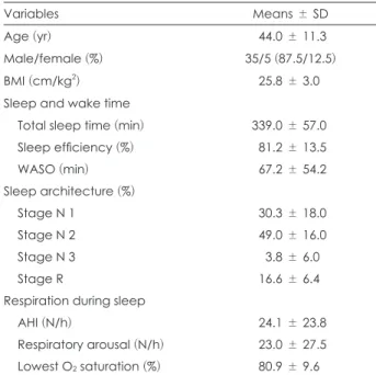
피험자들의 인구학적 특성과 수면다원검사의 결과는 Table
3에 기술하였다. 남자가 35명, 여자가 5명으로 평균연령은 44.0 ± 11.3세였다(Table 3). 피험자 중에 29명(72.5%)은 AHI가 5 이상으로 OSA로 진단이 가능하였으나 11명(27.5%)은 OSA 의 진단기준에 부합하지는 않았다. 수면다원검사 결과와 병 력상 OSA 외의 다른 수면장애로 진단이 가능한 피험자는 없 었다.
수면무호흡증 환자의 정립위 두부 계측 일반촬영 사진에서 더 높은 무호흡-저호흡 지수는, 더 먼 설골과 하악평면 간 거리 [mandibular plane(이하 MP)-hyoid](p = 0.023), 더 먼 C3과 설골 간 거리(C3-hyoid)(p = 0.014), 더 긴 혀의 길이[tongue length(이하 TL)](p = 0.003), 더 넓은 하부 혀의 넓이(Ton2) (p = 0.008), 더 심한 설골의 전방 전위(H-VL)(p = 0.024), 더 먼 후비극과 연구개 말단부 간 거리(PNS-P)(p = 0.021), 더 넓 은 연구개의 단면적[cross-sectional area of soft palate(이하 CS of SP)](p = 0.001)과 관련이 있었다.
또, 앙와위 일반촬영 사진에서는, 더 먼 설골과 하악평면 간 거리(MP-hyoid)(p = 0.008), 더 긴 혀의 길이(TL)(p = 0.037), 더 넓은 하부 혀의 넓이(Ton2)(p = 0.013), 더 두꺼운 목젖(UTH) Table 1. Definitions of cephalometric landmarks and reference lines
Landmarks
and reference lines Definitions
ANS Anterior nasal spine ; most anterior point of the nasal spine PNS Posterior nasal spine ; most posterior point of the nasal spine Or Orbitale ; deepest point on the infra-orbital margin Po Porion ; upper point on the bony ear opening
B Deepest anterior point in the concavity of the anterior mandible
Go Gonion ; a mid-plane point at the gonial angle located by bisecting the posterior and inferior borders of the mandible
Gn Gnathion ; most inferior point of the mandible in the midline Me Menton ; most inferior point of the bone chin
G Most posterior point on the symphysis of the mandible P Lowest point of the soft palate
C3 Most anterior and inferior point of the 3rd cervical vertebral body C4 Most anterior and inferior point of the 4th cervical vertebral body H Most antero-superior point of the hyoid
V Most antero-inferior point of the epiglottic fold TT Most anterior point of the tip of the tongue
Z Crossing point of the MP and a line connecting G and H FH Frankfort horizontal line ; a line through Or and Po NL Nasal line ; a line through ANS and PNS
MP Mandibular plane ; a plane constructed from Me through Go VL A line across C3 and C4
Hor True horizontal line
OPT Odontoid process tangent ; a line across the most superior and posterior point and the most inferior and posterior point of the axis
CVT Cervical vertebra tangent ; a line across the most superior and posterior point of the axis and the most inferior and posterior point of the 4th cervical vertebra
Table 2. Definitions of craniofacial and soft tissue parameters Craniofacial and
soft tissue parameters Definitions
MN angle Angle between FH and MP (°)
SPW Soft palatal width ; widest width along perpendicular line to PNS-P line (mm) MP-hyoid Linear distance along the perpendicular plane from H to MP (mm)
C3-hyoid Distance between C3 and H (mm)
TL Tongue length ; distance between V and TT (mm)
G-VL Linear distance along a perpendicular plane from G to VL (mm)
Ton Cross-sectional overall tongue area ; area of the tongue measured at its superior limits and the lines connecting TT, G, H, and V (mm2)
Ton2 Cross-sectional inferior tongue area ; part of the tongue outside the oral cavity, measured at its posterior limits and the lines connecting Go, Z, H, and V (mm2)
H-VL Linear distance along a perpendicular plane from H to VL (mm) FH/OPT Angle between FH and OPT (°)
NL/OPT Angle between NL and OPT (°) OPT/Hor Angle between OPT and Hor (°) CVT/Hor Angle between CVT and Hor (°)
PAS Posterior airway space ; linear measurement between base of tongue and posterior pharyngeal wall along the line B-Go (mm)
UTH Thickness of the uvula (mm) PNS-P Distance between PNS and P (mm) Go-Gn Distance between Go and Gn (mm) RGN to Hyo Distance between retrognathion and H (mm) CS of SP Cross-sectional area of soft palate (mm2) CS of OrP Cross-sectional area of oropharynx (mm2)
Fig. 1. Cephalometric landmarks and reference lines on lateral ceph- alometric radiograph.
Table 3. Demographic and polysomnographic results of the sub- jects
Variables Means ± SD
Age (yr) 044.0 ± 11.3
Male/female (%) 35/5 (87.5/12.5)
BMI (cm/kg2) 025.8 ± 3.0
Sleep and wake time
Total sleep time (min) 339.0 ± 57.0 Sleep efficiency (%) 081.2 ± 13.5
WASO (min) 067.2 ± 54.2
Sleep architecture (%)
Stage N 1 030.3 ± 18.0
Stage N 2 049.0 ± 16.0
Stage N 3 003.8 ± 6.0
Stage R 016.6 ± 6.4
Respiration during sleep
AHI (N/h) 024.1 ± 23.8
Respiratory arousal (N/h) 023.0 ± 27.5 Lowest O2 saturation (%) 080.9 ± 9.6
BMI : body mass index, WASO : wake after sleep onset, N : non- REM sleep, R : REM sleep (REM : rapid eye movement), AHI : ap- nea-hypopnea index, SD : standard deviation
(p = 0.004), 더 먼 후부턱끝융기점과 설골 간 거리(RGN to Hyo)(p = 0.025), 더 넓은 연구개의 단면적(CS of SP)(p = 0.001) 과 연관이 있었다. 이에 대한 구체적 결과는 Table 4와 5에 기 술하였다.
고 찰
수면다원검사는 폐쇄성수면무호흡증 환자의 진단에 있어 서 무호흡의 유형이나 무호흡지수, 동맥혈 산소 포화도 등의 객 관적이고 중요한 정보를 제공하며 수술의 적응뿐 아니라 환 자 예후의 예측에도 널리 이용되고 있다. 그러나, 수면다원검 사 결과의 판독은 많은 전극채널에 대해 수면시간 전체에 대 해 이루어져야 하기 때문에 상당한 시간과 노력이 소요된다. 또 한 나이, 수면시 자세 등에 의해 무호흡 횟수, 산소 포화도 등 여러 가지 요소가 변화하므로 임상적 의미를 복합적으로 해석 해야 하는 난점을 가지고 있다.7)
폐쇄성수면무호흡증 환자의 골격적 특성 및 연조직 형태에 대해 분석하는 방법으로는 두부 계측 일반촬영, 컴퓨터단층 촬영, 동적자기공명영상(dynamic magnetic resonance imag- ing), 내시경, 음향반사측정법, 방사선 조영 투시법, 압력계 검 사 등 여러 가지가 이용되고 있다. 이 중, 검사가 쉽고 저렴한 두부 계측 일반촬영이 일반적인 검사로 주로 이용되고 있다.8)
본 연구의 결과, AHI와 상관관계가 가장 큰 계측치는 정립 위와 앙와위에서의 연구개의 단면적이었다. 다음으로 상관관 계가 큰 계측치는 앙와위에서는 목젖의 두께, 설골과 하악평 면 간 거리, 하부 혀의 넓이, 후부턱끝융기점과 설골 간 거리, 혀의 길이의 순이었고, 정립위에서는 혀의 길이, 하부 혀의 넓 이, C3와 설골 간 거리, 후비극과 연구개 말단부 간 거리, 설골 과 하악평면 간 거리, 설골의 전방전위 순이었다.
과거 아시아인 OSA의 측면투영 두부계측치에 대한 연구 결과에 따르면, Li 등9)은 293명의 백인과 58명의 아시아인(이 중 한국인 3명) OSA 환자들을 비교하여 아시아인 OSA 환자 들은 상하악의 돌출, 좁은 두개저의 각도, 더 큰 후기도 폭경 보다 상방에 위치한 설골 등의 특징이 있다고 하였다. Tsuchiya 등10)은 비만도가 낮은 OSA 환자들이 하악이 후퇴되어 있고, 하악각이 큰 반면, 비만도가 높은 OSA 환자들은 설골의 위 치가 낮고 혀가 크며 연조직이 더 두껍다고 보고하였다. 한국 에서도 Hwang 등11)이 성인 남성 OSA 환자들에서 측모 두 부방사선계측치를 조사하였는데 비비만 OSA군에서는 설골 의 후방위치가, 비만 OSA군에서는 혀 길이가 OSA의 중요한 기여인자로 밝혀졌다.
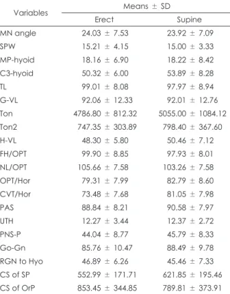
본 연구는 다른 연구들과 OSA의 진단기준, AHI의 판독기 준, 피험자군, 통계적 분석방법이 다르기 때문에 직접 비교하 Table 4. Cephalometry results of the subjects
Variables Means ± SD
Erect Supine
MN angle 0024.03 ± 7.53 0023.92 ± 7.09
SPW 0015.21 ± 4.15 0015.00 ± 3.33
MP-hyoid 0018.16 ± 6.90 0018.22 ± 8.42 C3-hyoid 0050.32 ± 6.00 0053.89 ± 8.28
TL 0099.01 ± 8.08 0097.97 ± 8.94
G-VL 0092.06 ± 12.33 0092.01 ± 12.76
Ton 4786.80 ± 812.32 5055.00 ± 1084.12
Ton2 0747.35 ± 303.89 0798.40 ± 367.60 H-VL 0048.30 ± 5.80 0050.46 ± 7.12 FH/OPT 0099.90 ± 8.85 0097.93 ± 8.01 NL/OPT 0105.66 ± 7.58 0103.26 ± 7.58 OPT/Hor 0079.31 ± 7.99 0082.79 ± 8.60 CVT/Hor 0073.48 ± 7.68 0081.05 ± 7.98
PAS 0088.84 ± 8.21 0090.58 ± 7.97
UTH 0012.27 ± 3.44 0012.37 ± 2.72
PNS-P 0044.04 ± 8.77 0045.79 ± 8.33
Go-Gn 0085.76 ± 10.47 0088.49 ± 9.78 RGN to Hyo 0046.89 ± 6.26 0045.46 ± 7.33 CS of SP 0552.99 ± 171.71 0621.85 ± 195.46 CS of OrP 0853.45 ± 344.85 0789.81 ± 373.91
Table 5. Correlation analysis between AHI and cephalometric variables
Variables Erect Supine
r p value r p value
MN angle 0.021 0.899 -0.072 0.661
SPW 0.149 0.360 0.267 0.096
MP-hyoid 0.359 0.023* 0.412 0.008*
C3-hyoid 0.387 0.014* 0.285 0.074
TL 0.462 0.003* 0.331 0.037*
G-VL 0.176 0.278 0.134 0.410
Ton 0.243 0.131 0.190 0.241
Ton2 0.414 0.008* 0.390 0.013*
H-VL 0.356 0.024* 0.239 0.138
FH/OPT 0.299 0.061 0.246 0.126
NL/OPT 0.230 0.152 0.293 0.067
OPT/Hor -0.262 0.103 -0.219 0.174
CVT/Hor -0.030 0.853 -0.224 0.165
PAS 0.157 0.333 0.017 0.918
UTH 0.290 0.069 0.449 0.004*
PNS-P 0.364 0.021* 0.309 0.052
Go-Gn -0.032 0.845 -0.061 0.706
RGN to Hyo 0.301 0.059 0.355 0.025*
CS of SP 0.512 0.001* 0.508 0.001*
CS of OrP 0.069 0.673 -0.050 0.759
* : significant variables. r : correlation coefficient, AHI : apnea hypopnea index
는 것에 무리가 있으나 본 연구에서는 Hwang 등11)과 Tsuchiya 등10)의 연구와 유사하게 혀 길이, 혀의 크기, 연조직의 두께 등 이 OSA와 연관성을 보였고, 설골의 위치와의 연관성에서는 Hwang 등11)의 연구결과와 상치되었다. 본 연구의 후속연구 에서는 비만군과 비 비만군의 분석을 추가하여 이 부분에 대 해 비교를 할 필요성이 있겠다.
본 연구의 강점은 한국인을 대상으로 하였고 환자의 수면 위치인 앙와위에서 얻은 사진을 대상으로 분석한 것이다. 앙와 위와 비교하여 정립위에서 상부기도의 폭경은 감소하고, 중력 에 의하여 혀와 연구개의 길이는 증가하게 된다.8) 환자의 수면 위치는 대부분 앙와위이므로, 정확한 측정을 위해서는 앙와 위에서 얻은 영상을 포함한 계측이 이루어져야 할 것이다.
본 연구의 한계점은 첫째, 본 연구는 예비연구로서 외국에 서 수행된 연구들보다 피험자 수가 적었고, 둘째, 수면클리닉 의 수면전문가들에게 찾아온 폐쇄성수면무호흡증 의심 환자 들만을 대상으로 하였기 때문에, 일반인구나 일차진료의 환 자군을 대표하지 못할 선택 오류의 가능성이다. 셋째, 피험자 중에서는 수면다원검사 결과 OSA가 아닌 피험자들도 있었다 는 점이다. 본 연구가 OSA 여부가 아닌 AHI와 두부방사선계 측치 간의 상관관계를 비교한 것이고 실제 수면클리닉에서 OSA가 의심되는 피험자들이 내원했을 때 AHI와 연관되는 계 측치가 무엇인지를 보는 측면에서는 임상상황에 보다 부합하 는 연구 세팅이라고 할 수도 있지만 연구자의 견해에 따라서 는 순수 OSA 환자군만을 대상으로 하는 것이 보다 정확한 결 론을 도출할 것이라는 반론이 있을 수 있다. 마지막으로, AHI 와 측모 두부계측치 간의 상관분석만을 시행하였고 다른 통 계분석이 시행되지 않아서 방사선계측치가 AHI나 OSA를 예 측하는 등의 임상적 가치는 제한된다.
결론으로, 본 연구는 폐쇄성수면무호흡증이 의심되는 환자 에서 앙와위 및 정립위 두부 측면 단순촬영사진에서 AHI와 유의한 연관을 보이는 계측치들을 발견하였으며, 이를 향후 대 규모의 피험자들을 대상으로 연구함으로써 OSA나 AHI를 예측할 수 있는 예측인자로서의 계측치를 발견하는 것이 필요 하겠다. 이를 통해 임상적으로 OSA가 의심되는 환자들의 수 면다원검사를 선별함에 있어서 보다 정확하고 객관적인 기준
을 제시하게 될 것으로 기대된다.
중심 단어:두계골계측・폐쇄성수면무호흡증.
Acknowledgments
이 논문은 2013년도 정부(교육부, 미래창조과학부)의 재원으로 한 국연구재단의 지원을 받아 수행된 기초연구사업임(No. NRF-2013 R1A1A2059105, No. NRF-2011-0013991).
Conflicts of interest
The authors have no financial conflicts of interest.
REFERENCES
1) Bresnitz EA, Goldberg R, Kosinski RM. Epidemiology of obstruc- tive sleep apnea. Epidemiol Rev 1994;16:210-227.
2) Ono T, Lowe AA, Ferguson KA, Fleetham JA. Associations among upper airway structure, body position, and obesity in skeletal Class I male patients with obstructive sleep apnea. Am J Orthod Dentofa- cial Orthop 1996;109:625-634.
3) Riley R, Guilleminault C, Herran J, Powell N. Cephalometric analy- ses and flow-volume loops in obstructive sleep apnea patients. Sleep 1983;6:303-311.
4) Tangugsorn V, Krogstad O, Espeland L, Lyberg T. Obstructive sleep apnea: a canonical correlation of cephalometric and selected demo- graphic variables in obese and nonobese patients. Angle Orthod 2001;
71:23-35.
5) Ong KC, Clerk AA. Comparison of the severity of sleep-disordered breathing in Asian and Caucasian patients seen at a sleep disorders center. Respir Med 1998;92:843-848.
6) Iber C, Ancoli-Israel S, Chesson ALJ, Quan SF. The AASM Manual for the Scoring of Sleep and Associated Events: Rules, Terminology and Technical Specifications. Westchester: American Academy of Sleep Medicine;2007.
7) Park J, Wang D, Kim WI, Lee SD, Nam SY, Lee YB. Clinical evalua- tion of polysomnography in snoring and obstructive sleep apnea syn- drome (OSA) patients. Korean J Otorhinolaryngol-Head Neck Surg 1994;37:369-376.
8) Kwon TG, Cho YW, Ahn BH, Suh YS. Cephalometric predictors of obstructive sleep apnea. J Korean Assoc Oral Maxillofac Surg 2003;
29:338-345.
9) Li KK, Powell NB, Kushida C, Riley RW, Adornato B, Guilleminault C. A comparison of Asian and white patients with obstructive sleep apnea syndrome. Laryngoscope 1999;109:1937-1940.
10) Tsuchiya M, Lowe AA, Pae EK, Fleetham JA. Obstructive sleep apnea subtypes by cluster analysis. Am J Orthod Dentofacial Orthop 1992;
101:533-542.
11) Hwang SH, Park IS, Nam KY, Kim JB, Cho YW, Suh YS, et al. Ceph- alometric differences in obstructive sleep apnea between obese and non-obese Korean male patients. Korean J Orthod 2008;38:202- 213.