215
Oral Biology
Actions of Group I Metabotropic Glutamate Receptor Agonist on Synaptic Transmission and Ionic Currents in Rat Medial Vestibular Nucleus
Neurons
Hae In Lee and Sang-Woo Chun*
Dept. of Oral Physiology, College of Dentistry, Institute of Wonkwang Biomaterial and Implant, Wonkwang University, Iksan 570-749 (received December 03, 2009 ; revised December 17, 2009 ; accepted December 22, 2009)
Medial vestibular nucleus (MVN) neurons are involved in the reflex control of the head and eyes, and in the recovery of vestibular function after the formation of peripheral vesti- bular lesions. In our present study, whole cell patch clamp recordings were carried out on MVN neurons in brainstem slices from neonatal rats to investigate the actions of a group I metabotropic glutamate receptor (mGluR) agonist upon synaptic transmission and ionic currents. Application of the mGluR I agonist (S)-3,5- dihydroxyphenylglycine (DHPG) increased the frequency of miniature inhibitory postsynap- tic currents (mIPSCs) but had no effect upon amplitude distributions. To then identify which of mGluR subtypes is responsible for the actions of DHPG in the MVN, we employed two novel subtype selective antagonists. (S)-(+)- α - amino-a-methylbenzeneacetic acid (LY367385) is a potent competitive antagonist that is selective for mGluR1, whereas 2-methyl-6-(phenylethynyl)-pyridine (MPEP) is a potent noncompetitive antagonist of mGluR5. Both LY367385 and MPEP antagonized the DHPG-induced increase of mIPSCs, with the former being more potent. DHPG was also found to induce an inward current, which can be enhanced under depolarized conditions. This DHPG- induced current was reduced by both LY367385 and MPEP.
The DHPG-induced inward current was also suppressed by the PLC blocker U-73122, the IP
3receptor antagonist 2- APB, and following the depletion of the intracellular Ca
2+pool by thapsigargin. These data suggest that the DHPG- induced inward current may be mainly regulated by the intracellular Ca
2+store via the PLC-IP
3pathway. In
conclusion, mGluR I, via pre- and postsynaptic actions, may modulate the excitability of the MVN neurons.
Key words: medial vestibular nucleus neurons, patch clamp, inward current, PLC-IP
3pathway
서 론
중추신경계에서 대사성
glutamate
수용체의 활성은 신경 흥분의조절
,
시냅스에서 전달물질의 분비,
시냅스가소성
(plasticity)
등에서 다양한 조절 작용을 가지고 있다(Conn & Pin, 1997; Anwyl, 1999).
대사성glutamate
수용체는
G-
단백질과 연결된 수용체로 아미노산 배열순서나 이차전달자 작용 방식에 따라 세 군으로 분류한다
.
즉
, I
군수용체는phospholipase C (PLC)
를이용하며ino- sitoltriphosphate (IP
3)
합성을 조절하는데mGluR1, 5
가해당되고
, II
군과III
군은adenosine 3',5'-cyclic mono- phosphate
전달계와 관련되는데II
군은mGluR2, 3, III
군은
mGluR 4, 6, 7, 8
이속한다(Pin & Bockaert, 1995;
Conn & Pin, 1997; Pin & Duvoisin, 1997).
I
군수용체는Gq
단백질을통하여PLC
가phosphatidy- linositol bisphosphate(PIP
2)
를diacylglycerol(DAG)
와inositol triphosphate(IP
3)
로 분해하는 경로를 통하여 세포반응을 매개하는데
,
만들어진IP
3는 세포내 칼슘 저장 소로부터세포질로칼슘을이동시킨다(Conn & Pin, 1997;
Fagni
et al, 2000; Valenti
et al, 2002).
최근의 연구에서 시냅스 말단에서 분비된
glutamate
에의해 대사성수용체를 통하여
IP
3경로로 세포내Ca
2+의 이동이 소뇌의Purkinje
세포와 해마의 세포에서관찰되었으며,
이는 장기강화
(long term potentiation; LTP)
나 장기약화(long
*Corresponding author: Sang-Woo Chun, Department of Oral
Physiology, College of Dentistry, Wonkwang University, 344-
2, Shinyong-Dong, Iksan 570-749, Korea Tel.: 82-63-850-
6932 E-mail: physio1@wonkwang.ac.kr
term depression; LTD)
와 같은 시냅스 가소성과 관련이된다
(Finch & Augustine, 1998; Bortolotto
et al, 1999;
Nakamura
et al, 2000; Bashir, 2003).
I
군 수용체의 칼슘신호 전달에 대한 작용 외에I
군 수용체의활성은 해마의
CA1
추체세포에서탈분극과활동전압의빈도증가등직접적인흥분효과를일으킨다
(Gereau
& Conn, 1995; Mannaioni
et al, 1999).
이는 칼슘활성 화 양이온 전류의 활성(Crepel
et al, 1994),
후과분극(afterhyperpolarization)
전류의억제(Charpak et al, 1990),
유출전류의 억제
(Guerineau
et al, 1994),
불활성화가 늦은전압의존성포타슘전류의억제(Luthi
et al, 1996)
등에 의하는데 이러한 전류의 변화가 세포내 칼슘양의 증가와관련되어 있다
(Mannaioni
et al, 2001; Kettunen
et al
, 2003; Morikawa
et al, 2003; Rae & Irving, 2004).
또한 시냅스 전달에 대한 작용으로는 억제성 개제 뉴론 에작용할경우
,
세포체의 수용체를활성화시켜 세포 흥 분성을증가시키거나(Llano & Marty, 1995; Poncer
et al, 1995; Zhou & Hablitz, 1997)
축삭 말단의 수용체에 작 용하여 시냅스전 촉진작용이 있음이 보고 되고 있다(Mannaioni
et al, 1999; Park
et al, 2003).
전정 신경핵에는
I
군수용체 중대사성glutamate
수용 체(mGluR) 1
과5, II
군 수용체인mGluR2
와3, III
군수용체인
mGluR7
이존재함이확인되었다(Shigemoto
etal
, 1992; Kinney
et al, 1993; Darlington & Smith, 1995; Neki
et al, 1996; Vidal
et al, 1999; Horii
et al, 2001). mGluR1, 2
효현제인1-aminocyclopentane -trans- 1,3-dicarboxylic acid (ACPD)
적용시 내측 전정핵 뉴론 의흥분발사율이 증가하거나감소하였으며(Darlington &
Smith, 1995),
전정구심신경자극에 의해 유발되는 흥분성시냅스후전압을 감소시켰다
(Kinney
et al, 1994).
또한,
최근에
Grassi
등(2002)
은II
군과III
군 수용체에 의해LTP
의 유발이 억제되는 반면mGluR1
에 의해서는 촉진됨을 보고하였고
Park
등(2003)
은mGluR1, 2
효현제인ACPD
와mGluR2, 3
효현제인(2S,2'R,3'R)-2-(2',3'-dicar boxycyclopropyl)glycine (DCG-IV)
가억제성시냅스의축색말단에서 신경전달물질 분비를 시냅스전 억제 작용으 로 감소시킴을 보고하였다
.
내측전정핵 세포에서
I
군 대사성glutamate
수용체의역할에대한이전연구에서
I
군수용체의효현제인DHPG
를 처리하였을때 막전압의 탈분극과 자발적 활동전압의 빈도 증가가 관찰되었고 이러한 흥분성의 변화가
K
+ 유출전류의차단에 의한 내향성 전류와 칼슘활성화 포타슘 전류의 억제를 통하여 발생하였음이 보고되었다
(Chun
et al
, 2004).
그러나DHPG
에 의한 탈분극이 어떠한 세포내 전달과정이 관여하여 발생하는지는 밝혀지지 않았 으므로이연구에서는내측전정핵에서
DHPG
의내향성전류의 발생과 세포내 칼슘와의 의존성
,
세포내 칼슘 농도변화를 일으키는 경로 등을
patch clamp
방법을 이용하여 규명하고자 하였다
.
실험재료 및 방법
뇌절편 제작
생후
10-17
일의Sprague-Dawley
흰쥐를 암수 구별 없 이 사용하였으며 이 연구는 원광대학교 동물실험윤리위 원회에서 승인을 얻었다(WKU09-076).
흰쥐를ether
로 깊게 마취한후단두하여소뇌와뇌간부위를 적출하고0- 4
oC
의 저Na
+ 농도의 절단용액에 넣어 보관한 후 꺼내 어 순간접착제(cyanoacrylate adhesive)
를 이용하여 뇌 조직을 고정시켰다. 95% O
2-5% CO
2를 공급하면서 조 직절편기(Vibroslicer, Campden 752M,
영국)
를 이용하 여 내측 전정신경핵부위를200
µm
두께로관상면 절단 하여 뇌절편을만들었다.
뇌절편은32
oC
의인공뇌척수액 용액에1
시간 정도 보관하여 정상상태로 회복시켰고,
이 후에는 실온에서 실험을시행하였다.
기록은 뇌절편을 현 미경(BX50WI, Olympus,
일본)
위의 기록용기(1 ml)
에옮긴 후 치실로 만든 그물로움직이지 않도록 고정한 후 시행하였고
,
실험기간동안 계속해서95% O
2-5% CO
2가 포함된 용액을 관류펌프(Minipuls 3, Gilson,
프랑스)
를이용하여 관류시켰다
(2-3 ml/min).
세포의 정확한 구분을위하여현미경에
differential interference contrast (DIC)
장치를부착했고
, 40
배의water immersion
대물렌즈로세포를 확인하였다
.
실험용액
뇌절편 제작에 사용했던 절단용액의 조성
(mM)
은252 Sucrose, 2.5 KCl, 0.1 CaCl
2, 2 MgCl
2, 10 glucose, 26 NaHCO
3, 1.25 NaH
2PO
4등으로 구성되었으며,
이온전류기록 및 세포내칼슘량의 변화를확인하기 위한 인공 뇌 척수액은
124 NaCl, 5 KCl, 1.2 KH
2PO
4, 1.3 MgSO
4, 2.4 CaCl
2, 10 glucose, 24 NaHCO
3이었고, 95% O
2-5%
CO
2를 공급하여pH
를7.4
로 유지하였다.
자발적 억제성시냅스후 전류 기록시 세포외용액에는 흥분성 시냅스 전 류를 억제시키기 위하여
AMPA
수용체 차단제인6,7- dinitroquinoxaline-2,3-dione (DNQX) 20
µM
과(
±)2-amino- 5-phosphono- pentanoic acid (AP5) 20
µM
을 포함시켰으며
,
미세시냅스후 전류기록시에는TTX 0.5
µM
을추가하였다
.
세포내용액은140 KCl, 1 MgCl
2, 0.1 CaCl
2,
10 HEPES, 3 MgATP, 0.3 NaGTP
를 사용하고, pH
는7.3
으로하였다.
실험에사용한AP5, DNQX, thapsigargin,
BAPTA
등은Sigma
사(
미국)
에서 구입하였고, (S)-3,5-
dihydroxyphenylglycine (DHPG), (S)-(+)-
α-amino-a-methy-
lbenzeneacetic acid (LY367385), 2-methyl-6-(phenylethynyl)-
pyridine (MPEP), U-73122, tetrodotoxin (TTX), nimodifine,
2-APB (2-aminoethoxydiphenylbolate), dantrolene sodium
은
Tocris
사(
영국)
에서구입하여사용하였다. AP5, DHPG, U-73122, LY367385, nimodifine
등은 물에 녹여사용하 였으며, thapsigargin, MPEP, 2-APB, dantrolene sodium
등은
DMSO (dimethyl sulfoxide)
에 먼저 녹인 후 최종 농도로실험직전에희석하여 사용하였다.
세포에대한 실 험용액의적용은 중력을 이용한관류장치(BPS-4SG, Ala Scientific Instruments,
미국)
를 이용하여 기록용기내 용 액을 교환하였다.
전기생리학적기록방법
미세 유리전극 제조기
(PP-830, Narishige,
일본)
와microforge (MF-830, Narishige,
일본)
를 이용하여 외경1.5 mm
의연질유리미세관(WPI,
미국)
을저항이5-8 M
Ω 이되도록기록전극을 제작하였다.
세포에전극을접근시 키는 과정은 보다 분명한 영상을 관찰하기 위하여 흑백CCD
카메라(Panasonic,
일본)
를이용한videomicroscopy
하에서시행하였다
.
전극에양압을 주면서세포에접근하 여내측 전정핵뉴론 세포막이 약간의변형을 보일 때 음 압을 가하여giga ohm seal
을이루었다.
전압 고정에 의한전류측정에는
Axopatch 200B
증폭기(Axon,
미국)
를사용하였고
,
이 증폭기는Digidata 1200B (Axon,
미국) AD
변환기를 통하여 컴퓨터에 연결하였으며, pCLAMP software (Version 8.0, Axon,
미국)
를 사용하여 실험수행의 명령과 얻어진 전기신호의 저장 및 분석에 이용하 였다
. Series resistance
는 보통3-8 M
Ω 정도였으며,
전류보정은 시행하지 않았고 발생된 전류는
low pass 8-
pole Bessel filter
로2 kHz
로여과하였다.
모든실험은 실온에서 시행하였다
.
실험자료의분석
억제성시냅스후전류의분석은
Mini Analysis program (version 6.0, Synaptosoft,
미국)
을 이용하였고 막전류의분석은
Clampfit (Version 8.0, Axon,
미국)
을 이용하였다
.
대조군과 처리군 사이에 통계적으로 유의한 차이가존재하는지의여부는
paired
혹은non-paired t-test
를 이용하였고
,
p <0.05
에서 통계적으로 유의하다고 판정하였다
.
통계자료의값은평균값 ± 표준오차(mean
±S.E.M.)
로표시하였다
.
결 과
억제성미세시냅스후전류
(miniature postsynaptic cu- rrent)
에대한 효과내측전정핵의시냅스 전달에는
non-NMDA, NMDA
수용체를통한 흥분성시냅스 전류보다는
GABA
수용체를통한 억제성 시냅스 전류가 신경흥분성 조절에 더 우세 하게작용하고있다고보고된바있어
(Chun
et al, 2003)
이 연구에서는 억제성미세시냅스후 전류에 대한
DHPG
의 효과를확인하였다
.
억제성시냅스후 전류를 기록하기위하여 세포외 용액에
AMPA
수용체 길항제인DNQX
20
µM
과NMDA
수용체 길항제인AP5 20
µM
을 추가 하여 흥분성 시냅스후 전류를 차단하였으며 유지전압을-70 mV
혹은-50 mV
로 고정하여 시냅스후 전류를 기록 하였다.
TTX 0.5
µM
을 처리하여 신경흥분 전달을 차단한 후미세시냅스후 전류에 대한
DHPG
의 효과를 확인하였다. DHPG
처리 후 빈도는2.5
±0.4 Hz
에서3.9
±0.5 Hz
로 유의하게 증가하였으며(P<0.05),
크기는22.7
±4.2 pA
에 서28.5
±4.6 pA
로증가하였으나유의하지 않았다(n = 7) (Fig. 1).
DHPG
의 작용이mGluR1
혹은mGluR5
에 선택적으로 일어나는지 확인하고자mGluR1
과mGluR5
에 길항작용 을 가지는LY367385
와MPEP
를DHPG
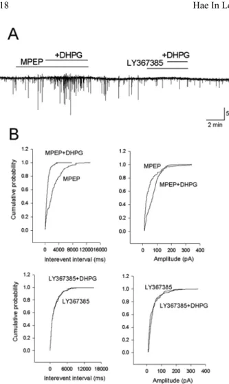
와 함께 투여하Fig. 1. Effects of DHPG on miniature inhibitory postsynaptic cur- rents (mIPSCs).
A; Current traces of mIPSCs show the effects of DHPG on the syn-
aptic activity of a MVN neuron. B; Cumulative fraction plot of
mIPSCs interevent interval and amplitude. Application of DHPG
increased the frequency of mIPSCs but the amplitude was not
changed. C; DHPG significantly increased the average mIPSCs
frequency.
였다
.
각각의 길항제가 포함된 용액에서DHPG
를 투여한 결과
MPEP
전처리 군에서는 유의한 빈도의 증가가관찰되었으나
(p
<0.05) (n = 5) LY 367385
전처리 군에서는 빈도의 증가가 유의하지 않았으며
(n = 5),
크기에서는두군에서유의한 변화를보이지 않았다
(Fig. 2, 3).
따 라서DHPG
에의한시냅스전류의빈도증가에는mGluR1
이주로 작용하는 것으로 사료된다
.
내향성전류의발생
DHPG
의 시냅스후 뉴론에 대한 효과를 알아보기 위하여 억제성 미세 시냅스후 전류를 기록하기 위한 용액
(DNQX, AP5, TTX
포함)
에서DHPG
에의한 전류의변 화를기록하였다.
유지전압을-50 mV
로고정하여DHPG
를처리하였을 때약물처리후
30
초- 2
분후부터내향성 전류가발생하였다(Fig. 1, 4).
전류의크기는61.0
±5.3 pA
이었으며
(n = 11),
약물이 포함되지 않은 용액으로 재관류시 서서히 원상태로회복되었다
.
유지전압을-70 mV
로고정하였을때
DHPG
에 의해 발생한 내향성전류는28.5
±3.4 pA
의(n = 7)
크기로 유의하게 감소하여(p
<0.05)
막 전압이 탈분극 됨에 따라 전류의 크기가 증가함을 확인하였다
. DHPG
에 의한 내향성 전류가 어느 수용체를 통하는가를 알아보기 위하여
LY367385
와MPEP
를전처리 한 후DHPG
를 투여하였다. DHPG
와mGluR5
의 억제제 인MPEP
가포함된용액으로관류시52.0
±6.5 pA
의내향 성전류를 기록하였다(n = 10).
그러나DHPG
와mGluR1
의억제제인
LY367385
가 포함된 용액으로 관류시에는 대조군에 비해유의하게감소하여
(p
<0.05), DHPG
에의한 내 향성전류는mGluR1
을통하여발생함을확인하였다(Fig. 4).
Fig. 2. Effects of MPEP and LY367385 on DHPG-induced in- crease in the frequency of mIPSCs.
A; Current traces of mIPSCs at a holding potential of -70 mV.
DHPG-induced increase in mIPSCs was remained by DHPG pre- incubation with MPEP, but decreased prominently by DHPG pre- incubation with LY367385. B; Cumulative fraction plot of mIPSCs interevent interval and amplitude induced by DHPG in the pres- ence of MPEP or LY367385.
Fig. 3. Bar graph showing the changes in the frequency and ampli- tude of mIPSCs induced by DHPG in the presense of LY367385 or MPEP. Note that significant increase of mIPSCs frequency by application of DHPG with MPEP.
Fig. 4. DHPG-induced inward current is mainly mediated by
mGluR
1. Bar graph showing the DHPG induced inward current in
the presence of LY367385 or MPEP.
내향성전류의칼슘의존성
DHPG
에 의해서 발생된 내향성 전류가 세포내 칼슘의변화와 관련 되는지를 알아보고 어느 세포내 신호전달 경로를통하는지를확인하고자
PLC
차단제U-73122,
세포내 칼슘
chelator BAPTA,
세포내 칼슘저장소의 칼슘 을 고갈시키는 작용이 있는thapsigargin,
칼슘저장소의IP
3 수용체차단제2-APB,
칼슘저장소의ryanodine
수용 체 차단제dantrolene sodium
등을 전처리 후DHPG
의 투여 효과를 확인 하였다.
U-73122 1
µM
을DHPG
적용2-3
분전에전처리한후DHPG
를투여 하였을 때9
개의 세포에서는내향성 전류가 발생 되지 않았고 두개의 세포에서
10-20 pA
의 작은전류가 기록되어
PLC
경로를 통한 세포내 칼슘의 증가가 내향성 전류의 발생에 중요하게 작용한 것으로 추측 된다
. PLC
의 활성은IP
3를 생성하여 세포내 칼슘 저장 소로부터 세포질로 칼슘 방출을 매개한다.
이 실험에서 세포내액의 칼슘을 제거시키기 위하여 세포내 용액에 칼 슘chelator
인BAPTA 10 mM
을 사용하였다. BAPTA
가포함된 전극으로 기록시
DHPG
에 의한 내향성 전류는4.2
±3.4 pA
로현저하게감소하였다(P
<0.05).
또한BAPTA
가 포함된 전극을 이용하여 막전압의 변화를 기록했을 때에도
DHPG
에의한탈분극을유발하지못하였다(n = 3).
Thapsigargin
은 세포내 칼슘저장소로Ca
2+을reuptake
하는 과정을 차단함으로써 칼슘저장소내의 칼슘을 고갈
시킨다
. Thapsigargin 1
µM
을45
분 이상 전처리하고DHPG
를 투여하였을 때8
개의 세포에서는 내향성 전류 가 완전히 차단되었고2
개의 세포에서20 pA
정도의 작 은 전류가 발생하였다(Fig. 5).
L
형 칼슘통로가 대사성 글루탐산염 수용체를 통한 칼 슘의 이동에 관여한다고 보고 되어 있어(Kettunen
et al, 2002) L
형 칼슘통로 차단제인nimodipine 10
µM
을 전처리 후DHPG
를 투여 하였다.
기록된10
개의 세포 모두에서 내향성 전류를 확인하였는데 크기는41.5
±4.7
pA
로DHPG
단독 투여에 비하여 유의한 감소를 보이지않았다
(Fig. 6).
세포내 저장소에서 칼슘을 방출 시키는데는
IP
3 수용체 와ryanodine
수용체가 관여한다. IP
3수용체 차단제인2- APB 20
µM
을 전처리 후DHPG
를 투여하였을 때4
개 의 세포 중1
개의 세포에서15 pA
의 내향성 전류를 기 록하였다. Ryanodine
수용체차단제인dantrolene sodium 10
µM
을 전처리 후DHPG
를 처리하였을 때는55
±6.1 pA
의 내향성 전류가 기록 되었다(n = 4) (Fig. 7).
고 찰
최근
I
군대사성glutamate
수용체인mGluR1
과5
가서로 다른세포반응을매개함이알려졌다.
하시상핵(subthalamic nucleus)
은mGluR1
과5
를 모두 가지고 있으나DHPG
에의한 탈분극에는
mGluR5
만이 작용한다고 하고(Awad
et al
, 2000),
흑질(substantia nigra)
의GABA
성 뉴론은두 가지 수용체를 모두 가지고 있으나
mGluR1
에 의하여 탈분극이 유발된다고 보고 되었으며
(Marino
et al, 1999),
해마의CA1
세포에서는mGluR1
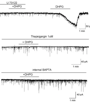
에 의해서는세Fig. 5. Inhibition of DHPG-induced inward current requires PLC activation and Ca
2+release from internal stores. DHPG-induced inward current was blocked with the PLC blocker U-73122, intrac- ellular calcium chelator BAPTA. Effect of DHPG was blocked when the internal Ca
2+stores were depleted with thapsigargin.
Fig. 6. Mean inward current amplitude induced by DHPG in con-
trol and in neurons pretreated with U-73122, BAPTA, tharpsigargin
and nimodipine. *denotes significant difference from the control
by independent t -test (*p < 0.05, **p < 0.01).
포내 칼슘 농도의 변화를 유발하였고
mGluR5
에 의해서 는칼슘활성화포타슘 전류(I
AHP)
의억제, NMDA
수용체전류의증가를 일으켰으며
(Mannaioni
et al, 2001),
이러 한 서로 다른 효과는 동물의 종에 따라 또는 세포의 종 류에따라차이를보인다.
내측전정핵에서도mGluR1, 5
수용체가모두존재하나그작용은서로 상이하다고보고 된 바 있다
(Grassi
et al, 2002).
즉, mGluR1
수용체를 통하여 고빈도 자극에 의해 유발되는LTP
를 증가시켰으 나mGluR5
수용체를 통하여는 억제된다고 하였다.
따라 서이 연구는 내측 전정핵에서I
군 대사성glutamate
수 용체 작용물질인DHPG
의효과가 어느 수용체를통하여 일어나는가를 확인하였다.
DHPG
의 처리에 의하여 미세 시냅스후 전류가 증가하였다
(Fig. 1).
이는DHPG
가 축삭말단에 존재하는 수용 체에 작용하여GABA
의quantal release
를 증가 시켰을것으로 생각된다
. DHPG
가 미세 시냅스후 전류의 빈도를 증가시키는 효과는 축삭말단에 존재하는
mGluR1
의활성화에 의해서
PLC
를 활성화시키고 이는IP
3 경로에 의해세포내 칼슘량을증가시켜 축삭말단에서GABA
분 비를 촉진한다고 알려져 있다(Stefani, 1994; Kawabata
et al
, 1996). DHPG
에의하여 미세 시냅스후 전위의 크기증가는 유의하지 않았으나일부의 세포에서 관찰되었 다
(Fig. 2, 3).
유지전압을
-50 mV
로 고정하여DHPG
를 처리 하였을때내향성전류가발생하였다
.
이는내측전정핵 뉴론에서는 안정막 전위상태에서도
I
군glutamate
수용체 활성이내향성 전류를발생시키고 이에 따라 막전위의 탈분극과 자발활성의 증가를 유발 할 수 있음을 의미한다
.
막전압을
-70 mV
로 고정 시켰을 때K
+ 이온의 기전력(driving force)
감소로 내향성 전류는 더 작게 발생되었다.
이전의 연구에서
DHPG
가 전압의존성 통로를 통한 전류는변화시키지 않았고
DHPG
에의한 전류의역전전압이K
+의평형전압이며세포내용액을
Cs
+으로교체하였을 때는 내향성전류가 발생하지 않아DHPG
는K
+ 유출전류를 감 소시킴에 의해서발생하였을 것으로추측하였다.(Chun
et al., 2004).
다른 세포에서도탈분극에따라내향성 전류의 증가가 관찰되었는데(Luthi
et al, 1997; Chuang
et al, 2000; Kettunen
et al, 2003),
내향성전류의 발생원인은 이 연구에서와 같이K
+ 유출 전류의 감소(Awad
et al, 2000; Mannaioni
et al, 2001)
외에도Na
+/Ca
2+교환기전 의 활성(Lee & Boden, 1997; Hirono
et al, 1998)
이나 비선택성양이온전류의활성(Crepel
et al, 1994; Chuang
et al
, 2000; Mannaioni,
et al, 2001; Ireland & Abraham, 2002)
등이 알려져 있다.
내측전정핵 뉴론에서
DHPG
에 의한 내향성 전류는 주로
mGluR1
을 통하여 발생하였는데 유사한 결과가 척수뉴론
(Kettunen
et al, 2003),
흑질의 억제성 개제뉴론(Marino
et al, 1999)
등에서 보고되었으나 하시상핵 뉴론에서는 반대로
mGluR5
에 의해서 내향성 전류가 발생됨이 보고 되었으며
(Awad
et al, 2000)
이러한 차이는세포의 종류와 동물의종 혹은 성장 정도에따라 다르게 나타난다고 알려져 있다
(Mannaioni
et al, 2001; Ireland
& Abraham, 2002; Kettunen
et al, 2003).
세포내에서 칼슘이이동하는 주요한 경로로는 일반적으 로
PLC-IP
3 경로이다. Ligand
가 세포막에 존재하는G
단백과 연결된 수용체와 결합하면
Gq/11
단백질이 활성화되고 활성화된
G
단백은PLC
β를 활성화시키고 활성화된
PLC
β는PIP
2를가수분해한다. PIP
2는 가수분해 되 어IP
3와diacylglycerol (DAG)
를 생성하는데IP
3는 세 포내 칼슘저장고인내형질세망(endoplasmic reticulum)
의막에 존재하는
IP
3 수용체에 결합하여 세포내로 칼슘을 분비시킨다.
그러나 이러한 세포내 칼슘의 분비 후 밖으로부터 칼 슘이유입되는데이를일으키는이온통로를이전부터
store- operated Ca
2+channel (SOCC)
혹은transient receptor potential channel (TRPC)
로 명명하였고 최근 많이 연구가 되어지고 있다
.
이SOCC
를 통해 들어온 칼슘은 여러 효현제
(agonist)
들에 의한 칼슘 반응을 조절할 뿐만아니라세포내저장고에칼슘을공급하는역할을하는것 으로 알려져 있다
(Parekh & Penner, 1997).
실험적으로연구자들이
TRP
통로를 구별하는 방법은 세포내 칼슘저장고를 고갈하였을 때 활성화되는 것을 기준으로 하는 데
,
즉thapsigargin
이나cyclopiazonic acid (CPA)
를 사용하여 전류가 활성화되거나 혹은 세포내 칼슘이 증가하 면
SOCC
가존재한다고하였다.
이연구에서도thapsigargin
을
45
분 이상 전저치하여 세포내칼슘저장고를 고갈시킨후
DHPG
를 처리하였을 때2
개의 세포에서20 pA
의작은 내향성 전류가 기록되었는데 이러한 변화가
SOCC
를통한 세포내로의 칼슘의 유입에 의해 발생했을 가능성을
Fig. 7. Effects of dantrolene sodium or 2-APB. DHPG-induced
current was markedly blocked in the presence of 2-APB.
배제할 수 없다
.
Cyclic ADP-ribose (cADPR)
은 포유류에서 보고된 또 다른 칼슘 방출 전달자이다(Petersen & Cancela, 1999).
cADPR
의칼슘이동에대한작용은신경계에서ryanodine
수용체를 통하여 칼슘에 의해 유발되는 칼슘 방출을 강 화하고
,
시냅스전말단에서 신경전달물질의 분비를증가 시키는 역할이 보고되었다(Empson & Galione, 1997;
Mothet
et al, 1998; Brailoiu & Miyamoto, 2000).
또한 최근중뇌의도파민뉴론에서 대사성glutamate
수용체에 의한칼슘의이동이IP
3경로와함께이cADPR
경로도같 이관여하고있음이 확인되었다(Morikawa
et al, 2003).
이 실험에서
I
군 대사성 수용체의 활성에 의한 세포내 칼슘의증가는 주로PLC
를 이용한IP
3 수용체를 통하여 발생하였다고 생각되나PLC
차단제인U-73122
를 사용 하였을 때 일부세포에서 약한 내향성 전류의 발생이 보 이고(Fig. 5), 2-APB
의 처리에 의해 세포내 칼슘저장소의
IP
3수용체를 차단하여도내향성전류가발생됨은(Fig.
7) cADPR
경로를통한ryanodine
수용체가작용했을 가 능성을 배제할 수 없으며 이는 차후의 연구에서 확인하 고자 한다.
이상의 연구 결과를종합하면
I
군 대사성glutamate
수 용체의활성은PLC
를 활성화 시켜 세포내 칼슘저장소의IP
3 수용체를 통하여 세포내 칼슘 농도의 증가를 일으키고 이는
K
+ 유출전류를 억제하여 내향성 전류를 발생시킨 것으로 사료된다
.
감사의 글
이 논문은
2007
년도 원광대학교의 교비지원에 의해서수행되었음