토천궁 정유 성분의 수종 사람 암 세포주에 대한 세포 독성
심연·신승원# 덕성여자대학교 약학대학
(Received August 31, 2011; Revised September 23, 2011; Accepted September 27, 2011)
Study on Cytotoxic Activities of the Essential Oil Compounds from Ligusticum chuanxiong against Some Human Cancer Strains
Youn Sim and Seungwon Shin#
College of Pharmacy, Duksung Women’s University, Seoul 132-714, Korea
Abstract — Ligusticum chuanxiong (Umbelliferae) is a perennial herb that has been used for invigoration of blood in Korean traditional medicine. It is especially important in gynecological therapy of amenorrhea and dysmenorrhea. In this study, the essential oil of L. chuanxiong was obtained by steam distillation and its main components of L. chuanxiong, Z-ligustilide and butylidene phthalide, were isolated by silica gel column chromatography. We investigated the cytotoxic effects of the essen- tial oil fraction of L. chuanxiong and its main components on MCF-7, HeLa and SK-Hep-1 cell lines by measuring the num- ber of surviving cancer cells after treatment through direct cell counting and MTT analysis, and by examining the morphological changes under the microscope. The essential oil from the rhizomes of L. chuanxiong and its main components showed significant cytotoxic activities for all three tested cell lines. We also observed morphological changes of shrinking and blebbing in the membranes of the three cell lines, depending on the concentration of L. chaunxiong oil or its main com- ponents.
Keywords □ Ligusticum chuanxiong, essential oil, Z-ligustilide, butylidene phthalide, MCF-7, HeLa, SK-Hep-1, MTT, mor- phology
토천궁(Ligusticum chuanxiong)은 미나리과에 속하는 다년생 초본으로, 일천궁(Cnidium officinale)와 함께 한방에서 불임, 월 경불순, 빈혈, 냉증 등과 같은 부인과 질환에 주로 사용되는 생 약인 천궁의 자원이다. 토천궁 근경의 생리활성 성분으로는 Z- ligustilide butyl phthalide, butylidene phthalide, neocnidilide, senkyunolide, cnidilide 등의 phthalide를 주로 함유하는 정유 1~2%와 ferulic acid, tetramethylpyrazine 등이 알려져 있다.1,2) 생리활성에 관하여는 주요 약효성분인 cnidilide, senkyunolide, Z-ligustilide에 중추성 근이완작용이, butylidene phthalide에는 진경작용이 있음이 확인되었다.3-10) 또한, senkyunolide, Z- ligustilide 및 butylidene phthalide가 mouse배양 대동맥 평활근 세포의 증식을 억제함이 보고되었으며, Z-ligustilide의 항콜린 작 용과 phthalide류의 항진균작용 및 항염증작용도 알려져 있다.11,12)
토천궁의 물 추출물은 임신동물의 자궁수축 항진, 소장 교감신 경의 흥분 억제작용, 혈압강하작용, 항비타민결핍증작용 등을 나 타내며, 일천궁의 경우에는 interferon 유도 항방사선작용, 항암 작용, 항균작용 등이 있다고 보고 되었다.13,14)
식물의 정유성분은 진정, 진경, 진통, 소염, 항균 등 화학구조 에 따라 여러 가지 다양한 생리활성을 나타내는 것으로 알려져 있어서 관련 질환의 치료제로 사용되어 왔으나, 근래에 와서는 다른 계열의 식물성분과 함께 천연항암제 개발의 중요 자원으로 주목 받게 되었다.15)식물정유는 특히 천연 향료 등으로 식품에 첨가되어 사용되기 쉬우므로, 식이와 연결된 천연 항암요법제의 대상으로 유망하다.16,17)이와 관련하여 초기에는 Citrus 속 열매 에 다량 함유된 limonene과 그 대사물질인 perillyl alcohol 및 기 타 geraniol 등의 monoterpene alcohol 들이 주 연구 대상이었 고,18-27) thymoquinone 등의 quinone류 및 기타 sesquiterpene 및 phenylpropane 계 물질들의 암세포 억제 효과가 입증된 바 있다.28,29)
본 연구에서는 한방에서 자궁기능 관련 질환에 대한 대표적인
#본 논문에 관한 문의는 저자에게로
(전화) 02-901-8384 (팩스) 02-901-8386 (E-mail) [email protected]
종설
치료제중 하나인 천궁의 정유분획 및 그 주성분인 Z-ligustilide 및 butylidene phthalide의 여성암 중 가장 사망률이 높은 유방 암 및 자궁경부암 그리고 유방 및 자궁의 기능의 조정에 중요한 역할을 하는 간에 발생하는 암의 억제 및 예방제로의 개발 가능 성을 알아보기 위하여, 인간 유방암세포(MCF-7), 자궁경부암세 포(HeLa) 및 간암세포(SK-Hep-1)의 세포증식 및 생존에 대한 천궁 정유분획 및 그 주성분인 Z-ligustilide 및 butylidene phthalide의 억제효과를 측정하였고, 이들 성분의 농도별 처리시 의 세포형태변화를 현미경으로 관찰, 비교하였다.
실험재료 및 방법
정유추출 및 분석
토천궁은 9~10월 강원도 진부, 경상북도 영주, 봉화에서 수확 하였고, 일천궁은 강원도 진부, 경상북도 봉화, 영양에서 수확하 여 근경의 흙을 제거하고 세척한 후 수증기 증류 장치(SDEA:
simultaneous steam distillation-extraction apparatus)를 이용하 여 근경을 5시간 동안 수증기 증류에 의하여 oil을 얻었는데, 토 천궁은 0.49%, 일천궁은 0.24%의 정유 수율을 나타냈다. 해당 표본(UM-LC1-2008)은 덕성여대 표본실에 보관되었다.
추출한 정유의 조성은 Hewlett-Packard 6890 GC와 Hewlett- Packard 5973 MSD 분석기기에 2% 용액(ether) 1 µl씩을 주입 하여 아래와 같은 조건에서 분석하였다. 측정된 각 peak의 mass spectrum은 computer에 저장된 mass data(PBM Search of Library file: Data: Wiley L.)와 비교하여 구조를 동정하였다.
Carrier gas: He(0.8 및 1.6 ml/min), column: HP-5MS(30 m×
0.25 mm×0.25µm) capillary column, oven temperature: 50 (10 min), 3oC/min(50~125oC), 1oC/min(125~145oC), 3oC/min (145~250oC) split (10 : 1).
토천궁 정유 주성분의 분리
전 보11)에서 보고한 바와 같은 방법으로, 토천궁 정유 분획 (2 g)을 silicagel 및 hexane-dicholormethane(8 : 2) 사용한 column chromatography를 하여 얻은 8개의 분획 중, 분획 2로 부터 butylidene phthalide를 분리하고, 분획 4를 silicagel과 hexane-ethylacetate 30 : 1~9 : 1 gradient로 2차 chromatography 를 하여 Z-ligustilide를 얻었다. 분리한 물질들은 spectral data에 의해 구조가 동정되었다.
세포주 배양
본 실험에 사용한 암세포주는 인체 간암 세포인 SK-Hep-1 (human hepatocellular carcinoma), 유방암 세포인 MCF-7 (human breast adenocarcinoma)은 덕성 여자 대학교 약학대학 생화학 연구실로부터, 또한 자궁경부암 세포인 HeLa(human cervicse adenocarcinoma)는 한국 세포주 은행에서 분양 받았다.
SK-Hep-1, HeLa 세포주는 DMEM(Dulbeco's modified eagle's medium), MCF-7 세포주는 RPMI medium 1640에서 10%의 FBS(fetal bovine serum)과 1% 100 units/ml의 penicillin- streptomycin이 함유된 것을 사용하여 37oC, 5% CO2 incubator 에서 monolayer로 배양하였다. 배양된 각각의 암세포는 일주일 에 1~2회 phosphate buffered saline(PBS, pH 7.0)으로 세척한 후, 세포형태와 성장양상을 관찰하였다. 세포가 plate 표면의 70%
정도 자라면 0.05% trypsin-EDTA(ethylene diamine tetraacetic acid)를 처리하여 바닥에 부착된 세포를 분리하여 배양액으로 암 세포가 골고루 분산되도록 희석하여 cell culture dish에 10 ml씩 분할하여 주입하고 계대 배양하면서 실험하였다. 계대 배양시 각 각의 passage number가 10회 이상일 때는 액체질소탱크로부터 새로운 세포를 꺼내어 다시 배양하여 실험하였다. 암세포주의 배 지는 SK-Hep 1와 HeLa 세포주는 DMEM(Dulbeco's modified eagle's medium, Gibco BRL), MCF-7 세포주는 RPMI medium 1640(Gibco BRL)에 10%의 FBS(fetal bovine serum)과 1%
100 units/ml의 penicillin-streptomycin을 배합하여 사용하였다.
적정 세포수 측정
단층 배양세포를 PBS로 세척한 후 0.05% trypsin-EDTA (ethylene diamine tetraacetic acid)를 처리하여 바닥에 부착된 세포를 분리하고, 배양액으로 세포가 골고루 분산되도록 가벼운 파이펫팅으로 분리하여 단세포 현탁액을 만든다. 부유된 세포를 취하여, hematocytometer로 현미경 하에서 세포 수를 측정하였 다. 이때 95% 이상이 단세포 임을 확인 하였다.
1×10³, 5×10³, 1×10⁴, 5×10⁴, 1×10⁵, 3×10⁵, 5×10⁵/ml의 세포를 각각 96 well에 100 µl씩 넣고 즉시 MTT assay를 시행 하여 세포수에 따른 흡광도를 측정하였다.
또한 12개의 well마다 MCF-7 및, SK-Hep 세포주는 1×10⁵, 3×10⁵, 5×10⁵/ml로, HeLa 세포주는 5×10⁴, 1×10⁵, 3×10⁵/ml 의 세포를 100 µl씩 넣고 72시간 동안 6~12시간마다 MTT assay를 시행하여 흡광도를 측정하여 세포성장 곡선을 얻었고, 가장 안정적으로 세포증식이 나타나는 세포수를 실험 세포수로 정하였다.
세포 형태 변화
세포 형태 변화 관찰을 위해 세포배양용 petridish에 MCF-7, SK-Hep-1 세포주는 3×10⁵/ml으로, HeLa 세포주는 1×10⁵/ml
세포를 2 ml씩 분주하고 24시간 동안 CO2 incubator에서 안정화 시킨 다음 정유를 농도별로 처리하여 24시간 동안 배양하였다.
위상차 현미경을 사용하여 각 농도에 따른 형태의 변화를 관찰 하였다.
MTT assay에 의한 천궁정유의 암세포 증식 억제효과 측정 토천궁 정유 성분의 암세포 증식 억제 효과는 3-(4,5-dimethyl- thiazol-2-yl)-2,5-diphenyltetrazolium bromide(MTT assay)를 사 용하여 행하였다. MCF-7, SK-Hep-1 세포주는 3×10⁵/ml, HeLa 세포주는 1×10⁵/ml의 농도로 맞추고 96well에 각각 100 µl씩 첨 가하여 24시간 동안 37oC, 5% CO2 incubator에 배양한 후 L.
chuanxiong oil, Z-ligustilide, butylidene phthalide를 ethanol에 녹여서 0.0125, 0.025, 0.05, 0.1, 0.2 mg/ml의 농도로 첨가하였 다. 6시간마다 각 well에 PBS 완충액에 녹인 MTT 용액을 25 µl 씩 첨가하여 4시간 동안 다시 배양시킨 후, well 바닥에 형성된 formazan이 흩어지지 않게 상등액을 제거하고 DMSO 100 µl에 녹여 ELIZA microplate reader(molecula Devices, Sunnyvale CA)로 540 nm에서 흡광도를 측정하였다. 정상 배지에서 배양된 암세포와 정유가 처리된 배지에서 배양된 세포들의 성장률을 비 교하였다. 6개 well의 평균 absorbance치를 결과로 사용하였으 며, 실험은 3회 반복하여 평균치를 사용하였다. 흡광도 측정치의 감소는 살아있는 세포수의 감소, 즉 암세포 증식 억제효과가 있 는 것으로 간주할 수 있다.시료 용해 및 분산에 사용한 시약은 세포성장에 영향이 없음을 대조실험을 통하여 확인하였다.
Fractional absorbance(%)는 다음 공식을 이용하여 계산하였다.
통계처리
본 연구의 실험 성적은 평균±표준편차(mean±SD)로 나타내 었으며, 각 실험균간의 평균의 차이를 검정할 때에는 Student's t-test로 검정하여 p 값이 0.05 미만일 때 유의한 차이가 있는 것 으로 판정하였다.
실험결과 및 고찰
천궁의 항암작용과 관련하여 손 등30)은 천궁(Cnidii Rhizoma) 의 물 추출액이 간암효소계에 대한 저해효과가 있음을 보고하였 고, 남 등31)은 천궁(Cnidium officinale)의 열수추출물이 유방암 세포의 증식을 억제함을 확인하였다. 그러나 천궁에서 주 약효 성분 계열로 알려진 정유분획 및 정유 주성분의 단일성분 차원 에서의 항암작용에 대해서는 보고된 바가 없다.
이에 본 연구에서는 한방 부인과에서 빈용되는 토천궁(L.
chuanxiong)의 정유가 대표적인 여성암인 유방암과 자궁암, 또한
이들 기관의 기능유지에 중요한 역할을 하는 간장 관련 암세포 에 대한 영향을 알아 보기 위하여, 토천궁 근경으로 부터 추출한 정유분획 및 column chromatography로 순수 분리한 천궁정유의 주성분 Z-ligustilide 및 butylidene phthalide를 간암세포주인 SK-Hep-1, 자궁경부암 세포주 HeLa 및 유방암세포주인 MCF-7 에 처리하여 배양하고 토천궁 정유가 이들 암세포주에 미치는 영 향을 관찰하여 다음과 같은 결과를 얻었다.
일차적으로 토천궁 정유 처리 농도에 따른 각 암세포 주 억제
Mean of absorbance in the test wells Mean of absorbance in the control wells --- 100×
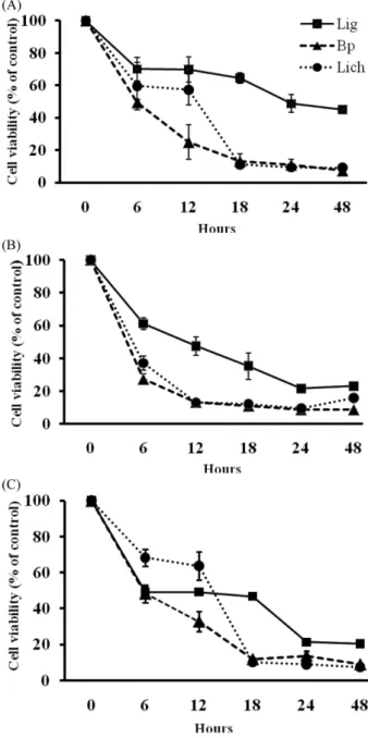
Fig. 1− Changes of cell viability(%) of SK-Hep-1 (A), HeLa (B) and MCF-7 (C) depending on time of treatment with Z- ligustilide (Lig), butylidene phthalide (Bp) and the essential oil fraction of L. chuanxiong (Lich). The values are mean±SD (n=3).
효과 실험에서 적용할 적정 처리 시간을 확정하기 위하여, 3종 의 암세포 배양액에 토천궁 정유 및 그 주성분을 예비실험에서 90% 이상의 세포생장 억제율을 나타낸 0.1 mg/ml로 처리한 후 6시간 간격으로 48시간까지, 암세포주의 시간별 숫자 감소를 MTT의 방법으로 측정 했을 때, 암세포 종류나 처리물질에 따라 암세포 수 변화의 양상은 조금씩 달랐으나 전반 적으로 시료 처 리 6시간까지 가장 급격한 감소율을 나타냈으며, 이 후에도 시 간에 따른 세포 수의 현저한 감소가 진행되어, 24시간 경과 시 최고의 감소율을 나타내고, 이후에는 거의 변화가 없었다(Fig. 1).
종합적으로 살펴보면, 처리시간 경과에 따른 세포 생존율 감소 는 실험한 3종 암세포에서 모두 butylidene phtahlide가 가장 가 파른 생장억제 곡선을 나타냈고, 다음으로 토천궁 정유 분획에 의한 암세포 수 감소 속도가 빠르게 나타났으며, Z-ligustilide 처 리 시 가장 완만한 곡선이 관찰되었다.
이와 같은 실험 결과를 기초로 하여, 토천궁 정유의 농도에 따 른 암세포증식 억제에 대한 비교실험을 하였다. 각 시료를 0.2 mg/
ml로부터 단계적으로 배수 희석하여 0.015 mg/ml까지 만들어서 각 암세포 주 배양액에 처리하여 24시간 배양한 후 MTT의 방
법으로 세포 생존율을 측정하여 비교하고, 각 농도 시료 처리결 과에 의한 암세포주의 상태를 위상차 현미경으로 관찰한 결과는 다음과 같다.
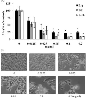
SK-Hep-1 세포 배양액에 토천궁 정유 분획 및 주성분을 각각 처리했을 때, 농도 의존적으로 세포 증식율이 감소함이 확인되 었다(Fig. 2A). Z-ligustilide의 경우 0.05 mg/ml 처리군은 대조군 에 비하여 약 40% 이상 세포 증식이 억제되었으며, 0.2 mg/ml 에서는 약 50% 이상 억제 되었다. Butylidene phthalide의 경우 는 0.025 mg/ml 처리군에서 대조군에 비하여 약 49% 이상 세포 증식이 억제되었으며, 0.1 g/ml에서는 생존율이 10% 정도로 암 세포가 정상적인 생존을 하지 못하였음을 알 수 있다. SK-Hep 1 세포의 생장은 L. chuanxiong 정유 0.025 mg/ml 처리에 의해 약 50% 정도 억제 되었고, 0.1 mg/ml 및 0.2 mg/ml 처리 시 약 90% 이상 억제되어 butylidene phthalide가 보다 강한 암세포 억 제작용을 나타내었다. 또한 토천궁 정유분획 및 그 주 성분의 처 리에 의한 암세포들의 형태변화를 위상차 현미경으로 관찰했을 때 처리시간 경과에 따라, 실험한 3종 암세포들 모두 세포질이 적어지면서 움츠려 들어(shrinking) 크기가 작아지고, 주위의 세
Fig. 3− (A) Effects of Z-ligustilide (Lig), butylidene phthalide (BP) and the essential oil fraction of L. chuanxiong (Lich) on the proliferation of HeLa cells The values are mean±SD (n=3). Significant differences (*p<0.05) between the means were determined by Student's t-test. (B) Changes of cell morphology after treatment of Z-ligustilide on various concentrations (0~0.2 mg/ml) for 24 h were observed by light microscope. Magnification, ×200.
Fig. 2− (A) Effects of Z-ligustilide (Lig), butylidene phthalide (BP) and the essential oil fraction of L. chuanxiong (Lich) on the proliferation of SK-Hep-1 cells. The values are mean±SD (n=3). Significant differences (*p<0.05) between the means were determined by Student's t-test. (B) Changes of cell morphology after treatment of Z-ligustilide on various concentrations (0~0.2 mg/ml) for 24 h were observed by light microscope. Magnification, ×200.
포들과 경계가 분명해 지는 변화가 일어 났고, 일부 세포에서는 부분적으로 형태가 둥글게 변하고 약간 튀어 나오는 blebbing 현 상이 보였다. 정유의 농도가 증가할수록 이러한 형태 변화가 생 긴 세포들이 증가하였고, 부유세포 수도 증가 하였다. 시료에 따 른 뚜렷한 형태 변화의 차이는 관찰되지 않아서 암세포의 형태 적 변형의 정도에는 시료의 종류 보다는 처리 농도가 더욱 큰 영 향을 미침이 확인되었다(Fig. 2B).
HeLa 세포는 Z-ligustilide 0.025 mg/ml 처리에 의해 약 68%
이상 세포 증식이 억제되었으며, 0.1 mg/ml에서는 약 79% 이상 억제 되었다(Fig. 3A). Butylidene phthalide의 경우는 0.0125 mg/ml 처리 군에서 대조 군에 비하여 약 49% 이상 세포증식이 억제되었으며, 0.05 mg/ml에서는 생존율이 9% 정도로 암세포가 정상적인 생존을 하지 못하였음을 알 수 있었다. L. chuanxiong 정유의 경우는 0.025 mg/ml에서 약 69% 정도 억제 되었고, 0.1 mg/ml에서는 약 90% 이상 억제되었다. 결과적으로 HeLa 세포 에 대하여 butylidene phthalide가 가장 강한 암세포 증식 억제 작용을 나타내었다. 또한 Z-ligustilide 처리 후의 HeLa 세포의 형태변화는 MCF-7 의 경우와 유사하게 세포가 부정형 또는 둥 글게 변화하고, 시료의 고농도 처리 시에는 세포파편과, 배양용
기 바닥에서 분리되어 부유하는 세포가 증가함이 관찰되었다 (Fig. 3B).
한편 MCF-7 배양액에 Z-ligustilide 0.025 mg/ml를 처리했을 때 대조군에 비하여 약 57% 이상 세포 증식이 억제되었으며, 0.2 mg/ml에서는 약 80% 이상 억제 되었다(Fig. 4A). Butylidene phthalide의 경우는 0.05 mg/ml 처리군에서는 대조군에 비하여 약 59% 이상 세포증식이 억제되었으며, 0.1 mg/ml에서는 생존 율이 13% 정도로 암세포가 정상적인 생존을 하지 못하였음을 알 수 있었다. L. chuanxiong 정유를 0.025 mg/ml 처리 시 세포증 식이 약 45% 정도 억제 되었고, 0.1 mg/ml에서는 약 90% 이상 억제되었다. 결과적으로 0.025~0.05 mg/ml 농도를 기준으로 볼 때, MCF-7 유방암 세포에 대하여 Z-ligustilide가 L. chuanxiong 정유와 butylidene phthalide 보다 강한 암세포 증식 억제작용을 나타내었으나, 상대적으로 고농도인(0.1~0.2 mg/ml) 처리시에는 반대로 L. chuanxiong 정유와 butylidene phthalide의 억제작용 이 더 강한 것으로 나타났다. Z-ligustilide 농도에 따는 세포형태 변화는 앞의 2종 암세포의 경우와 유사하였으나, 특히 다수의 파 편이 관찰되어, 실험한 암세포 중 가장 심한 세포의 손상을 나타 내었다(Fig. 4B).
이상의 결과를 종합해 보면 butylidene phthalide와 L.
chuanxiong 정유는 MCF-7, 간암세포주인 SK-Hep-1 및 자궁경 부암 세포주 HeLa에 뚜렷한 억제효과 나타내며, 이들 정유가 Z- ligustilide 보다 강한 암세포 증식 억제효과가 있음이 확인되었 다. 특히, 0.1~0.2 mg/ml의 고농도에서는 6시간 이후서부터 뚜 렷한 암세포 억제 효과를 보여 주었다. 이와 같은 결과는 토천궁 의 정유 분획 및 그 주성분 들이 천연 항암제로 개발될 수 있을 가능성을 시사하였다.
참고문헌
1) Cui, F., Feng, L. and Hu, J. : Factors affecting stability of Z- ligustilide int eht volatile of Radix Angelicae sinensis and Ligusticum chuanxiong and its stability prediction. Drug Dev.
Ind. Pharm. 32, 747 (2006).
2) Zschocke, S., Klaiber, I., Bauer, R. and Vogler, B. : HPLC- coupled spectroscopic techniques IUV, IR, NMR) for the structure elucidation of phthalides in Ligusticum chuanxiong.
Mol. Divers. 9, 33 (2005).
3) Lu, Q., Qiu, T.-Q. and Yang, H. : Z-ligustilide inhibits vascular smooth muscle cell proliferation. Eur. J. Pharm. 542, 136 (2006).
4) Hou, Y.-Z., Zhao, G.-R., Yuan, Y.-J., Zhu, G.-G. and Hiltunen, R. : Inhibition of rat vascular smooth muscle cell proliferation by extract of Ligusticum chuanxiong and Angelica sinensis. J.
Ethnopharmacol. 100, 140 (2005).
5) Ozaki, Y., Sekita, S. and Harada, M. : Centrally acting muscle Fig. 4− (A) Effects of Z-ligustilide (Lig), butylidene phthalide (BP)
and the essential oil fraction of L. chuanxiong (Lich) on the proliferation of MCF-7 cells. The values are mean±SD (n=3). Significant differences (*p<0.05) between the means were determined by Student's t-test. (B) Changes of cell morphology after treatment of Z-ligustilide on various concentrations (0~0.2 mg/ml) for 24 h were observed by light microscope. Magnification, ×200.
relaxant effect of phthalides (Z-ligustilide, cnidilide and senkyunolide) obtained from Cnidium officinale Makino. J.
Pharm. Soc. Jap. 109, 402 (1989).
6) Kuang, X., Yao, Y., Du, J. R., Liu, Y.-X., Wang, C.-Y. and Qian, Z. M. : Neuroprotective role of Z-ligustilide against forebrain ischemic injury in ICR mice. Brain Res. 1102, 145 (2006).
7) Zhang, L.-C., Hu, J.-H., Li, L., Gao. L.-H., Zhu, Q.-G., Li, Z. and Wang, Z.-Z. : In Vivo and in Vitro Evaluation of Essential Oils from Ligusticum chuanxiong HORT on the Transdermal Delivery of Flurbiprofen in Rabbits. Biol. Pharm. Bull. 29, 1217 (2006).
8) Tian, J.-W., Fu, F.-H., Jiang, W.-L., Wang, C.-Y., Sun, F. and Zhang, T.-P. : Protective effect of Ligusticum chuanxiong phthalides on focai cerebral ischemia in rats and its related mechanism of action. Chin. Chin. Mat. Med. 30, 466 (2005).
9) Sunny, S.-C., Angela, O.-C., Robert, L.-J. and Ge, L. : Mechanisms underlying the vasorelaxing effects of butylidene- phthalide, an active constituent of Ligusticum chuanxiong, in rat isolated aorta. Eur. Pharmacol. 537, 111 (2006).
10) Matsumoto, K., Kohno, S.-I., Ojima, K., Tezuka, Y., Kadota, S.
and Watanabe, H. : Effects of methylenechloride-soluble fraction of Japanese angelica root extract, Z-ligustilide and butylidenephthalide, on pentobarbital sleep in group housed and socially isolated mice. Life Sci. 62, 2073 (1998).
11) Sim, Y. and Shin S. : Combinatorial anti-Trichophyton effects of Ligusticum chuanxiong essential oil components with antibiotics. Arch. Pharm. Res. 31, 497 (2008).
12) Lim, H. and Shin, S. : Effects of the essential oil components from Ligusticum chuanxiong on proinflammatory mediators of RAW 264.7 macrophage cells. Nat. Prod. Sci. 16, 259 (2010).
13) Sasieni, P. P. : Chemoprevention of cervical cancer. Best Pract.
Res. Clin. Obstet. Gynaecol. 20, 295 (2006).
14) De Flora, S. S. and Ferguson, L.-R. : Overview of mechanisms of cancer chemopreventive agents. Mutat. Res. 591, 8 (2005).
15) Baser, K. H. C. and Buchbauer, G. : Essential Oils, Boca Raton, p. 236 (2010).
16) Zheng, G.-Q., Kenney, P.-M., Zhang, J. and Lam, L. K. T. : Chemoprevention of benzo[a]pyrene-induced forestomach cancer in mice by natural phthalides from celery seed oil.
Nutrition and Cancer 19, 77 (1993).
17) Woods, J.-A., Jewell, C. and O'Brien, N.-M. : Sedanolide, a natural phthalide from celery seed oil: effect on hydrogen peroxide and tert-butyl hydroperoxide-induced toxicity in HepG2 and CaCo-2 human cell lines. In Vitro Mol. Toxicol. 14, 233 (2001).
18) Shoff, S.-M., Grummer, M., Yatvin, M.-B. and Elson, C.-E. : Concentration dependent increase of murine P388 and B 16 population doubling time by the acyclic monoterpene geraniol.
Cancer Res. 51, 37 (1991).
19) Reddy, B.-S., Wang, C.-X., Samaha, H., Lubet, R. R., Steele, V. E., Kelloff, V. E. and Rao, C. V. : Chemoprevention of colon carcinogenesis by dietary perillyl alcohol. Cancer Res. 57, 420 (1997).
20) Crowell, P.-L. : Prevention and therapy of cancer by dietary monoterpenes. J. Nutr. 129, 775 (1999).
21) Phillips, L.-R., Malspeis, L. and Supko, J.-G. : Pharmacokinetics of active drug metabolites after oral administration of perillyl alcohol, an investigational antineoplastic agent, to the dog.
Drug Metab. Dispos. 23, 676 (1995).
22) Lantry, L. E., Zhang, Z., Gao, F., Crist, K. A., Wang, Y. Y., Kelloff, G. J., Lubet, R. A. and You, M. : Chemopreventive effect of perillyl alcohol on 4-(methylnitrosamino)-1-(3- pyridyl)-1-butanone induced tumorigenesis (C3H.HeJ X A/
J)F1 mouse lung. J. Cell. Biochem. Suppl. 27, 20 (1997).
23) Löw-Baselli, A., Huber, W.-W., Käfer, M., Bukowska, K., Schulte-Herrmann, R. and Grasl-kraupp, B. : Failure to demonstrate chemoprevention by the monoterpene oerillyl alcohol during early rat heptocarcinogenesis. Carcinogenesis 21, 1869 (2000).
24) Stark, M.-J., Burke, Y.-D., Mckinzie, J.-H., Ayoubi, A.-S. and Crowell, P.-L. : Chemotherapy of pancreatic cancer with the monoterpene perillyl alcohol. Cancer Lett. 4, 15 (1995).
25) Jahangir, T. and Sultana, S. : Perillyl alcohol protects against Fe- NTA-induced nephrotoxicity and early Tu, or promotional events in rat experimental model. Evid. Based complement.
Altern. Med. 4, 439 (2007).
26) Wattenberg, I.-W. : Inhibition of azoxymethane-induced neoplasia of the large bowel by 3-hrdroxy-3,7,11-trimethyl- 1,6,10-dodecatriene (nerolidol). Carcinogenesis 12, 151(1991).
27) Yu, S.-G., Hildebrandt, L.-A. and Elson, C.-E. : Geraniol, an inhibitor of mevalonate biosynthesis, suppresses the growth of hepatomas and melanomas transplanted to rats and mice. J.
Nutr. 125, 2763 (1995).
28) Badary, O. A. and El-Din, A.M.G. : Antitumor activity of thymoquinine against fibrosarcoma tumorgeneis. Cancer Mol.
Biol. 7, 1515 (2000).
29) Zeitinoglu, H., Incesu, Z. and Baser, K. H. C. : Inhibition of DNA synthesis by crvacrol in mouse myoblast cells bearing a human N-RAS oncogene. Phytomedicine 10, 292 (2003).
30) Shon, Y.-H., Kim, H.-G. and Nam, K.-S. : Effect of Cnidii Rhizoma water extract on chemopreventive enzymes for hepatocarcinoma. Kor. J. Pharmacogn. 34, 297 (2003).
31) Nam, K.-S., Son, O.-L., Lee, K.-H., Cho, H.-J. and Shon, Y.-H. : Effect of Cnidii Rhizoma on proliferation of breast cancer cell, nitric oxide production and ornithine decarboxylase activity.
Kor. J. Pharmacogn. 35, 283 (2004).