Original Paper
Horm Res Paediatr 2015;83:242–251 DOI: 10.1159/000368657
Identification of Candidate Gene Variants in Korean MODY Families by Whole-Exome Sequencing
Ye Jee Shim Jung Eun Kim Su-Kyeong Hwang Bong Seok Choi Byung Ho Choi Eun-Mi Cho Kyoung Mi Jang Cheol Woo Ko
Department of Pediatrics, Kyungpook National University School of Medicine, Daegu , Republic of Korea
dictive program, Thr207Ile in PTPRD was considered patho- genic. Conclusions: Whole-exome sequencing is a valuable method for the genetic diagnosis of MODY. Further evalua- tion is necessary about the role of PTPRD, SYT9 and WFS1 in normal insulin release from pancreatic beta cells.
© 2015 S. Karger AG, Basel
Introduction
Maturity-onset diabetes of the young (MODY) is one of monogenic diabetes mellitus caused by a single-gene defect [1–3] . MODY is characterized by a young age onset before 25 years and autosomal dominant inheritance and shows an impaired insulin secretion from pancreatic beta cell without autoimmune cause. To date, 13 MODY genes have been identified worldwide – HNF4A, GCK, HNF1A, PDX1, HNF1B, NEUROD1, KLF11, CEL, PAX4, INS, BLK , ABCC8 and KCNJ11 [1–5] .
Key Words
Maturity-onset diabetes of the young · Type 2 diabetes mellitus · Whole-exome sequencing · PTPRD · SYT9 · WFS1
Abstract
Aims: To date, 13 genes causing maturity-onset diabetes of the young (MODY) have been identified. However, there is a big discrepancy in the genetic locus between Asian and Caucasian patients with MODY. Thus, we conducted whole- exome sequencing in Korean MODY families to identify causative gene variants. Methods: Six MODY probands and their family members were included. Variants in the dbSNP135 and TIARA databases for Koreans and the variants with minor allele frequencies >0.5% of the 1000 Genomes database were excluded. We selected only the functional variants (gain of stop codon, frameshifts and nonsynony- mous single-nucleotide variants) and conducted a case-con- trol comparison in the family members. The selected vari- ants were scanned for the previously introduced gene set implicated in glucose metabolism. Results: Three variants c.620C>T:p.Thr207Ile in PTPRD , c.559C>G:p.Gln187Glu in SYT9 , and c.1526T>G:p.Val509Gly in WFS1 were respectively identified in 3 families. We could not find any disease-caus- ative alleles of known MODY 1–13 genes. Based on the pre-
Received: June 16, 2014 Accepted: September 22, 2014 Published online: March 7, 2015
HOR MON E RE SE ARCH I N PÆDIATRIC S
This article has been accepted for poster presentation and has been nominated for a President Poster Award at the 53rd Annual Meet- ing of the European Society of Paediatric Endocrinology, September 18–20, 2014, Dublin, Ireland.
In the Caucasian population, HNF1A, GCK , HNF4A and HNF1B mutations comprise more than 95% of causes of MODY, and the others are known to be very rare [1, 3] . There are close genotype-phenotype correlations be- tween the genetic locus of mutation and the clinical man- ifestation in MODY patients. Thus, an accurate diagnosis is important for special implications in the individualized treatment for a generic counselling of the family and for the prognosis.
However, there is a big discrepancy in the disease- causing genetic locus between the Asian and Caucasians MODY patients. In Korea, only 10% of 40 MODY or ear- ly-onset type 2 diabetes patients showed a known MODY gene defect ( HNF1A 5%, GCK 2.5% and HNF1B 2.5%) among MODY 1–9 genes [6, 7] . Also in Japan and China were only 10–20% of known MODY gene defects report- ed [8–11] . In other words, there are much more ‘MODY X’ patients in Asia, whose range is 80–90%.
Recently, next generation sequencing methods have been applied for MODY X patients to discover unknown genetic variants [12, 13] . Whole-exome sequencing is a useful method to discover unknown rare germline mutations of mendelian disorders [14] . Thus, in this study, we conducted exome sequencing in Korean MODY patients to identify new candidate gene variants and to compare the results with Caucasian patients. This research was intended for MODY probands and their family members with available blood samples.
Materials and Methods
Ethics Statement
This research was approved by the relevant Ethics Committees (The Institutional Review Board in Kyungpook National Univer- sity Hospital, approval No. 2013-07-037). This study was per- formed in accordance with the Declaration of Helsinki. Informed consent was obtained from all study subjects before blood sam- pling.
Subjects
Six MODY probands and their family members with available blood samples were included for whole-exome sequencing. Five MODY subjects were selected using the following inclusion crite- ria: (1) fasting serum C-peptide >0.6 ng/ml, (2) negative islet cell antibody (1A-2) test, glutamic acid decarboxylase (GAD) antibody test and insulin autoantibody (IAA) test, (3) early age at diabetes onset before 25 years, (4) autosomal dominant inheritance and (5) family history of diabetes in at least 3 generations. The fasting se- rum C-peptide in one proband was 0.5 ng/ml; however, we in- cluded her family in our study because she satisfied all other inclu- sion criteria. Genetic testing for MODY is also recommended for patients classified as type 1 diabetes and with strong family history because those patients may have a progression to severe hypergly-
cemia and insulin dependence as time goes on in the case of HNF1A or HNF4A alteration [1] . The partial pedigrees for the par- ticipants are described in figure 1 . The clinical characteristics of all 6 probands and their family members with diabetes are described in table 1 . We followed the classification of body mass index (BMI) and weight status for the Asian population for adults [15] because Asian populations including Korean have higher diabetes preva- lence at lower cutoff values of BMI [16] . In the case of children and adolescents (age from 2 to 20 years), we followed the criteria as follows: underweight (<5th percentile), normal weight ( ≥ 5 and
<85 percentile), overweight ( ≥ 85 and <95 percentile), and obesity ( ≥ 95 percentile) [17, 18] . The growth curve of BMI percentile was based on Korean pediatric data.
DNA Extraction and Quality Estimation
Genomic DNA was extracted from the subject’s peripheral blood using the Wizard DNA purification Kit (Promega Corpora- tion, Madison, Wisc., USA) based on the manufacturer’s manual.
The DNA quantity measurement was done by Picogreen (Invitro- gen, Calif., USA) method using Victor 3 fluorometry. The mea- surement of DNA purity was done by NanoDrop instrument.
DNA condition assessment was done by the gel electrophoresis method.
Whole-Exome Sequencing
Capturing of whole exome was performed from the subjects’
genomic DNA using SureSelect Human All Exon V4 (Agilent Technologies, Calif., USA). Sequencing was undertaken by HiSeq2000 (Illumina, Calif., USA). The generated reads were mapped against UCSC hg19 (http://genome.ucsc.edu/) using a mapping program Burrow-Wheeler Alignment tool (http://bio- bwa.sourceforge.net/). The PCR duplicates were removed by the PICARD tool (http://picard.sourceforge.net/). The single nucleo- tide variants (SNVs) and insertions-deletions (Indels) were detect- ed by SAMTOOLS (http://samtools.sourceforge.net/). The vari- ants were filtered to include only those with >8 read depth and >30 mapping quality, and annotated by ANNOVAR (http://www.
openbioinformatics.org/annovar/).
Identification of Disease-Causative Candidate Variants Variants were filtered out in the dbSNP135 and TIARA data- base for Koreans (http://tiara.gmi.ac.kr/) to identify the disease- causing mutations. Variants with minor allele frequencies >0.5%
of the 1000 Genomes database (http://www.1000genomes.org/) were also excluded. We selected only the functional variants (gain of stop codon, frameshifts and nonsynonymous SNVs) as patho- genic mutations. Then, we conducted a case-control comparison in the family members and selected those variants commonly found in both the MODY proband and the family member with diabetes which were not found in a healthy family member. Fi- nally, the selected variants were scanned for the previously intro- duced gene set implicated in glucose metabolism [12] . These are the genes with an essential role in pancreatic beta cells, genes pre- viously known to cause monogenic diabetes or associated syn- dromes and genes from the genome-wide association data of type 2 diabetes [19–26] . The list of genes of interest is shown in table 2 . The finally selected candidate variants were verified by the in silico analysis database, PROVEAN (http://provean.jcvi.org/index.
php/) [27] , SIFT (http://sift.jcvi.org/) [28] , and PolyPhen-2 (http://
genetics.bwh.harvard.edu/pph2/) [29] . The final corresponding
Table 1.
Characteristics of the 6 clinical MODY probands and their family members with diabetes in this study
Family 1 2 3 4 5 6
proband her father
her mother
prob and proband her mother’s sister
proband her
mother
proband his mother
proband his father
his mother
Sex/age, years F/17 M/45 F/43 F/23 F/16 F/38 F/21 F/44 M/13 F/47 M/18 M/50 F/46
Age at diagnosis, years 15 35 33 14 14 26 12 30 11 33 16 34 30
BMI, kg/m2 22.2a 20.6b 27.1b 18.4a 29.0a 26.7a 13.1a 18.8b 23.1a 23.7b 23.9a 23.4b 27.1b
BMI percentile 75th 25–50th >95th <5th 90–95th 75–85th
Weight status normal normal obesity normal obesity overweight underweight normal overweight normal normal normal obese
HbA1c, % 10.1 11.3 10.4 13.7 10.0 9.9 9.6 8.7 12.2 n.a. 7.9 n.a. n.a.
Fasting C-peptide, ng/dl 1.73 1.86 2.02 3.20 4.41 0.80 0.50 0.80 2.39 n.a. 2.84 n.a. n.a.
Diabetic ketoacidosis no n.a. n.a. no no no no no no n.a. no n.a. n.a.
Total cholesterol, mg/dl 160 n.a. n.a. 130 n.a. 143 202 186 140 n.a. 131 n.a. n.a.
CRP, mg/dl 0.00 n.a. n.a. n.a. 0.23 0.55 0.06 0.00 1.05 n.a. 0.03 n.a. n.a.
Treatment insulin insulin No No OHA insulin and
OHA
insulin insulin and OHA
insulin OHA OHA OHA insulin
Complication no no no no no yes no yes no n.a. no no no
Last HbA1c, % 6.3 n.a. n.a. 5.3 8.6 9.5 8.5 7.7 8.4 n.a. 8.9 n.a. n.a.
n.a. = Not available; OHA = oral hypoglycemic agent. a Initial BMI at diagnosis of diabetes. b These values are the last BMI because initial BMI values at diagnosis were not avail- able in family members.
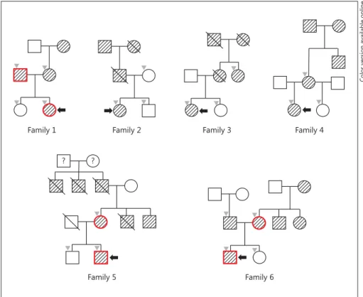
Fig. 1.
Partial pedigrees of the 6 clinical MODY families involved in this study.
Shaded symbols with deviant crease lines indicate the individuals with diabetes.
Crossed symbols denote the deceased indi- viduals. Black arrows indicate respective probands in each family. Gray inverted tri- angles denote the family members whose blood sampling was available. Red-bor- dered symbols means the subjects who shared a common variant among the genes of interest for MODY and not in other fam- ily members (color refers to the online ver- sion only).
Color version available online
Family 1 Family 2 Family 3 Family 4
Family 5
? ?
Family 6
genomic regions were amplified using an automatic genetic ana- lyzer Dr. MAX DNA Polymerase (MG Taq-HF DNA Polymerase) kit. The PCR products were confirmed by Sanger sequencing in both forward and reverse directions.
Results
Whole-Exome Sequencing and Filtering
We performed whole-exome sequencing in 6 MODY probands and 14 family members. The number of total reads [mean (range)] was 91,756,007 (75,996,864 ∼ 148,983,998) and the observed variants were 68,801 (67,791 ∼ 70,073).
After filtering out the variants with a frequency greater than
0.5% in the 1000 Genomes database and in the dbSNP135 and TIARA databases, residual exonic mutations were 464 (428 ∼ 481) in each MODY proband, and 236 (224 ∼ 250) were functional variants of them. After a case-control com- parison between probands and family members, the num- ber of variants with disease-causing possibility was de- creased to 50 (31 ∼ 63) in each MODY proband in one-side pedigree. After scanning regarding the specific gene list implicated in the glucose metabolism, 3 variants in PTPRD , SYT9 and WFS1 were identified in families 1, 5 and 6, respectively. Three variants were verified by PROVEAN, SIFT, and PolyPhen-2. Because there was no disease- causing variant in family 4 and the proband had insulin dependent manifestations, we checked the respective function of residual 52 genes in family 4. Finally, one variant in PTPRN2 , the well-known type 1 diabetes gene, was identified in family 4. We were not able to detect the disease- causing mutation in families 2 and 3. A summary of the results of exome sequencing and the process of variant reduction are described in table 3 for all MODY probands.
Identification of Potential Disease-Causative Variants of MODY
PTPRD in Family 6
In family 6, one nonsynonymous mutation, exon6:
c.620C>T:p.Thr207Ile, in PTPRD was found in both the proband and his mother. The proband visited our hospital because of a fasting hyperglycemia (168 mg/dl) during the regular medical checkup. He had no diabetes-specific symptoms. His HbA1c was 7.9% and fasting C-peptide 2.84 ng/ml. IA-2, GAD antibody and IAA were negative.
His parents were both diabetics and he had a strong ma- ternal family history (mother, mother’s brother, mother’s sister and maternal grandmother). He was treated with an oral hypoglycemic agent, and his last follow-up HbA1c was 8.9%. His mother was diagnosed with diabetes at 30 years of age. She has been treated with insulin by her physician. In the predictive program, Thr207Ile in PTPRD was deleterious/damaging/possibly damaging (table 4).
PTPRD (protein tyrosine phosphatase receptor type delta) is a member of a receptor type IIA subfamily, which also includes receptor type F (PTPRF) and sigma (PTPRS).
A genetic alteration of PTPRD is associated with multifactorial forms of diabetes according to a genome- wide association study [25, 30] . Especially in Han Chinese, PTPRD alteration seems to cause a progression to diabetes through insulin resistance [31] . In addition, an enhanced phosphatase activity can cause defective insulin secretion by a reduced ATP/ADP ratio or the phosphorylation of
Table 2.
The list of genes of interest for MODY used in this study MODY ABCC8, BLK, CEL, GCK, HNF1A, HNF1B, HNF4A, INS, KCNJ1, KLF11, NEUROD1, PAX4, PDX1
Genome-wide association studies
ADAM30, ADAMTS9, ADCY5, ADRA2A, ARAP1, BCL11A, C2CD4B, CAMK1D, CDC123, CDKAL1, CDKN2A, CDKN2B, CHCHD9, CRY2, DGKB, DUSP8, DUSP9, FADS1, FTO, G6PC2, GCKR, GIPR, GLIS3, HHEX, HMGA2, HNF1A, HNF1B, IDE, IGF1, IGF2BP2, IRS1, ITGB6, JAZF1, KCNJ11, KCNQ1, KLF14, LGR5, MADD, MTNR1B, NOTCH2, PPARG, PRC1, PROX1, PTPRD, RBMS1, SLC2A2, SLC30A8, SRR, TCF7L2, THADA, TMEM195, TP53INP1, TSPAN8, VEGFA, VPS13C, WFS1, ZBED3, ZFAND6 Neonatal diabetes
mellitus
ABCC8, GCK, INS, INS, KCNJ11, NEUROG3, RFX6
Congenital hyperinsulinism of infancy
ABCC8, GCK, GLUD1, HADH, HNF4A, INSR, KCNJ11, SLC16A1
Syndrome AGPAT2, AKT2, ALMS1, BSCL2, CAV1, CISD2, EIF2AK3, FXN, HFE, LMNA, LMNB2, WFS1, ZAC
Candidate APPL1, FOXA1, FOXA2, FOXA3, GATA4,
GATA6, INSM1, ISL1, LMX1A, MAFA,
MAFB, MNX1, MYT1, NKX2-2, NKX6-1,
ONECUT1, PAX6, PBX1, PTF1A, SOX2,
SOX4, SOX9, SREBF1, SYT9, UCP2
This is the previously introduced gene set implicated in glucose
metabolism with an essential role in pancreatic beta cells, genes
previously known to cause monogenic diabetes or associated syn-
dromes and genes from the genome-wide association data of type
2 diabetes. Adapted from references [1–5, 12, 19–26].
proteins regulating the insulin release in pancreatic beta cells [32] . Considering the structural similarity of recep- tor type IIA subfamily members, we kept in mind the pos- sibility that PTPRD mutation should concern not only insulin resistance but also insulin release from pancreatic beta cells.
SYT9 in Family 5
In family 5, another nonsynonymous SNV, exon3:
c.559C>G:p.Gln187Glu, in SYT9 was found in both the proband and his mother. The proband visited our hospi- tal at the age of 11 years because of a random hyperglyce- mia (295 mg/dl) noted at a regular medical checkup. He had persistent polyphagia, polydipsia, polyuria and weight loss for 1 year. His initial HbA1c was 12.1% and
fasting C-peptide 4.67 ng/ml. IA-2, GAD antibody and IAA were all negative. He had a strong maternal family history of diabetes (mother, 2 mother’s brothers, mater- nal grandfather and 2 maternal grandfather’s brothers).
His last follow-up HbA1c was 8.4% during insulin infu- sion. His mother was diagnosed with diabetes at 33 years.
Although his mother has been treated with an oral hypo- glycemic agent, additional insulin was recommended by her physician. Because Gln187Glu in SYT9 was neutral/
tolerated/benign in the predictive program (table 4), the role of SYT9 in MODY needs more study.
Synaptotagmin 9 (SYT9) is one of the synaptotagmin family proteins known to play a role as an important Ca
2+sensor in exocytosis. The glucose-induced hormone release was decreased by the reduction of the expression
Table 3.
Results of whole-exome sequencing of 6 MODY probands and the process of variant reduction Family of the proband
1 2 3 4 5 6
Total reads 75,996,864 81,447,800 91,148,066 148,983,998 101,749,122 84,199,586
Mappable reads 75,264,066 81,154,224 90,840,274 147,621,080 100,805,966 83,962,588 On-target reads 55,621,495 59,864,707 66,864,766 106,053,901 74,527,885 60,389,279
Coverage of target region (>10×) 97.7% 98.0% 98.1% 98.6% 98.3% 98.0%
Mean read depth of target regions 93.5 100.6 112.7 179.7 125.4 101.6
Total SNPs 69,063 68,637 69,341 68,858 69,200 67,791
Exonic regions 20,178 19,891 20,044 18,981 19,859 19,971
After filtering
a468 470 428 481 476 463
Functional variants
b250 224 224 235 238 243
After case-control comparison 98 (43 paternal and 55 maternal)
51 31 52 63 120 (60 paternal
and 60 maternal) In genes of interest 1 (WFS1,
paternal side)
0 0 0 1 (SYT9) 1 (PTPRD,
maternal side)
a
The variants with a frequency of ≤0.5% in the 1000 Genomes database and not in the dbSNP135 or the TIARA database.
bGain of stop codon, frameshifts, and nonsynonymous SNVs.
Table 4.
Finally identified variants in MODY families and the results of the predictive program
Family Gene Chr:position Variant Frequency in 1000
Genomes/dbSNP135/
TIARA
PROVEAN SIFT Polyphen-2
1
WFS1
4:6303048 exon8:c.T1526G:p.Val509Gly 0/0/0 deleterious (–3.35) tolerated (0.086) benign (0.002) 5SYT9
11:7334687 exon3:c.C559G:p.Gln187Glu 0/0/0 neutral (0.02) tolerated (0.715) benign (0.059) 6PTPRD
9:8524975 exon6:c.C620T:p.Thr207Ile 0/0/0 deleterious (–3.80) damaging (0.003) possibly damaging(0.796)
Figures in parentheses indicate scores.
of both SYT5 and 9 isoforms, indicating that they are directly involved in the Ca
2+-dependent stimulation of exocytosis [33] . In addition, the insulin release induced by glucose was decreased in the pancreas islets of rats after an adenovirus-mediated silencing of SYT9 [34] .
WFS1 in Family 1
In family 1, another nonsynonymous SNV, exon 8:
c.1526T>G:p.Val509Gly, in WFS1 was identified in both the proband and her father. The proband visited the clinic at the age of 15 years due to glucosuria noticed at a regular checkup in her school. She had a general weakness and polydipsia. Her initial HbA1c was 10.1%, and fasting C-peptide was 1.73 ng/ml. IA-2, GAD antibody and IAA were all negative. Her last follow-up HbA1c was 6.3%
following an intermittent low-dose insulin treatment (0.5 IU/kg, once a day, 3 times per week). She did not need insulin injections after physical activity. Her father, with the same variant in WFS1 , was diagnosed as having diabetes at the age of 35 years. His HbA1c was 11.3% and fasting C-peptide 1.86 ng/ml. Although he initially had been treated with insulin by his physician, he was not on any medication at the time of the study. In the predictive program PROVEAN, Val509Gly in WFS1 was deleterious (table 4).
WFS1 is the gene which encodes Wolframin, the transmembrane protein of the endoplasmic reticulum [23] . A homozygous mutation of WFS1 is associated with an autosomal recessive inheritance of Wolfram syndrome, which means the complex of diabetes mellitus, diabetes insipidus, hearing impairment and optic atrophy [35] , while the heterozygous mutation of WFS1 causes an ear- ly onset of autosomal dominant diabetes without other syndromic appearances [36] . The proband and her father in family 1 had no specific symptoms of Wolfram syn- drome except diabetes mellitus. Although family 1 shows a strong maternal history of diabetes (mother and mater- nal grandmother), we suppose that p.Val509Gly in WFS1 as a potential variant caused MODY because the proband and her father had very similar clinical manifestations.
An offspring study will be necessary in the future.
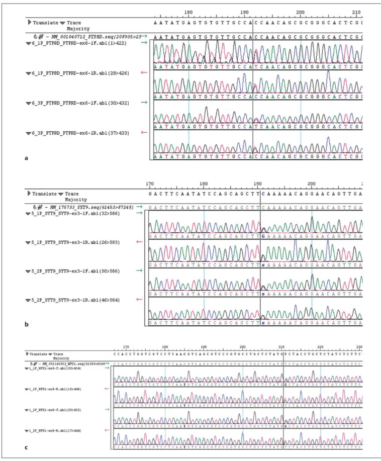
Validation by Sanger Sequencing
The final 3 candidate gene variants for MODY, p.
Thr207Ile in PTPRD , p.Gln187Glu in SYT9 and p.Val509Gly in WFS1 were confirmed by Sanger sequenc- ing and are shown in figure 2 . The primers used are as follows: ATTGAATCGACGTTGAGTGG (forward) and CAAACTCCAAGCCTCAGGAC (reverse) in PTPRD , AAAGGCCCACAGATCTAAGC (forward) and GCC
AAGTCTAGGAAGTGATCC (reverse) in SYT9 , CTT TACCGTGACCAGCTACC (forward) and TCTTGG TGAGCTCCAGAGAC (reverse) in WFS1 .
Exclusion from MODY PTPRN2 in Family 4
In family 4, another nonsynonymous SNV, exon 1:c.85A>C:p.Arg29Ser, in PTPRN2 was found in both the proband and her mother. The proband was referred to our hospital at the age of 12 years because of a fasting hy- perglycemia (259 mg/dl) that was noticed in the clinic.
She had polydipsia, polyuria, polyphagia and general weakness for 2 weeks. Her initial HbA1c was 9.6%, and the fasting C-peptide was 0.05 ng/ml. IA-2, GAD anti- body, and IAA were all negative. She had a strong mater- nal family history of diabetes (mother, maternal grandfa- ther and maternal grandmother). Her last follow-up HbA1c was 8.5% during insulin treatment. Her mother was diagnosed with diabetes at the age of 30 years and has been treated with insulin and oral hypoglycemic agent.
She had an early diabetic peripheral neuropathy. The clinical manifestations of the proband and her mother are very similar. Both of them had Hashimoto’s thyroiditis and took oral thyroid hormone.
PTPRN2 is a well-verified gene causing type 1 diabetes and is also known as IA-2beta or phogrin . Both, IA-2beta and IA-2 are precursors for autoantigens of pancreatic islet cells [37] , and their intracellular domains are very similar (73% identical) [38] . An overexpression of IA- 2beta reduced the insulin secretion stimulated by glucose in insulinoma cells [39] . The clinical manifestations in family 4 were in accordance with a PTPRN2 alteration considering their relatively low C-peptide level and accompanying autoimmune thyroid disease with negative laboratory tests for IA-2, GAD antibody and IAA. Thus, family 4 was ruled out to have MODY by genetic testing.
Family 4 seems to belong to the rare group of autosomal dominantly inherited type 1 diabetes, which is known to occur in <10% of diabetes patients [3] .
Discussion
We conducted whole-exome sequencing in 6 clinical
MODY families to identify MODY genetic variants in
Korea for the first time. Surprisingly, we could not find
any disease-causative alleles among known MODY
1–13 genes. One synonymous exonic mutation, exon
2:c.294G>A:p.Gln98Gln, in PAX4 (MODY9) was iden-
tified in both the proband and her mother in family 1.
Fig. 2.
Validation by Sanger sequencing for final potential disease-causative gene variants for MODY.
ac.620C>T:
p.Thr207Ile in PTPRD .
bc.559C>G:p.Gln187Glu in SYT9 .
cc.1526T>G:p.Val509Gly in WFS1 .
Color version available online
a
b
c
However, it was excluded through the filtering step con- sidering its prioritization because it is nonfunctional (silent).
Exome sequencing is a potential, economical method to identify novel genetic variants of rare monogenic disorders. However, it cannot be a panacea for all the rare monogenic disorders. The application of exome sequencing has both an advantage and disadvantage for MODY cases. Because MODY cases have an autosomal dominant inheritance, we can filter many meaningless variants which exist in healthy controls. On the other hand, it is a disadvantage that MODY is a heterogeneous group of monogenic diabetes. It is difficult to narrow the range of variants by filtering across only multiple unrelated, affected probands. We may overcome this problem by acquiring the blood of multiple affected fam- ily members from within a pedigree and by the applica- tion of a known interested gene list.
In this study, we mostly conducted the case-control method for family members of two generations due to practical issues like death of grandparents or failure to obtain consent from distant relations. We could reduce the number of functional variants from 220–250 to 30–60 in one side pedigree. In the case of two-generation fami- lies, we could reduce the variants from 1/4–1/5. In the case of family 3, it was possible to obtain the blood of the sister of the proband’s mother, and so we could reduce the functional variants to 1/7. However, it was not enough to identify novel variants which do not exist in the genes of interest list. We closely follow them with the idea of further genetic evaluation like linkage analysis with other distant relations.
In a normal pancreatic beta cell, the glucose transpor- tation by a GLUT-2 transporter allows glucokinase to phosphorylate glucose. ATP generated via glycolysis and Kreb cycle in mitochondria closes the potassium channel resulting in membrane depolarization and opening of the calcium channel. Ca
2+entry results in an exocytosis of insulin from the endoplasmic reticulum through secre- tory granules [1, 3] . The known MODY genes are in- volved in each step of exocytosis of insulin from the pan- creatic beta cell. For example, the relatively frequent MODY genes HNF4A , HNF1A and HNF1B encode tran- scriptions factors. A GCK mutation reduces the glucoki- nase activity and glucose phosphorylation. The latest MODY genes, ABCC8 and KCNJ11 , are the genes coding the protein subunit of the beta cell potassium channel [1, 3, 23] .
In this study, 3 potential candidate gene variants for MODY were identified in PTPRD, SYT9 and WFS1 . Con-
sidering the established roles of these genes or their fam- ily genes, we suppose the respective roles of them in the pancreatic beta cell as follows: (1) PTPRD should involve the generation of ATP or phosphorylation of proteins regulating the insulin release, (2) SYT9 alteration should cause an impairment of the a Ca
2+channel resulting in decreased exocytosis of insulin stimulated by glucose, (3) WFS1 mutation causes stress to the endoplasmic reticu- lum of the pancreatic beta cell resulting in a reduced in- sulin secretion.
PTPRD is a member of a receptor type IIA subfamily, which also includes PTPRF and PTPRS. They are known to be implicated in neural growth and regeneration, metabolic regulation and cancer [40] . PTPRF knockout mice showed both lower fasting insulin and glucose, suggesting a heightened level of insulin sensitivity [41] . Moreover, an increased expression of PTPRF in the muscle causes whole-body insulin resistance in mice most likely due to the dephosphorylation of specific reg- ulatory phosphotyrosines on insulin receptor substrate proteins [42] . In PTPRS knockout mice, pancreatic islets were hypoplastic, and the immunoreactivity of insulin was decreased [43] . The glucose homeostasis is altered in mice lacking PTPRS [44] . An overexpression of PTPRS in beta cells suggests an increased consumption or degradation of ATP resulting in an impaired glucose- induced insulin secretion in hereditary diabetic rats [32] . Considering the structural similarity of receptor type IIA subfamily members, we also suggest that PTPRD is thought to have a role in a decreased insulin secretion and beta cell failure [30] .
SYT is involved in a Ca
2+-regulated secretion and has been suggested to perform a general Ca
2+sensor on the membrane of secretory vesicles in neuronal cells [33, 45, 46] . The inhibition of SYT9 by direct antibodies decreased the calcium-induced norepinephrine release from PC12 cells in rats [45] . An acute deletion of SYT9 in striatal neurons severely impaired a fast synchronous release [46] . The pancreatic beta cell is another example of Ca
2+channel-mediated exocytosis. Iezzi et al. [34] reported that the glucose- or tolbutamide-induced insulin release was decreased in pancreas islets of rats after the adenovirus-mediated silencing of SYT9. On the other hand, there was no decline of a glucose-induced insulin secretion in genetic knockout SYT9 mice [47] . Because there are differences in the glucose metabolism between rats, mice and human, more studies are needed about the role of SYT9 in diabetes mellitus.
WFS1 null mice and genetic association studies sug-
gest a role for the WFS1 gene in insulin secretion [48–50] .
In a previous study, another novel variant, c.2017T>C:
p.Arg703Cys, in WFS1 was also found in a Norwegian MODY family; however, the authors suggested further evaluation of the role of WFS1 in MODY considering an inheritance pattern [12] . The localization of the WFS1 protein at the endoplasmic reticulum suggests that it has physiological functions in membrane trafficking, secre- tion, processing or regulation of calcium homeostasis.
Disturbances or overloading of these functions induce stress of the endoplasmic reticulum [51] . This hypothesis was demonstrated by a functional study [36] .
In conclusion, whole-exome sequencing is a valuable method for the genetic diagnosis of MODY. We suggest further evaluation of PTPRD , SYT9 and WFS1 in glucose metabolism and normal insulin release from pancreatic beta cell in other Asian countries. A large-scale control
study in a local population cohort was not performed in this study. However, this research is valuable despite this limitation because all the 3 variants have a 0% frequency in the 1000 Genomes, dbSNP135 and TIARA databases for Koreans, and they were not found in diabetes-nega- tive family members.
Acknowledgement
This work was supported by Kyungpook National University Industry Academic Cooperation Foundation (2014).
Disclosure Statement
The authors declare that they have no conflict of interest.
References
1 Fajans SS, Bell GI, Polonsky KS: Molecular mechanisms and clinical pathophysiology of maturity-onset diabetes of the young. N Engl J Med 2001; 345: 971–980.
2 Bonnefond A, Froguel P, Vaxillaire M: The emerging genetics of type 2 diabetes. Trends Mol Med 2010; 16: 407–416.
3 Henzen C: Monogenic diabetes mellitus due to defects in insulin secretion. Swiss Med Wkly 2012; 142:w13690.
4 Bowman P, Flanagan SE, Edghill EL, Dam- huis A, Shepherd MH, Paisey R, Hattersley AT, Ellard S: Heterozygous ABCC8 muta- tions are a cause of MODY. Diabetologia 2012; 55: 123–127.
5 Bonnefond A, Philippe J, Durand E, Dechau- me A, Huyvaert M, Montagne L, Marre M, Balkau B, Fajardy I, Vambergue A, Vatin V, Delplanque J, Le Guilcher D, De Graeve F, Lecoeur C, Sand O, Vaxillaire M, Froguel P:
Whole-exome sequencing and high through- put genotyping identified KCNJ11 as the thir- teenth MODY gene. PLoS One 2012; 7:e37423.
6 Hwang JS, Shin CH, Yang SW, Jung SY, Huh N: Genetic and clinical characteristics of Korean maturity-onset diabetes of the young (MODY) patients. Diabetes Res Clin Pract 2006; 74: 75–81.
7 Hwang JS: MODY syndrome. J Korean Soc Pediatr Endocrinol 2010; 15: 1–6.
8 Iwasaki N, Oda N, Ogata M, Hara M, Hinokio Y, Oda Y, Yamagata K, Kanematsu S, Ohga- wara H, Omori Y, Bell GI: Mutations in the hepatocyte nuclear factor-1alpha/MODY3 gene in Japanese subjects with early- and late- onset NIDDM. Diabetes 1997; 46: 1504–1508.
9 Nishigori H, Yamada S, Kohama T, Utsugi T, Shimizu H, Takeuchi T, Takeda J: Mutations in the hepatocyte nuclear factor-1 alpha gene (MODY3) are not a major cause of early-on-
set non-insulin-dependent (type 2) diabetes mellitus in Japanese. J Hum Genet 1998; 43:
107–110.
10 Tonooka N, Tomura H, Takahashi Y, Oniga- ta K, Kikuchi N, Horikawa Y, Mori M, Takeda J: High frequency of mutations in the HNF- 1alpha gene in non-obese patients with diabe- tes of youth in Japanese and identification of a case of digenic inheritance. Diabetologia 2002; 45: 1709–1712.
11 Xu JY, Dan QH, Chan V, Wat NM, Tam S, Tiu SC, Lee KF, Siu SC, Tsang MW, Fung LM, Chan KW, Lam KS: Genetic and clinical char- acteristics of maturity-onset diabetes of the young in Chinese patients. Eur J Hum Genet 2005; 13: 422–427.
12 Johansson S, Irgens H, Chudasama KK, Molnes J, Aerts J, Roque FS, Jonassen I, Levy S, Lima K, Knappskog PM, Bell GI, Molven A, Njølstad PR: Exome sequencing and genetic testing for MODY. PLoS One 2012; 7:e38050.
13 Tanaka D, Nagashima K, Sasaki M, Funako- shi S, Kondo Y, Yasuda K, Koizumi A, Inaga- ki N: Exome sequencing identifies a new can- didate mutation for susceptibility to diabetes in a family with highly aggregated type 2 dia- betes. Mol Genet Metab 2013; 109: 112–117.
14 Bamshad MJ, Ng SB, Bigham AW, Tabor HK, Emond MJ, Nickerson DA, Shendure J:
Exome sequencing as a tool for Mendelian disease gene discovery. Nat Rev Genet 2011;
12: 745–755.
15 Wildman RP, Gu D, Reynolds K, Duan X, He J: Appropriate body mass index and waist cir- cumference cutoffs for categorization of over- weight and central adiposity among Chinese adults. Am J Clin Nutr 2004; 80: 1129–1136.
16 Jih J, Mukherjea A, Vittinghoff E, Nguyen TT, Tsoh JY, Fukuoka Y, Bender MS, Tseng W, Kanaya AM: Using appropriate body mass in-
dex cut points for overweight and obesity among Asian Americans. Prev Med 2014; 65:
1–6.
17 Sketon JA, Rudolph CD: Overweight and obe- sity: nutrition; in Kliegman RM, Jenson HB, Behrman RE, Stanton BF (eds): Nelson Text- book of Pediatrics, ed 18. Philadelphia, Saun- ders, 2007, pp 234–236.
18 Gahagan S: Overweight and obesity: nutri- tion; in Kliegman RM, Stanton BF, St Geme III JW, Schor NF, Behrman RE (eds): Nelson Textbook of Pediatrics, ed 19. Philadelphia, Saunders Elsevier, 2011, pp 179–181.
19 Zeggini E, Scott LJ, Saxena R, Voight BF, et al:
Meta-analysis of genome-wide association data and large-scale replication identifies ad- ditional susceptibility loci for type 2 diabetes.
Nat Genet 2008; 40: 638–645.
20 Oliver-Krasinski JM, Stoffers DA: On the ori- gin of the beta cell. Genes Dev 2008; 22: 1998–
2021.
21 Edghill EL, Minton JA, Groves CJ, Flanagan SE, Patch AM, Rubio-Cabezas O, Shepherd M, Lenzen S, McCarthy MI, Ellard S, Hatter- sley AT: Sequencing of candidate genes select- ed by beta cell experts in monogenic diabetes of unknown aetiology. JOP 2010; 11: 14–17.
22 Dupuis J, Langenberg C, Prokopenko I, Sax- ena R, et al: New genetic loci implicated in fasting glucose homeostasis and their impact on type 2 diabetes risk. Nat Genet 2010; 42:
105–116.
23 Molven A, Njølstad PR: Role of molecular ge- netics in transforming diagnosis of diabetes mellitus. Expert Rev Mol Diagn 2011; 11: 313–
320.
24 Voight BF, Scott LJ, Steinthorsdottir V, Mor- ris AP, et al: Twelve type 2 diabetes suscepti- bility loci identified through large-scale asso- ciation analysis. Nat Genet 2010; 42: 579–589.
25 McCarthy MI: Genomics, type 2 diabetes, and obesity. N Engl J Med 2010; 363: 2339–2350.
26 Grarup N, Sparsø T, Hansen T: Physiologic characterization of type 2 diabetes-related loci. Curr Diab Rep 2010; 10: 485–497.
27 Choi Y, Sims GE, Murphy S, Miller JR, Chan AP: Predicting the functional effect of amino acid substitutions and indels. PLoS One 2012;
7:e46688.
28 Kumar P, Henikoff S, Ng PC: Predicting the effects of coding non-synonymous variants on protein function using the SIFT algorithm.
Nat Protoc 2009; 4: 1073–1081.
29 Adzhubei I, Jordan DM, Sunyaev SR: Predict- ing functional effect of human missense mu- tations using PolyPhen-2. Curr Protoc Hum Genet 2013; 76: 7.20.1–7.20.41.
30 Tsai FJ, Yang CF, Chen CC, Chuang LM, Lu CH, Chang CT, Wang TY, Chen RH, Shiu CF, Liu YM, Chang CC, Chen P, Chen CH, Fann CS, Chen YT, Wu JY: A genome-wide asso- ciation study identifies susceptibility variants for type 2 diabetes in Han Chinese. PLoS Gen- et 2010; 6:e1000847.
31 Chang YC, Chiu YF, Liu PH, Shih KC, Lin MW, Sheu WH, Quertermous T, Curb JD, Hsiung CA, Lee WJ, Lee PC, Chen YT, Ch- uang LM: Replication of genome-wide asso- ciation signals of type 2 diabetes in Han Chi- nese in a prospective cohort. Clin Endocrinol 2012; 76: 365–372.
32 Ostenson CG, Sandberg-Nordqvist AC, Chen J, Hällbrink M, Rotin D, Langel U, Efendic S:
Overexpression of protein-tyrosine phospha- tase PTP sigma is linked to impaired glucose- induced insulin secretion in hereditary dia- betic Goto-Kakizaki rats. Biochem Biophys Res Commun 2002; 291: 945–950.
33 Iezzi M, Kouri G, Fukuda M, Wollheim CB:
Synaptotagmin V and IX isoforms control Ca 2+ -dependent insulin exocytosis. J Cell Sci 2004; 117: 3119–3127.
34 Iezzi M, Eliasson L, Fukuda M, Wollheim CB:
Adenovirus-mediated silencing of synaptotag- min 9 inhibits Ca 2+ -dependent insulin secre- tion in islets. FEBS Lett 2005; 579: 5241–5246.
35 Rigoli L, Lombardo F, Di Bella C: Wolfram syndrome and WFS1 gene. Clin Genet 2011;
79: 103–117.
36 Bonnycastle LL, Chines PS, Hara T, Huyghe JR, Swift AJ, Heikinheimo P, Mahadevan J, Peltonen S, Huopio H, Nuutila P, Narisu N, Goldfeder RL, Stitzel ML, Lu S, Boehnke M, Urano F, Collins FS, Laakso M: Autosomal dominant diabetes arising from a Wolfram syndrome 1 mutation. Diabetes 2013; 62:
3943–3950.
37 Lu J, Li Q, Xie H, Chen ZJ, Borovitskaya AE, Maclaren NK, Notkins AL, Lan MS: Identifi- cation of a second transmembrane protein tyrosine phosphatase, IA-2beta, as an auto- antigen in insulin-dependent diabetes mel- litus: precursor of the 37-kDa tryptic frag- ment. Proc Natl Acad Sci U S A 1996; 93:
2307–2311.
38 Cui L, Yu WP, DeAizpurua HJ, Schmidli RS, Pallen CJ: Cloning and characterization of is- let cell antigen-related protein-tyrosine phos- phatase (PTP), a novel receptor-like PTP and autoantigen in insulin-dependent diabetes. J Biol Chem 1996; 271: 24817–24823.
39 Doi A, Shono T, Nishi M, Furuta H, Sasaki H, Nanjo K: IA-2beta, but not IA-2, is induced by ghrelin and inhibits glucose-stimulated in- sulin secretion. Proc Natl Acad Sci U S A 2006; 103: 885–890.
40 Chagnon MJ, Uetani N, Tremblay ML: Func- tional significance of the LAR receptor pro- tein tyrosine phosphatase family in develop- ment and diseases. Biochem Cell Biol 2004;
82: 664–675.
41 Ren JM, Li PM, Zhang WR, Sweet LJ, Cline G, Shulman GI, Livingston JN, Goldstein BJ:
Transgenic mice deficient in the LAR protein- tyrosine phosphatase exhibit profound de- fects in glucose homeostasis. Diabetes 1998;
47: 493–497.
42 Zabolotny JM, Kim YB, Peroni OD, Kim JK, Pani MA, Boss O, Klaman LD, Kamatkar S, Shulman GI, Kahn BB, Neel BG: Overexpres- sion of the LAR (leukocyte antigen-related) protein-tyrosine phosphatase in muscle causes insulin resistance. Proc Natl Acad Sci U S A 2001; 98: 5187–5192.
43 Batt J, Asa S, Fladd C, Rotin D: Pituitary, pan- creatic and gut neuroendocrine defects in protein tyrosine phosphatase-sigma-deficient mice. Mol Endocrinol 2002; 16: 155–169.
44 Chagnon MJ, Elchebly M, Uetani N, Dom- browski L, Cheng A, Mooney RA, Marette A, Tremblay ML: Altered glucose homeostasis in mice lacking the receptor protein tyrosine phosphatase sigma. Can J Physiol Pharmacol 2006; 84: 755–763.
45 Fukuda M, Kowalchyk JA, Zhang X, Martin TF, Mikoshiba K: Synaptotagmin IX regulates Ca 2+ -dependent secretion in PC12 cells. J Biol Chem 2002; 277: 4601–4604.
46 Xu J, Mashimo T, Südhof TC: Synaptotag- min-1, -2, and -9: Ca 2+ sensors for fast release that specify distinct presynaptic properties in subsets of neurons. Neuron 2007; 54: 567–581.
47 Gustavsson N, Wang X, Wang Y, Seah T, Xu J, Radda GK, Südhof TC, Han W: Neuronal calcium sensor synaptotagmin-9 is not in- volved in the regulation of glucose homeosta- sis or insulin secretion. PLoS One 2010; 5:
e15414.
48 Sandhu MS, Weedon MN, Fawcett KA, Was- son J, Debenham SL, Daly A, Lango H, Fray- ling TM, Neumann RJ, Sherva R, Blech I, Pharoah PD, Palmer CN, Kimber C, Taven- dale R, Morris AD, McCarthy MI, Walker M, Hitman G, Glaser B, Permutt MA, Hattersley AT, Wareham NJ, Barroso I: Common vari- ants in WFS1 confer risk of type 2 diabetes.
Nat Genet 2007; 39: 951–953.
49 Cheurfa N, Brenner GM, Reis AF, Dubois- Laforgue D, Roussel R, Tichet J, Lantieri O, Balkau B, Fumeron F, Timsit J, Marre M, Vel- ho G: Decreased insulin secretion and in- creased risk of type 2 diabetes associated with allelic variations of the WFS1 gene: the Data from Epidemiological Study on the Insulin Resistance Syndrome (DESIR) prospective study. Diabetologia 2011; 54: 554–562.
50 Cheng S, Wu Y, Wu W, Zhang D: Association of rs734312 and rs10010131 polymorphisms in WFS1 gene with type 2 diabetes mellitus: a meta-analysis. Endocr J 2013; 60: 441–447.
51 Ueda K, Kawano J, Takeda K, Yujiri T, Ta- nabe K, Anno T, Akiyama M, Nozaki J, Yoshi- naga T, Koizumi A, Shinoda K, Oka Y, Tani- zawa Y: Endoplasmic reticulum stress induc- es Wfs1 gene expression in pancreatic beta-cells via transcriptional activation. Eur J Endocrinol 2005; 153: 167–176.