INTRODUCTION
Osteoarthritis refers to a chronic condition with symptoms of joint pain and functional impairment due to stiffness and edema (Castrogiovanni
& Musumeci, 2016). Because the prevalence rate is especially high in joints that bear more weight, osteoarthritis is most common in the knee joint (Juhl, Christensen, Roos, Zhang, & Lund, 2014). Additionally, the osteoarthritis prevalence rate and symptoms increase with age, and the prevalence rate is higher in elderly women compared to elderly men (Fransen et al., 2015). Because of inflammation at the articular capsule, symptoms of knee osteoarthritis include reduce muscle strength and stability, limitation of joint range of motion, and difficulty with gait, which is a basic activity of daily living. Additionally, reduced muscle strength and decreased gait speed lead to joint instability and defor- mation; this not only limits basic activities of daily living including gait, but also increases the risk of falling (Melikoglu & Kul, 2017; Ko, Ling, Schreiber, Nesbitt, & Ferrucci, 2011). Therefore, as one of the primary diseases related to aging of the knee joint, it is the cause of movement limitations (Dillon, Rasch, Gu, & Hirsch, 2006) and is discussed as a con- dition that decreases general quality of life and contributes to anxiety,
depression, decrease in self-efficacy, and chronic fatigue (Chapple, Abbott,
& Tumilty, 2018; Sabashi et al., 2018).
The mechanical characteristic of osteoarthritis patients' gait is a large left-and-right movement of the pelvis and hip joint. This can not only cause stress and muscle imbalance, but also induce physical imbalance.
It is also caused by a compensatory mechanism for knee pain (Ko, Hong, Lee, & An, 2013; Moon et al., 2013; Ryu, 2008; Park & Kim, 2008).
Patients with meniscal osteoarthritis have the characteristics of low knee flexor-extensor moment to minimize knee pain when walking, and often use the flexor-extensor moment of the hip joint as a com- pensatory mechanism (Holsgaard-Larsen et al., 2018). In addition, the muscles are often weakened because osteoarthritis patients fear bearing weight on the painful joint. Even in elderly persons who do not feel meniscal pain, it also has a close relationship with the weakening of the flexor-extensor in the quadriceps femoris and meniscus. Thus, the weakening of lower extremity muscle strength is simultaneously a result of, and risk factor for, meniscal osteoarthritis (Yu, 2007).
There are non-pharmacological treatments, pharmacological treat- ments, and surgical treatments for knee osteoarthritis. These treatment methods are used for temporary relief of pain and contracture (Lauche,
KJSB
http://e-kjsb.org eISSN 2093-9752ORIGINAL
Effects of a 12-week Combined Exercise Program on Gait Parameters in Elderly Women with Osteoarthritis
Jin Lee
Department of Sports & Health Science, Hanbat National University, Daejeon, South Korea
Received : 21 November 2018 Revised : 28 December 2018 Accepted : 03 January 2019
Corresponding Author Jin Lee
Department of Sports & Health Science, Hanbat National University, 125, Dongseodaero, Yuseong-gu, Daejeon, 34158, South Korea Tel : +82-42-828-8551 Fax : +82-42-828-8889 Email : [email protected]
Objective: The purpose of this study was to investigate the effects of a 12-week combined exercise program on gait parameters in elderly women with osteoarthritis.
Method: The subjects of this study were 11 elderly women (age: 67.09±2.47, height: 157.35±4.30 cm, weight:
62.49±6.36 kg) with knee osteoarthritis. The combined exercise program of this study was divided into aerobic exercise and lower extremity strengthening exercises. The exercises were performed for 60 minutes per session, three times a week, for a total of 12 weeks. The maximum joint moments of the hip, knee, and ankle joints with walking were obtained with 8-3D cameras (MX-T20, Vicon, USA) and 2-force plate (AMTI OR6-7-400, AMTI, USA). SPSS Windows version 23.0 was used for statistical analysis. A paired t-test was used for pre-post comparison. An alpha level of .05 was utilized in all tests.
Results: The maximum extension moment was significantly higher in the hip joint after P1 on the X axis.
The maximum joint moment of P3 in extension was statistically significantly lower after intervention. On the Z axis, the maximum joint moment was significantly lower after the exercise intervention at P3. There was a statistically significant increase in the extension moment of the left and right knee joints in the X axis after exercise intervention. In the right ankle joint, P1 (plantar flexion moment) showed a statistically significant high moment after exercise intervention.
Conclusion: These results suggest that combined exercise, including lower limb and aerobic exercise, may have a positive effect on mobility and walking moments in patients with osteoarthritis of the knee.
Keywords: Osteoarthritis, Gait, Joint moment, Combined exercise, Elderly women
Langhorst, Dobos, & Cramer, 2013). However, in the case of knee osteo- arthritis, the symptoms are chronic rather than temporary; thus, a long- term care plan with the goal of alleviating symptoms rather than curing pain is needed. Therefore, the combination of pharmacotherapy treat- ment and exercise therapy is very important for controlling the symp- toms of knee osteoarthritis (Lee & So, 2015).
Exercise supplies nutrients to the joint cartilage and increases the synthesis of proteoglycan to quickly recover from a damaged joint (Yu, 2007). In addition, it increases joint mobility and strengthens the muscles surrounding the joint, which plays a role in protecting the joint and reducing pain (Philbin et al., 1996).
Exercise, which is a non-pharmacological treatment method, can improve osteoarthritis patient's symptom control and activities of daily living to prevent falling. Exercise programs including joint range of motion exercise, muscle strength exercise, dry land exercise, and muscle strength exercise are helpful (Choi, 2018; Callahan et al., 2009). Choi (2011) reported that exercise is effective in reducing pain, strengthening endurance, reducing medication use, increasing physical activity, increa- sing life satisfaction, and increasing range of motion of the joint. How- ever, despite the advantages of exercise, Thorstensson, Roos, Petersson,
& Ekdahl (2005) reported that in patients with moderate-severity knee arthritis, performing moderate (60% HR max) weight-bearing exercise and home exercise for 6 weeks resulted in no particular changes in activities of daily living or physical fitness at the conclusion of the 6- week program or at the 6-month follow up. Therefore, it was deter- mined that weight-bearing exercise beyond moderate intensity does not improve the pain or functional abilities of patients with moderate- severity osteoarthritis.
The results of a study of osteoarthritis patients performing an exercise using water-resistance equipment, which utilizes the principle of water buoyancy, demonstrated improvement in balance, lower extremity muscle strength, and gait ability, suggesting that it can help prevent falls (Katsura et al., 2010; Kim & Song, 2010). Further, research by Takacs, Krowchuk, Garland, Carpenter, & Hunt (2016) showed that the main problem of osteoarthritis patients is loss of muscle strength combined with the proprioceptive reduction, which results in diminished balance skills and further negatively affects gait. Therefore, they stated that ex- ercises are needed for lower extremity muscle and ankle joint strength using various tools such as a balance pad and Swiss ball to stimulate proprioceptive sensation.
Because weight loss can reduce the symptoms of osteoarthritis, treating obesity is important. Since osteoarthritis patients have reduced physical activity due to joint pain, it is difficult for them to lose weight (Singh et al., 2016). Jung (2017) recommended aerobic exercise for osteoarthritis patients, which increases the flexibility of the joints and strengthens the muscles around the joints. Aerobic exercise should be performed 3 times per week at the intensity of 70-85% of the maximum heartrate. Additionally, he recommended avoiding hiking or climbing stairs as much as possible because these activities have the possibility of worsening the symptoms by increasing the joint burden. Focusing on aerobic exercises such as swimming, indoor biking, and walking are recommended.
There are many studies showing that an exercise program can regain
the musculoskeletal function that has been diminished by osteoarthritis, and an exercise program also reduces pain (Holsgaard-Larsen et al., 2018; McIlroy, Sayliss, Browning, & Bearne, 2017; Bieler, Siersma, Magnusson, Kjaer, & Beyer, 2016; Castrogiovanni & Musumeci, 2016;
Kim, Seo, Lim, Kim, & Kim, 2012; Choi, Kim, & Kim, 2009). As such, there is significant interest in the effect that exercise has on pain because various exercise programs, such as muscle strengthening exercise, aerobic exercise, water exercise, and tai chi, have been applied as an intervention for osteoarthritis patients. Research has mostly focused on the changes in the thigh muscles, changes in the range of motion of the joint, and changes in the activities of daily living and functional fitness, such as standing up following exercise. Although mechanical improvements in walking and joint function can be anticipated with an exercise inter- vention for osteoarthritis patients, a previous study investigating the kinetic characteristics of arthritis patients before and after exercise in- tervention was insufficient. Therefore, using combined exercise in elderly women with osteoarthritis and measurement of the maximum moment of each joint during walking, this study aims to present quantitative information about the mechanical compensatory mechanisms that occur during walking, to test the effectiveness of exercise intervention on osteoarthritis, and to elucidate the effect that it has on osteoarthritis and gait parameters by introducing a combined exercise program as the intervention method.
METHODS
1. ParticipantsThe subjects of this study were elderly women over 65 years old. The final subjects were eleven women who sufficiently understood the pur-
Table 1. Characteristics of participants Participants
(N=11) Age
(years) Height
(cm) Weight (kg)
K/L scale Left Right
Subject 1 66 153.6 63.5 1 3
Subject 2 67 155.3 67.8 2 2
Subject 3 74 164.6 68.1 3 2
Subject 4 67 160.8 66.5 3 3
Subject 5 67 151.4 53.4 0 2
Subject 6 66 157.5 52.9 2 2
Subject 7 65 151.4 60.6 3 2
Subject 8 67 160.9 70.5 4 3
Subject 9 66 161.2 68.5 2 1
Subject 10 68 158.4 59.4 3 3
Subject 11 65 155.7 56.2 2 1
M ± SD ±2.47 67.09 157.35 ±4.30 ±6.36 62.49 -
pose and contents of this study and agreed to voluntarily participa- tion. The study subjects were those who were diagnosed with chronic meniscus osteoarthritis of at least 3 months' duration and had a level of pain greater than 4 on the visual analogue scale (VAS). Generally, in the case of meniscus osteoarthritis patients, the severity is categorized with the Kellgren-Lawrence Scale (K/L Scale). Patients are categorized between Grade 0 (Normal) and Grade 4 (Very Severe). If rated greater than Grade 2 on the Kellgren-Lawrence Scale, osteoarthritis is classified as radiographic osteoarthritis. If rated greater than Grade 2 and there is meniscal pain, it is classified as a symptomatic osteoarthritis (Kellgren
& Lawrence, 1957).
In addition, we selected subjects who could independently walk and stand, those who did not have difficulty with exercises, and those without complications such as deformities or neurological abnormalities.
The physical characteristics of the subjects were as follows: age 67.09±
2.47 years, height 157.35±4.30 cm, and weight 62.49±6.36 kg (Table 1).
2. Measurements
1) Combined exercise program
The combined exercise program of this study comprised an aerobic exercise and a lower extremity muscle strengthening program. The aerobic exercise was done using a treadmill and bicycle ergometer to
improve the flexibility around the joint and the walking function of the subjects (30 minutes). The lower extremity muscle strengthening exercise was revised from the osteoarthritis rehabilitation program of Petterson et al. (2009) to strengthen the muscle around the joint (30 minutes).
This program was performed for a total of 60 minutes per session, 3 times per week, for 12 weeks. The specific exercise program of this study is as shown in Table 2.
2) Measuring tools and experimental procedure
The experiment commenced after the subjects received detailed ex- planation and sufficient training to accurately place their feet on 2 ground reaction machines. Subjects were asked to walk comfortably in a straight line across the laboratory at the speed the researcher in- structed. This process was repeated 5 times to achieve error free data and the result was used.
In order to obtain imaging data, 8 Motion Master 100 Cameras (VICON, USA) were used at 250 Hz per second. Measurement of the ground reaction force was collected through two AMTI OR6-7-400 (AMTI, USA) set at 100 Hz sampling (Table 3).
Each segment was classified as a linear segment (two points) or a plane segment (three points or more). A total of 46 reflective markers (diameter 10 mm) were attached to the joints and surfaces of the body (Figure 1).
Table 3. Measuring tools
Equipment Model name Production company
Body composition analyzer X-Scan PLUS 950 ACCUNIC
3D motion analyzer 8 Motion master 100 Camera VICON
Data collecting system Trigger master A/D Sync Box VICON
Force plate 2 AMTI OR6-7-400 AMTI
Software for analysis Kwon 3D XP Version 3.1 VISOL
Laptop 15ZD980-GX70K LG
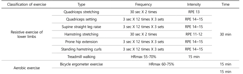
Table 2. Combined exercise program
Classification of exercise Type Frequency Intensity Time
Resistive exercise of lower limbs
Quadriceps stretching 30 sec X 2 times RPE 13
30 min Quadriceps setting 3 sec X 12 times X 3 sets RPE 14~15
Supine straight leg raise 3 sec X 12 times X 3 sets RPE 14~15
Hamstring stretching 30 sec X 2 times RPE 11-12
Prone hip extension 3 sec X 12 times X 3 sets RPE 14~15 Standing hamstring curls 3 sec X 12 times X 3 sets RPE 14~15
Treadmill walking HRmax 55-70% 15 min
Aerobic exercise Bicycle ergometer exercise HRmax 60-75% 15 min
15 min
The NTL (nonlinear transformation) method was used to determine the spatial coordinates. To determine the standards for both the space in which motion occurs and the image, in terms of the laboratory frame of reference, the frontal direction of a subject while walking was set as the X axis, the lateral directions of a subject were set as the Y axis, and the perpendicular direction was set as the Z axis. For the local frame of reference, left-and-right was set as the X-axis, anterior-posterior as the Y-axis, and perpendicular as the Z axis.
3. Data processing
The moment of the lower extremity joint was calculated through both kinematic data and ground reaction force data using an inverse dynamics analysis. Data obtained from each subject were standardized with the body mass, and the joint moments of the subject's bilateral hip joints, meniscus, and ankle joints were calculated. As shown in Table
4, the hip joint is a 3-axial joint because it is a ball and socket joint that can move along all 3 axes. Thus, longitudinal axis (X), horizontal axis (Y), and vertical axis (Z) data were collected for the hip joint. The meniscus and ankle joint movement is centered around one motor axis since they are hinge joints in which one type of rotation movement is possible.
Thus, X-axis data were selected and collected for these joints.
The one-time data of stance phase and swing phase of each foot at the mid-point during walking were standardized with a time. For the axis of each joint, the time at which the direction of momentum of each joint switches was defined from Peak 1 (P1) to Peak 3 (P3) to compare the maximum moment value.
4. Statistical analysis
The Kwon 3D XP Ver 3.1 (VISOL, Inc., Korea) was used to analyze the 3-dimensional coordinates and the kinematic and kinetic data for the gait parameters collected in this study following exercise intervention.
In order to analyze the pretest and posttest differences in parameter values obtained here, Windows SPSS Ver 23.0 was used to calculate means and standard deviations. In order to statistically verify the pretest- posttest mean difference values, an independent t-test was conducted.
All statistical significance was set at 0.05.
RESULTS
1. Result of peak hip joint moment
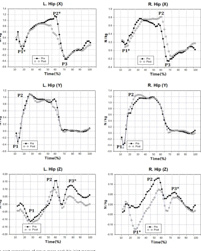
The results of comparing the maximum joint moment during walking before and after the 12-week combined exercise intervention for osteo- arthritis patients in this study are as shown in Table 5 and Figure 2.
In the left hip joint, the horizontal axis (x-axis)'s Peak 1 (P1) showed a statistically significant difference between 0.02±0.01 N/kg pretest and 0.13±0.06 N/kg posttest (t=-6.264, p=.000). Additionally, the maximum Table 4. Definition of joint motion
Joint/Segment Axis Joint motion
Positive, + Negative, -
R. hip
X Flexed Hyperextended
Y Adducted Abducted
Z Internally rotated Externally rotated
L. hip
X Flexed Hyperextended
Y Abducted Adducted
Z Externally rotated Internally rotated
R. L. knee X Hyperextended Flexed
R. L. ankle X Dorsi-flexed Plantar-flexed Figure 1. Reflective marker setting and experimental equipment
Table 5. Pre-post comparison of peak hip joint moment (unit: N/kg)
Joint/Segment Axis Pre Post
t p
M ± SD M ± SD
L. hip X
P1 0.02±0.01 0.13±0.06 -6.264* .000
P2 1.15±0.74 0.88±0.32 .934 .372
P3 -0.34±0.06 -0.25±0.03 -4.467* .001
Figure 2. Pre-post comparison of group mean peak hip joint moment
joint moment of the P3 in the extensor direction also showed a statisti- cally significant difference between -0.34±0.06 N/kg pretest and -0.25
±0.03 N/kg posttest (t=-4.467, p=.001). In the vertical axis (z-axis), P3 showed a statistically significant difference between 0.13±0.06 N/kg pretest and 0.05±0.01 N/kg posttest (t=4.084, p=.002).
In the right hip joint, the maximum joint moment had a statistically significant difference in the P1 (extensor moment) of the X-axis between 0.15±0.02 N/kg and 0.08±0.01 N/kg (t=10.076, p=.000). In addition, the maximum joint moments at the P1 (supination moment) and P3 (pronation moment) points in the Z-axis showed statistically significant differences between -0.04±0.01 N/kg and 0.06±0.03 N/kg pretest and -0.12±0.01 N/kg and 0.04±0.01 N/kg posttest (t=18.138, p=.000, t= 2.888, p=.016).
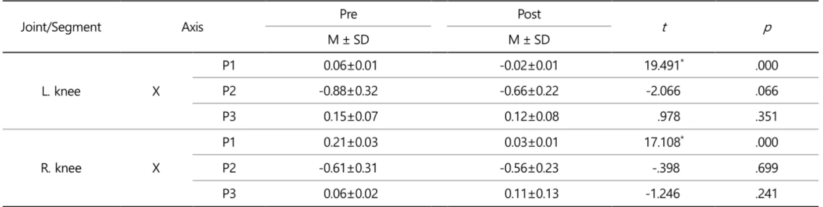
2. Result of peak knee joint moment
The comparison results of meniscus moment before and after exercise intervention in this study are as shown in Table 6 and Figure 3.
In the left and right meniscus, P1 (extensor moment) of the x-axis had statistically significantly higher moments at posttest, at 0.06±0.01 N /kg and 0.21±0.03 N/kg pretest and -0.02±0.01 N/kg and 0.03±0.01 N/
kg posttest (t=19.491, p=.000, t=17.108, p=.000). There were no sta- tistically significant differences in P2 and P3.
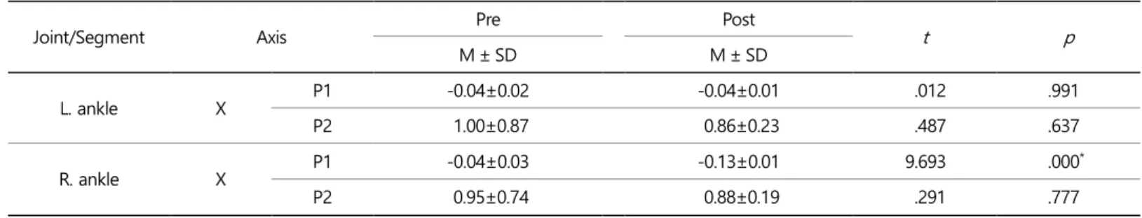
3. Result of peak ankle joint moment
The results of comparing the ankle joint moments before and after the exercise intervention in this study are as shown in Table 7 and Figure 4.
Table 5. Pre-post comparison of peak hip joint moment (Continued) (unit: N/kg)
Joint/Segment Axis Pre Post
t p
M ± SD M ± SD
L. hip
Y P1 -0.30±0.15 -0.36±0.13 .937 .371
P2 1.09±0.78 1.08±0.65 .022 .983
Z
P1 -0.11±0.05 -0.14±0.04 .925 .377
P2 0.16±0.07 0.10±0.04 2.032 .070
P3 0.13±0.06 0.05±0.01 4.084* .002
R. hip
X
P1 0.15±0.02 0.08±0.01 10.076* .000
P2 0.77±0.23 0.81±0.18 -.510 .621
P3 -0.25±0.13 -0.15±0.07 -1.946 .080
Y P1 -0.38±0.19 -0.49±0.20 1.142 .280
P2 1.19±0.98 1.26±0.84 -.151 .883
Z
P1 -0.04±0.01 -0.12±0.01 18.138* .000
P2 0.13±0.11 0.07±0.21 .779 .454
P3 0.06±0.03 0.04±0.01 2.888* .016
*L. hip X axis: (+) Flexed, (-) Hyperextended, L. hip Y axis: (+) Adducted, (-) Abducted, L. hip Z axis: (+) Internally rotated, (-) Externally rotated, R.
hip X axis: (+) Flexed, (-) Hyperextended, R. hip Y axis: (+) Abducted, (-) Adducted, R. hip Z axis: (+) Externally rotated, (-) Internally rotated
Table 6. Pre-post comparison of peak knee joint moment (unit: N/kg)
Joint/Segment Axis Pre Post
t p
M ± SD M ± SD
L. knee X
P1 0.06±0.01 -0.02±0.01 19.491* .000
P2 -0.88±0.32 -0.66±0.22 -2.066 .066
P3 0.15±0.07 0.12±0.08 .978 .351
R. knee X
P1 0.21±0.03 0.03±0.01 17.108* .000
P2 -0.61±0.31 -0.56±0.23 -.398 .699
P3 0.06±0.02 0.11±0.13 -1.246 .241
*X axis: (+) Hyperextended, (-) Flexed
In the left ankle joint, no statistically significant differences were observed in the P1 and P2 of the x-axis between pretest and posttest.
In the right ankle joint, P1 (plantarflexion moment) had a statistically significantly higher moment in the posttest at -0.04±0.03 N/kg and -0.13±0.01 N/kg (t=9.693, p=.000).
DISCUSSION
This study involved a comparative analysis of joint moments during walking after a 12-week combined exercise intervention in elderly women with osteoarthritis aged 65 years old or older.
First, in the left hip joint, the maximum extensor moment appeared to be statistically significantly larger in the posttest in the P1 (initial heel
Table 7. Pre-post comparison of peak ankle joint moment (unit: N/kg)
Joint/Segment Axis Pre Post
t p
M ± SD M ± SD
L. ankle X P1 -0.04±0.02 -0.04±0.01 .012 .991
P2 1.00±0.87 0.86±0.23 .487 .637
R. ankle X P1 -0.04±0.03 -0.13±0.01 9.693 .000*
P2 0.95±0.74 0.88±0.19 .291 .777
*X axis: (+) Dorsi-flexed, (-) Plantar-flexed
Figure 3. Pre-post comparison of group mean peak knee joint moment
Figure 4. Pre-post comparison of group mean peak ankle joint moment
supporting phase) of the horizontal axis (x-axis) (t=-6.264, p=.000).
Further, the maximum joint moment of the P3 in the extensor direction appeared to be statistically significantly lower after intervention (t= -4.467, p=.001). In the vertical axis (z-axis), P3 showed an even lower maximum joint moment after exercise intervention (t=4.084, p=.002).
In the right hip joint, the maximum joint moment showed a low maxi- mum moment in the P1 (extensor moment) of the x-axis after inter- vention (t=10.076, p=.000). Moreover, P1 (supination moment) at the time of stance phase on the Z-axis appeared to be larger in the posttest.
At P3 (pronation moment), the maximum joint moment showed a lower maximum moment after intervention (t=18.138, p=.000, t=2.888, p=.016). This shows that, after exercise intervention, the maximum ex- tensor moment at stance phase increases, and it appears to be a compensatory mechanism to minimize the knee flexor moment. Add- itionally, it appears that the supination moment in the vertical axis (z- axis) decreases. This is in line with a previous study reporting that the left-and-right movement of the pelvis and hip joint can cause stress and muscular imbalance which can induce physical imbalance. Furthermore, the large left-and-right pelvis movements are caused by a compen- satory mechanism due to the knee pain of patients with osteoarthritis and chronic backache (Ko et al., 2013; Moon et al., 2013; Ryu, 2008; Park
& Kim, 2008).
Statistically, a high extensor moment after exercise intervention was found in both the left and right meniscus in P1 (extensor moment) of the x-axis (t=19.491, p=.000, t=17.108, p=.000). It is thought that the activation of the extensor at the initial stance phase after exercise in- tervention caused a large extensor moment. This is in line with the results of a previous study, which found that, in comparison to normal controls, meniscus osteoarthritis patients have greatly reduced peak moment and peak power. Therefore, movement of the knee joint is within a smaller range, and the hip joint is strengthened from increased bending as a compensatory mechanism for the weakening of this meniscus extensor (Lee, Kwon, & Park, 2013).
Comparing pretest-posttest in the left ankle joint did not show a statistically significant difference between P1 and P2 in the x-axis. How- ever, in the right ankle joint, a statistically high moment was found in P1 (plantar flexor moment) after exercise intervention (t=9.693, p=.000).
The large plantar flexor moment at the initial stance phase can signify the strengthening of momentum when walking and is thought to sug- gest the possibility of improved walking ability after exercise intervention.
There are various treatment methods for knee osteoarthritis. Surgical methods, pharmacological methods, and non-pharmacological methods are used as interventions (Chang et al., 2017). Although surgical and non-pharmacological methods are effective at alleviating pain, there are limitations such as high cost, risk of anesthesia, thrombus, and side effects of medications (Kim et al., 2018; Ahn & Bae, 2018). Therefore, an exercise intervention that alleviates pain in arthritis patients through strengthening of muscles around the joint and weight loss is being presented as an effective method (Choi, 2018; Kang, Na, Jang, Lee, &
Kim, 2010). However, there is an opinion that excessive exercise that puts stress on the meniscus can actually exacerbate osteoarthritis; thus, sug- gesting an appropriate exercise intervention method is very important (Bricca et al., 2018; Ernstgard, PirouziFard, & Thorstensson, 2017; Focht
et al., 2017).
Examining previous studies on exercise interventions, Messier et al.
(2000) reported that the aerobic exercise group showed the greatest improvement in dynamic balance skill in a study that compared groups after 18 weeks of intervention. In that study, arthritis patients were divided into 3 groups as follows: aerobic exercise group, health edu- cation group, and muscle strengthening exercise group. In addition, Cho (2016) reported that when elastic band exercise and muscle streng- thening exercise were performed by 26 patients with meniscus osteo- arthritis, walking ability and dynamic balance skills increased. Iwamoto, Takeda, & Sato (2007) showed that meniscus extensor muscles im- proved after exercise intervention on osteoarthritis patients using lower extremity muscle strengthening equipment once per week for 3-6 months. Moreover, Al-Khlaifat, Herrington, Hammond, Tyson, & Jones (2016) reported that, through 6 weeks of lower extremity muscle streng- thening exercise, the muscle strength, pain, and functional level of the hip joint and knee joint significantly improved.
Ahmed (2011) conducted muscle strengthening exercise and balance exercise with 20 female meniscus osteoarthritis patients for 6 weeks and found that balance skills, walking ability, and activity level signifi- cantly increased. Allen et al. (2006) conducted lower extremity muscle strengthening exercise with female patients with meniscal osteoarthritis and found positive effects on the joint function. Additionally, Park &
Kim (2017) divided the osteoarthritis patients into the lower extremity muscle strengthening exercise group and the muscle strengthening exercise in addition to exercise group, and conducted the exercises for four weeks. They found that both groups showed improvements in pain, joint range of motion, lower extremity muscle strength, and balance level, but that no differences were observed in the balancing exercise addition group. Based on the analysis of results by combining the pre- vious studies and this study, it is thought that, in the combined exercise applied in our research, the lower extremity muscle strengthening ex- ercise promoted the activity of extensor and flexor muscles in patients with meniscus osteoarthritis and induced high extensor moment of the meniscus during walking, which increased the muscle strength of the meniscus and improved the level of walking ability.
CONCLUSION
This study was conducted to examine the changes in maximum joint moment during walking after a 12-week combined exercise inter- vention comprising lower extremity muscle strengthening exercise and aerobic exercise in 11 elderly women with meniscus osteoarthritis aged 65 years or older. The results showed that the maximum extensor moment was statistically significantly large in the posttest of P1 in x- axis in the hip joints. Additionally, the maximum joint moment of the P3 in the in the direction of extensor was found to be statistically significantly lower after intervention. This seems to be a compensatory mechanism to minimize the knee flexor moment. In the vertical axis (z-axis), P3 showed an even lower maximum joint moment after exercise intervention. This is thought to be a compensatory mechanism to decrease knee pain after exercise intervention.
In each of the left and right menisci, statistically significantly high
extensor moment in the x-axis was found after exercise intervention.
This seems to be caused by the activation of extensor during the initial stance phase after exercise intervention.
Pretest-posttest comparison of the left ankle joint showed no statisti- cally significant differences between P1 and P2 in the x-axis. However, a statistically significantly high moment appeared in the P1 (plantar flexor moment) of the right ankle joint after exercise intervention. The large plantar flexor moment in the initial stance phase can be inter- preted to mean that the momentum when walking was strengthened.
It is thought that the muscle strength of the meniscus and the level of walking ability have improved after exercise intervention.
Through these results, it was confirmed that a combined exercise program comprising lower extremity muscle strengthening exercise and aerobic exercises in patients with meniscus osteoarthritis can have a positive impact on walking ability and joint moment when walking.
Because observation of the intervention effects in a randomized con- trolled trial design is generally desirable for clinical application studies, an investigative study that adds a control group is needed in the future.
REFERENCES
Ahmed, A. F. (2011). Effect of Sensorimotor Training on Balance in Elderly Patients with Knee Osteoarthritis. Journal of Advanced Research, 2(4), 305-311.
Ahn, G. Y. & Bae, S. C. (2018). Strategies for the safe use of non-steroidal anti-inflammatory drugs. Journal of the Korean Medical Association, 61(6), 367-375.
Al-Khlaifat, L., Herrington, L. C., Hammond, A., Tyson, S. F. & Jones, R. K.
(2016). The effectiveness of an exercise programme on knee loading, muscle co-contraction, and pain in patients with medial knee osteo- arthritis: A pilot study. The Knee, 23(1), 63-69.
Allen, K. D., Arbeeva, L., Schwartz, T., Callahan, L., Golightly, Y., Goode, A. & Coffman, C. (2018). Identifying Subgroups of Patients with Differential Benefit from Physical Therapy or Internet-based Exercise Training for Knee Osteoarthritis. Osteoarthritis and Cartilage, 26, 334-335.
Bieler, T., Siersma, V., Magnusson, S. P., Kjaer, M. & Beyer, N. (2016).
OP0006-HPR Even in The Long Run Nordic Walking Is Superior To Strength Training and Home Based Exercise for Improving Physical Function in Older People with Hip Osteoarthritis-An RCT. Annals of the Rheumatic Disease, 75(2), 55.
Bricca, A., Struglics, A., Larsson, S., Steultjens, M., Juhl, C. B. & Roos, E. M.
(2018). Impact of exercise therapy on molecular biomarkers related to articular cartilage and inflammation in people at risk of, or with established, knee osteoarthritis: a systematic review and meta- analysis of randomized controlled trials. Osteoarthritis and Cartilage, 26, 314-315.
Callahan, L. F., Wiley-Exley, E. K., Mielenz, T. J., Xiao, C., Currey, S. S., Sleath, B. L., Sloane, P. D., DeVellis, R. F., Brady, T. J. & Sniezek, J. (2009). Peer Reviewed: Use of Complementary and Alternative Medicine Among Patients With Arthritis. Preventing Chronic Disease,6(2), A44.
Castrogiovanni, P. & Musumeci, G. (2016). Which is the best physical treatment for osteoarthritis?. Journal of Functional Morphology and
Kinesiology, 1(1), 54-68.
Chang, A. H., Chmiel, J. S., Hayes, K. W., Almagor, O., Moisio, K. C., Zhang, Y. & Sharma, L. (2017). Baseline Hip and Knee Muscle Strength and Improvement in Function and Disability 5 Years Later in Persons with Knee Osteoarthritis. Osteoarthritis and Cartilage, 25, 337.
Chapple, C. M., Abbott, J. H. & Tumilty, S. (2018). How does frequency of manual therapy influence outcome for people with knee osteo- arthritis? A feasibility study. Osteoarthritis and Cartilage, 26, 339-340.
Cho, I. S. (2016). The Effects of Visual Feedback Balance Training on Pain, Physical Function, Gait, Balance in Chronic Knee Osteoarthritis Patients. Unpublished doctoral thesis, Daegu University.
Choi, H. K., Kim, N. S. & Kim, H. S. (2009). Effects of Water Exercise Program on Physical Fitness, Pain and Quality of Life in Patients with Osteoarthritis. Journal of Muscle and Joint Health, 16(1), 55-56.
Choi, J. H. (2018). Effect of Taping on a Home Program of Hip Abductor Exercise on Pain and Quadriceps Muscle Strength in Elderly Women with Knee Osteoarthritis, Journal of the Korean Society of Physical Medicine, 13(3), 61-66.
Choi, P. B. (2011). Long-term Combined Exercise has Effect on Regional Bone Mineral Density and Cardiovascular Disease Risk Factors of the Elderly with Osteoporosis. Journal of Korea Gerontological Society, 31(2), 355-369.
Dillon, C. F., Rasch, E. K., Gu, Q. & Hirsch, R. (2006). Prevalence of knee osteoarthritis in the United States: arthritis data from the Third National Health and Nutrition Examination Survey 1991-94. The Journal of Rheumatology, 33(11), 2271-2279.
Ernstgard, A., PirouziFard, M. & Thorstensson, C. A. (2017). Health en- hancing physical activity in patients with hip or knee osteoarthritis- an observational intervention study. BMC Musculoskeletal Disorders, 18(1), 42.
Focht, B. C., Garver, M. J., Lucas, A. R., Devor, S. T., Emery, C. F., Hackshaw, K. V. & Rejeski, W. J. (2017). A group-mediated physical activity intervention in older knee osteoarthritis patients: effects on social cognitive outcomes. Journal of Behavioral Medicine, 40(3), 530-537.
Fransen, M., McConnell, S., Harmer, A. R., Van der Esch, M., Simic, M. &
Bennell, K. L. (2015). Exercise for osteoarthritis of the knee. Cochrane Database of Systematic Reviews, 1(1), 1-144.
Holsgaard-Larsen, A., Christensen, R., Clausen, B., Søndergaard, J., Andriacchi, T. P. & Roos, E. M. (2018). One year effectiveness of neuromuscular exercise compared with instruction in analgesic use on knee function in patients with early knee osteoarthritis: the EXERPHARMA randomized trial. Osteoarthritis and Cartilage, 26(1), 28-33.
Iwamoto, J., Takeda, T. & Sato, Y. (2007). Effect of muscle strengthening exercises on the muscle strength in patients with osteoarthritis of the knee. The Knee, 14(3), 224-230.
Juhl, C., Christensen, R., Roos, E. M., Zhang, W. & Lund, H. (2014). Impact of exercise type and dose on pain and disability in knee osteo- arthritis: a systematic review and meta‐regression analysis of rando- mized controlled trials. Arthritis & Rheumatology, 66(3), 622-636.
Jung, K. H. (2017). Diagnosis and Treatment of Arthritis. Journal of the Korean Neurological Association, 35(4), 25-30.
Kang, J. H., Na, J. Y., Jang, J. H., Lee, K. I. & Kim, K. Y. (2010). Isokinetic Test
and the Effect of Exercise Therapy of Ipsilateral Knee Osteoarthritis.
Korean Journal of Sport Biomechanics, 20(1), 75-81.
Katsura, Y., Yoshikawa, T., Ueda, S. Y., Usui, T., Sotobayashi, D., Nakao, H., Sakamoto, H., Okumoto, T. & Fujimoto, S. (2010). Effects of aquatic exercise training using water-resistance equipment in elderly.
European Journal of Applied Physiology, 108(5), 957-964.
Kellgren, J. H. & Lawrence, J. S. (1957). Radiological assessment of osteo- arthrosis. Annals of the Rheumatic Diseases, 16(4), 494.
Kim, D. H., Lee, D. E., Noh, J. W., Ahn, Y. M., Ahn, S. Y. & Lee, B. C. (2018).
A Study of the Co-Administration of Herbal and Western Medi- cines to Hospitalized Patients with Osteoarthritis. The Journal of Internal Korean Medicine, 39(2), 97-106.
Kim, S. M. & Song, J. M. (2010). The efficacy of community-based reha- bilitation exercise to improve physical function in old women with knee arthritis. The Journal of Korean Physical Therapy, 22(1), 9-17.
Kim, Y. J., Seo, N. S., Lim, Y. N., Kim, H. S. & Kim, S. J. (2012). Effects of a Taekwondo Exercise Program in Women with Osteoarthritis. Journal of Muscle and Joint Health, 19(2), 210-222.
Ko, E. A., Hong, S. Y., Lee, K. K. & An, K. O. (2013). Effect of Active Change of the Foot Progression Angle on Lower Extremity Joint During Gait. Korean Journal of Sport Biomechanics, 23(1), 85-90.
Ko, S. U., Ling, S. M., Schreiber, C., Nesbitt, M. & Ferrucci, L. (2011). Gait patterns during different walking conditions in older adults with and without knee osteoarthritis-results from the Baltimore Longitudinal Study of Aging. Gait & Posture, 33(2), 205-210.
Lauche, R., Langhorst, J., Dobos, G. & Cramer, H. (2013). A systematic review and meta-analysis of Tai Chi for osteoarthritis of the knee.
Complementary Therapies in Medicine, 21(4), 396-406.
Lee, I. H., Kwon, K. H. & Park, S. Y. (2013). Biomechanical Properties of the Anterior Walker Dependent Gait of Patients with Knee Osteo- arthritis. The Journal of Korean Society of Physical Therapy, 25(5), 239-245.
Lee, S. S. & So, Y. S. (2015). The Effects of Aquatic Rehabilitation Exercise on Pain, Physical Function, Walking Ability and Falls Efficacy in Patients with Knee Osteoarthritis. The Korean Society of Sports Science, 24(5), 1241-1251.
McIlroy, S., Sayliss, L., Browning, P. & Bearne, L. M. (2017). Aquatic therapy for people with persistent knee pain: A feasibility study.
Musculoskeletal Care, 15(4), 350-355.
Melikoglu, M. A. & Kul, A. (2017). AB0809 Fall risk and related factors in knee osteoarthritis. Annals of the Rheumatic Diseases, 76, 1340.
Messier, S. P., Royer, T. D., Craven, T. E., O'toole, M. L., Burns, R. & Ettinger, W. H. (2000). Long‐term exercise and its effect on balance in older, osteoarthritic adults: Results from the Fitness, Arthritis, and Seniors Trial (FAST). Journal of the American Geriatrics Society, 48(2), 131 -138.
Moon, H. J., Choi, K. H., Kim, D. H., Kim, H. J., Cho, Y. K., Lee, K. H. &
Choi, Y. J. (2013). Effect of lumbar stabilization and dynamic lumbar strengthening exercises in patients with chronic low back pain.
Annals of Rehabilitation Medicine, 37(1), 110-117.
Park, J. E. & Kim, S. Y. (2017). Effects of Lower Extremity Strengthening Exercise Combined with Balance Exercise on Lower Extremity Funtion, Range of Motion, Muscle Strength, and Balance in Patients with Knee Osteoarthritis. Journal of the Korean Society of Physical Medicine, 12(4), 147-158.
Park, Y. S. & Kim, S. H. (2008). The Relationship of Elderly Female with Chronic Low Back Pain during Their Gait and Function Fitness Test.
Journal of Korean Association of Physical Education and Sport for Girls and Women, 22(2), 163-174.
Petterson, S. C., Mizner, R. L., Stevens, J. E., Raisis, L. E. O., Bodenstab, A., Newcomb, W. & Snyder‐Mackler, L. (2009). Improved function from progressive strengthening interventions after total knee arthroplasty:
A randomized clinical trial with an imbedded prospective cohort.
Arthritis Care & Research, 61(2), 174-183.
Philbin, E. F., Ries, M. D., Groff, G. D., Sheesley, K. A., French, T. S. &
Pearson, T. A. (1996). Osteoarthritis as a determinant of an adverse coronary heart disease risk profile. Journal of Cardiovascular Risk, 3(6), 529-533.
Ryu, J. S. (2008). Dynamic Stability Analysis of Patients with Degenera- tive Osteoarthritise during Walking. Korean Journal of Sport Bio- mechanics, 18(1), 21-30.
Sabashi, K., Yamanaka, M., Chiba, T., Samukawa, M., Saitoh, H., Yuri, M.
& Tohyama, H. (2017). Association of functional the reach test with quality of life in patients with knee osteoarthritis. Osteoarthritis and Cartilage, 25, 398-399.
Singh, J. A., Saag, K. G., Bridges Jr, S. L., Akl, E. A., Bannuru, R. R., Sullivan, M. C. & Curtis, J. R. (2016). 2015 American College of Rheuma- tology guideline for the treatment of rheumatoid arthritis. Arthritis
& Rheumatology, 68(1), 1-26.
Takacs, J., Krowchuk, N. M., Garland, S. J., Carpenter, M. G. & Hunt, M.
A. (2017). Dynamic balance training improves physical function in individuals with knee osteoarthritis: A pilot randomized controlled trial. Archives of Pysical Medicine and Rehabilitation, 98(8), 1586 -1593.
Thorstensson, C. A., Roos, E. M., Petersson, I. F. & Ekdahl, C. (2005). Six- week high-intensity exercise program for middle-aged patients with knee osteoarthritis: A randomized controlled trial. BMC Musculo- skeletal Disorders, 6(1), 27.
Yu, Y. J. (2007). The Study of Exercise Interventions for Developing Exercise Program in Patient with Osteoarthritis. Journal of Coaching Development, 9(3), 127-140.