Effects of Lycopene on Endothelial Protein C Receptor Shedding In Vitro and In Vivo
Hayoung Yoo
1†, Hyun-Shik Lee
2†, Wonhwa Lee
1,3and Jong-Sup Bae
1,3*
1
College of Pharmacy, Research Institute of Pharmaceutical Sciences, Kyungpook National University, Daegu 702-701 Korea
2
School of Life Sciences, College of Natural Sciences, Kyungpook National University, Daegu 702-701, Korea
3
Department of Biochemistry and Cell Biology, Cell and Matrix Research Institute, School of Medicine, Kyungpook National University, Daegu 700-422, Korea
Received April 26, 2013 /Revised May 12, 2013 /Accepted May 22, 2013
Endothelial protein C receptor (EPCR) plays a pivotal role in augmenting Protein C activation through the thrombin-thrombomodulin complex. EPCR activity is markedly changed by ectodomain cleavage and released as the soluble protein (sEPCR). EPCR shedding is mediated by tumor necrosis factor-α converting enzyme (TACE). Lycopene found in tomatoes and tomato products has anti-oxidant, an- ti-cancer and anti-inflammatory effects. However, little is known about the effects of lycopene on EPCR shedding. We investigated this issue by monitoring the effects of lycopene on the phor- bol-12-myristate 13-acetate (PMA), tumor necrosis factor (TNF)-α, interleukin (IL)-1β and on the cecal ligation and puncture (CLP)-mediated EPCR shedding. Data showed that lycopene potently inhibited the PMA, TNF-α, IL-1β and CLP-induced EPCR shedding by suppressing TACE expression.
Furthermore, lycopene reduced PMA-stimulated phosphorylation of p38, extracellular regulated kin- ases (ERK) 1/2 and c-Jun N-terminal kinase (JNK). Given these results, lycopene should be viewed as a candidate therapeutic agent for the treatment of various severe vascular inflammatory diseases via inhibition of the EPCR shedding.
Key words : Lycopene, endothelial protein C receptor (EPCR), shedding, phorbol-12-myristate 13-acetate (PMA), cecal ligation and puncture (CLP)
†
Two authors equally contributed to this work.
*Corresponding author
*Tel:+82-53-950-8570, Fax:+82-53-950-8557
*E-mail : baejs@knu.ac.kr
This is an Open-Access article distributed under the terms of the Creative Commons Attribution Non-Commercial License (http://creativecommons.org/licenses/by-nc/3.0) which permits unrestricted non-commercial use, distribution, and reproduction in any medium, provided the original work is properly cited.
Journal of Life Science 2013 Vol. 23. No. 5. 650~656 DOI : http://dx.doi.org/10.5352/JLS.2013.23.5.650
서 론
토마토에 함유되어 있는 라이코펜은 항산화효과, 항암효과, 세포막 보호효과 그리고 항염증효과가 있다고 알려져 있다[2, 10, 14]. 그리고 토마토 또는 토마토를 재료로 한 음식을 섭취 하는 것은 만성혈관질환, 염증질환 및 암을 예방하는 것으로 보고되어 있다[10]. 최근에 라이코펜이 혈관내피세포의 투과 성, 세포부착단백질의 발현 및 백혈구의 혈관내피세포에 대한 부착과 이동을 억제하고 그 기전으로써 염증을 유발하는데 중요한 두 가지 사이토카인(tumor necrosis factor-α, nuclear factor-κB)의 발현을 억제하는 것이 보고되었다[2, 14].
내피 세포 단백질 C 수용체(EPCR)는 46 kDa의 크기를 가지 며 제1형 막관통 당단백질로서 클래스I 단백질의 주요 조직적 합성 복합체에 대해서 비슷한 성질을 가진다[5]. EPCR 단백질 (전체 221-아미노산으로 구성)은 25개의 아미노산으로 이뤄진
막관통 도메인인 세포 외 도메인과 3개의 아미노산으로 구성 된 세포질 내 단기 염기서열로 구성되어 있고[5] 이 유전자는 20번 염색체에 위치한다[9]. EPCR은 단백질 C (PC)와 활성화 된 단백질 C (APC)에 높은 친화력(Kd=30 nM)으로 부착한다 [5]. EPCR의 주요한 역할은 단백질 C에 높은 친화력으로 결합 되고 트롬빈-트롬보모듈린 복합체에 의한 단백질 C의 활성화 를 증가시키기 때문에 항응고 작용이다[19]. 방출된 활성화 단 백질 C (APC)는 보조인자인 단백질 S에 부착하고 응고인자 Va와 VIIIa를 감소시킨다[19].
EPCR의 노출은 EPCR의 분열에 강하게 의존하고 수용성
형태(sEPCR)로 방출된다[4, 7, 16, 23]. EPCR은 metal-
loproteinase이 매개하는 탈락(shedding)을 통해 그 활성과 기
능이 조절되는 세포 표면 수용체이다[5, 24]. In vitro 상에서의
여러 가지 처리를 통해 EPCR의 탈락을 증가시키고 단백질
C 활성화 비율을 감소시키며[23], 방출된 수용성 EPCR
(sEPCR)은 EPCR과 연관된 막에서 APC/PC 부착 억제를 통
해 단백질 C (PC)와 활성화된 단백질 C (APC)의 기능을 감소
시킨다[13]. 수용성 EPCR (sEPCR)은 막 EPCR 탈락의 결과로
혈장 내에서 측정할 수 있고 그 농도는 대략 100 ng/ml이며,
전신성 염증 질환에서 높은 농도의 수용성 EPCR이 발견된다
[12]. 뿐만 아니라 혈액응고인자 VII (FVII)과 그것의 활성화
형태인 FVIIa와 수용성 EPCR의 상호작용은 EPCR과 sEPCR
이 FVII/FVIIa의 부착 장소로써 역할을 하고 FVIIa-Tissue
Factor (TF) 복합체의 응혈촉진 활성을 억제한다는 것을 잘 알려진 사실이다[6, 17]. 그리고, 여러 In vitro 연구에서 다양한 염증성 중재자들(IL-1β, H
2O
2, phorbol myristate acetate)과 트롬빈이 혈관내피에서 EPCR 탈락을 급격히 증가시킨 것과 EPCR 탈락이 미세소관 방해인자인 nocadazole에 의해 더 증 가됨이 증명되었다[24].
최근 우리는 라이코펜이 인간 내피 세포에서 항 염증효과와 장벽 보호효과를 가진다는 것을 보고하였다[2, 14]. 그래서, 이 번 연구에서 우리는 라이코펜이 수용성 EPCR 탈락을 억제할 것이라는 가정 하에 인간 내피 세포와 cecal ligation and puncture (CLP)에 의해 유도된 패혈증 쥐에서 라이코펜의 EPCR 탈락 억제 효과와 근본적인 분자적 기전을 조사하였다.
재료 및 방법
재료
수용성 EPCR과 TNF-α는 Abnova (Taiwan)에서 구입하였 다. Phorbol-12-myristate 13-acetate (PMA), IL-1β와 항생제 (penicillin G, streptomycin)는 Sigma (St.Louis, MO)에서 구 입하여 사용하였다. 라이코펜(Fig. 1)은 이전에 설명한 대로 준비되었다[2, 14].
세포 배양
혈관내피세포(Primary human umbilical vein endothelial cells, HUVECs)는 Cambrex Bio Science (Charles City, IA) 에서 구입하였고 이전에 설명한 대로 배양하였다[1]. 즉, 세포 들은 Fetal Bovine Serum (FBS, Cambrex Bio Science)가 첨가 된 EBM-2 기저 배지에 37°C, 5% CO
2조건을 유지시키면서 배양되었다. Passage Number 3 또는 4번으로 분주된 HUVECs이 실험에 사용되었다.
동물과 사육
C57BL/6 수컷 쥐(6-7주 된, 18-20 g)는 Orient Bio Co.
(Sungnam, KyungKiDo, Republic Korea)에서 구입하였고, 12 일 적응시킨 후에 사용하였다. 동물들은 polycarbonate 케이 지에서 조건(20-25°C/RH 40-45%)을 조절하고 12:12시간 낮/
밤 주기로 5마리씩 사육하였고, 일반적인 설치류 먹이와 물을 자유식으로 공급하였다. 모든 동물들은 경북대학교에서 발행 한 Guidelines for the Care and Use of Laboratory Animals에 부합되게 처리되었다.
세포의 EPCR 발현측정을 위한 효소면역측정법(Enzyme- linked immuno sorbent assay, ELISA)
개선된 whole-cell ELISA는 HUVECs에서 EPCR의 발현 정 도의 측정을 위해 이전에 설명한대로 수행되었다[11]. 즉, 단층 의 HUVEC은 라이코펜을 6시간 동안 처리한 것과 처리하지 않은 군으로 분리하고 PMA, 종양괴사인자(tumor necrosis
factor (TNF)-α, TNF-α) 혹은 인터류킨(interleukin, IL)-1β를 1시간 처리하여 실험하였다. 그 후 배지는 제거하고 세포들은 phosphate buffered saline (PBS)에 씻어주고 상온에서 15 동 안 50 μl의 1% paraformaldehyde로 고정시킨다. 세포를 씻어 준 후 100 μl의 EPCR 항체(Abonova)를 첨가하고 1시간(37°C, 5% CO
2) 후에 3번 씻어주고 peroxidase가 결합된 항 토끼 IgG 항체(Sigma, Saint Louis, MO)를 1:2,000으로 100 μl 처리한다.
1시간 후에 3번에 걸쳐 씻어주고 O-phenylenediamine 기질 (Sigma, St.Louis, MO)을 넣고 490 nm에서 흡광도를 측정한 다. 모든 측정은 3개 well에서 수행되었다. EPCR의 변이계수 (coefficient of variability, CV); inter-assay는 8-12%보다 적었 고, intra-assay는 6-8%보다 적었다.
세포 Viability 측정
라이코펜이 세포에 미치는 viability를 측정하기 위해 MTT 를 사용하였다. 혈관내피세포(5x10
3개/well)을 하루 동안 배 양한 후, 농도별 라이코펜을 처리하였다. 48 시간 후, 세포를 씻은 후, 100 μl의 MTT (1 mg/ml)을 넣어주고 4 시간 배양하 였다. 100% DMSO 3 ml를 첨가하여 세포에 흡수된 MTT for- mazan을 녹여내고 96-well 배양접시에 100 μl씩 분주하여 540 nm에서 흡광도를 측정하였다.
수용성 EPCR과 TACE 측정을 위한 경쟁적 효소면역측정 법(ELISA, Competitive ELISA)
96well microtiter plate (Corning, NY, USA)는 0.02% so- dium azide가 포함된 20 mM carbonate-bicarbonate 완충액 (pH 9.6)에서 하룻밤 동안 4
oC에서 수용성 EPCR 또는 TACE 단백질로 코팅되었다. 그 후 Plate는 PBS-0.05% Tween 20 (PBS-T)에서 3번 씻어주고, 4
oC에 두었다. 수용성 EPCR 측정 을 위해 세포 배양 배지와 쥐 혈장으로부터 준비된 샘플 또는 TACE 측정을 위해 세포 용해물로부터 얻은 샘플은 항 EPCR 항체(Abnova; PBS-T에서 1:500으로 희석) 또는 항 TACE 항 체(Santa Cruz; PBS-T에서 1:500으로 희석)와 함께 96well 둥 근 microliter plate에 90분 동안 37
oC에서 배양하였고 전 처 리된 plate에 옮겨서 상온에서 30분 동안 배양하였다. 그 후 PBS-T로 3번 씻어주고 peroxidase와 결합된 항 토끼 또는 항 염소 IgG 항체(diluted 1:2000 in PBS-T, Amersham Pharmacia Biotech)를 처리하고 상온에서 90분 동안 배양하 였다. 다시 PBS-T로 3번 씻어주고 암상태에서 기질(100 μg/
ml o-phenylenediamine, 0.003% H
2O
2)을 200 μl씩 넣어 상온 에서 60분 동안 배양하였다. 반응은 반응중지액인 H
2SO
4(8N, 50 μl)을 사용하여 중지시키고 490 nm에서 흡광도를 측 정한다. 수용성 EPCR의 변이계수(CV); inter-assay는 6-10%
보다 적었고, intra-assay는 4-6%보다 적었다. TACE의 변이
계수(CV); inter-assay는 7-10%보다 적었고, intra-assay는
5-7%보다 적었다.
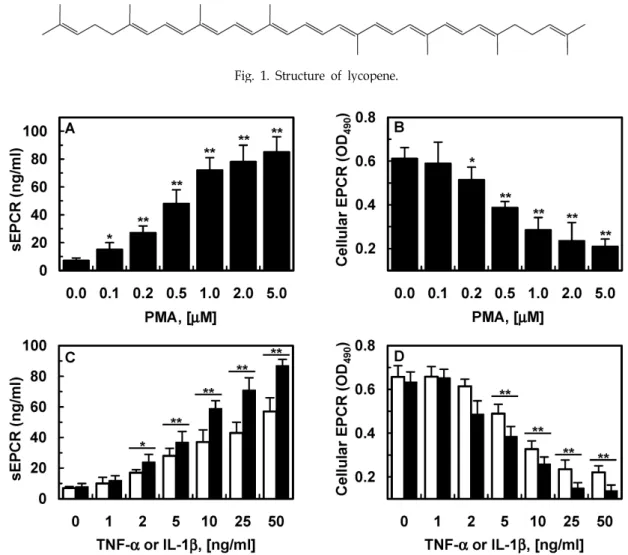
Fig. 1. Structure of lycopene.
Fig. 2. Effect of PMA, TNF-α and IL-1b on EPCR shedding. HUVECs were treated with indicated concentration of PMA (A, B), TNF-α and IL-1β (C, D) for 1 h. (A, C) The conditioned media was collected and sEPCR was measured by ELISA. (B, D) Expression of cellular EPCR was determined by whole cell ELISA. All results indicate the mean±SD of three separate experiments. * p <0.05 or ** p <0.01 vs. 0.
Cecal ligation and puncture (CLP)
패혈증을 유발하기 위해, 수컷 쥐를 zoletil 50과 rompun으 로 마취시킨다. Wang, H. et al.의 방법에 따라 패혈증 쥐 모델 을 제작했다[22]. 즉, 쥐의 복부 중간에 2 cm 정도 절개 후 장과 인접한 맹장을 꺼낸 후, 맹장의 끝에서부터 5.0 mm 부위를 3.0- 실크 봉합사로 결찰한 후 22 게이지 주사바늘로 한번 뚫어준 후 다시 4.0-실크 봉합사로 절개 부위를 봉합한다. Sham mouse 는 절개 후 맹장을 묶거나 뚫어주지 않고 그대로 봉합한다. 이 실험 방법은 경북대학교 동물 관리위원회로부터 승인 받았다.
인산화된 p-38, ERK1/2와 JNK의 효소면역측정법(ELISA) 세포 내 총/인산화된 p38 발현(Cell Signaling Technology, Danvers, MA, USA)과 총/인산화된 ERK1/2와 JNK (R&D Systems, Minneapolis, MN)의 측정은 ELISA 키트를 이용하 여 수행하였다.
Statistical analysis
각 실험은 최소 3번 이상 검정하였고, 실험 결과는 평균±표 준오차로 표시하였고 non-paired Student’s t test로 검정하여 P값이 5% 미만일 때 통계적으로 유의하다고 간주하였다.
결과 및 고찰
라이코펜이 PMA, TNF-α 또는 IL-6가 유도하는 EPCR 탈락에 미치는 영향
이전의 연구에서 PMA가 혈관내피세포로부터 EPCR의 탈
락을 유발한다고 알려져 있다[20, 21]. 이전의 연구 결과와 동
일하게, 우리는 PMA 1 μM이 혈관내피세포에서 EPCR 탈락을
완전히 유발한다는 점과 PMA에 의해 농도의존적으로 EPCR
이 감소함을 확인했다(Fig. 1A, B). TNF-α 또는 interleukin
(IL)-1β에 의해 EPCR 탈락이 이전의 연구와 동일하게 증가되
Fig. 3. Effect of lycopene on PMA, TNF-α and IL-6-induced EPCR shedding. The effects of various concentrations of lycopene on PMA (1 μM, 1 h)-induced EPCR shedding were monitored by measuring sEPCR (A) or cellular EPCR on HUVECs (B).
(C and D) The same as A and B except that HUVECs were incubated with TNF-α (25 ng/ml for 1 h, white bar) or IL-1β (25 ng/ml for 1 h, black bar). Results indicate the mean±SEM of three separate experiments. ** p <0.01 vs. PMA alone (A, B) or TNF-α/IL-1β alone (C, D).
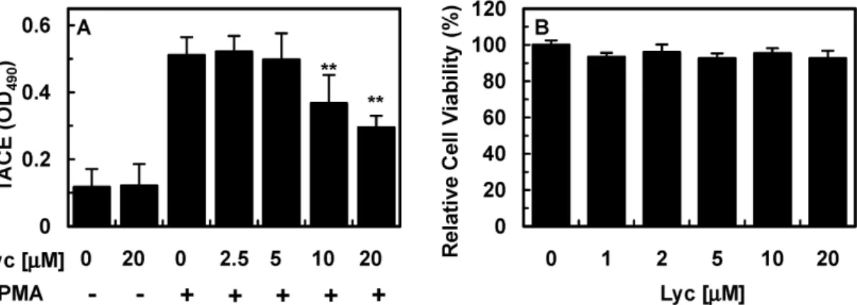
Fig. 4. Effect of lycopene on PMA-stimulated TACE expression. The effects of various concentrations of lycopene on PMA (1 μM, 1 h)-induced TACE expression were monitored by measuring TACE ELISA. All results indicate the mean±SEM of three separate experiments. * p <0.01 vs. PMA alone.
었다(Fig. 1C, D) [18].
PMA가 매개하는 EPCR 탈락에서 라이코펜의 효과를 검증 하기 위해, 혈관내피 세포에 라이코펜(Fig. 1)을 농도 별로 6시 간 동안 처리를 한 다음 PMA 1 μM로 한 시간 동안 처리하였
다. 그 결과 라이코펜 10-20 μM이 내피세포에서 PMA가 유도
하는 EPCR 탈락을 저해함을 보였다(Fig. 3A, B). 그러나 라이
코펜만 처리했을 경우, EPCR 탈락에는 영향을 주지 않았다
(Fig. 3A, B). 라이코펜은 또한 혈관내피세포에서 TNF-α와
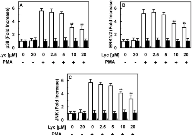
Fig. 6. Effect of lycopene on PMA-induced phosphorylation of p38, ERK1/2 and JNK. PMA (1 μM, 1 h)-mediated phosphorylation of phospho-p38 (A, white bar) or total p38 (A, black bar), phospho-ERK1/2 (B, white bar) or total ERK1/2 (B, black bar) and phospho-JNK (C, white bar) or total JNK (C, black bar) were analyzed after treating cells with the indicated concentrations of lycopene. Results are expressed as fold increase over control values. All results indicate the mean±SEM of three separate experiments. ** p <0.01 vs. PMA alone.
IL-1β가 매개하는 EPCR 탈락을 저해하였다(Fig. 3C, D).
라이코펜이 PMA가 유발하는 TACE 발현과 세포 viability 에 미치는 영향
이전의 연구에서 PMA가 유발하는 EPCR 탈락은 tumor ne- crosis factor-α converting enzyme/ADAM17 (TACE)에 의해 매개한다고 보고되었다[20]. 라이코펜이 활성화 된 TACE 발 현을 저해하는지 밝히기 위해, 내피세포에 라이코펜을 농도 별로 6시간 처리한 후, PMA 1 μM로 한 시간 동안 처리하였다.
그 결과, 라이코펜이 혈관내피세포에서 PMA가 유도한 TACE 발현을 저해하였다(Fig. 4A). 뿐만 아니라, 라이코펜은 세포 viability에는 아무런 영향을 미치지 않았다(Fig. 4B).
Effect of lycopene on CLP-induced EPCR shedding In vivo 상에서 라이코펜의 EPCR 탈락 저해 효과를 검증하 기 위해, 우리는 인간의 패혈증과 가장 유사한 CLP 쥐 모델을 사용했다[3, 25]. 라이코펜을 CLP 12시간 후 21.5 μg으로 일회 투여한 경우에는 CLP가 유도한 EPCR 탈락을 막지 못했다.
그렇지만, CLP 12, 50 시간 후 21.5 μg으로 2회 투여한 경우,
Fig 5. Effect of lycopene on CLP-induced EPCR shedding.
Serum was obtained from sham-operated (white bar), CLP-induced septic mice (gray bar) at indicated day af- ter CLP surgery (n=5). The effects of lycopene (black bar, 21.5 μg/mouse, i.v.) on CLP-induced EPCR shed- ding were monitored by measuring sEPCR. All results indicate the mean±SEM of three separate experiments.
** p <0.01 vs. CLP alone.
EPCR의 탈락을 저해하는 결과를 나타내었다(Fig. 5). 쥐의 무
게는 평균 20 g이고, 총 혈액양의 평균은 2 ml 이므로 혈액 내 처리한 라이코펜의 농도는 대략 20 μM 정도이다. 이를 통 해 라이코펜 투여가 EPCR의 탈락을 억제함으로써 중증 혈관 질환의 치료 전략이 될 것이라 예상된다.
라이코펜이 PMA가 유발하는 p38, ERK1/2와 JNK의 인 산화에 미치는 영향
이전 연구에서 p38, ERK1/2와 JNK는 사이토카인이 유발 한 EPCR 탈락에서 관련이 있으며, PMA 자극에 의해 p38, ERK1/2와 JNK의 인산화는 증가된다고 알려져 있다[8, 15, 18]. 그래서 라이코펜이 PMA가 유도하는 EPCR 탈락의 저해 기전을 규명하기 위해, PMA가 유도한 p38, ERK1/2와 JNK의 인산화를 라이코펜이 저해하는지 실험하였다. 그 결과 라이코 펜이 PMA가 유발한 p38 (Fig. 6A), ERK1/2 (Fig. 6B)와 JNK (Fig. 6C)의 인산화를 감소시켰다.
최근 우리는 토마토에서 분리한 활성물질 라이코펜이 혈관 내피세포에서 혈관장벽을 보호하고, 염증 사이토카인, 세포부 착 단백질, 백혈구의 부착과 이동을 저해함을 보고하였다[2, 14]. EPCR 탈락이 혈관염증 질환에서 병태 생리적 기전에 관 련이 있고, 라이코펜이 혈관 혐증 치료제 후보물질로서 사용 될 수 있다는 가설을 우리의 이전 연구와 현재의 연구 결과를 통해 검증하였다.
결과적으로, 이번 연구는 라이코펜이 p38, ERK1/2와 JNK 를 저해하여 TACE의 발현을 저해시킴으로써 PMA, TNF-α, IL-1β와 CLP가 매개하는 EPCR 탈락을 감소시킨다는 결과를 보여준다. 세포막에 결합된 수많은 단백질들이 TACE에 의해 탈락되고, 이 연구에서는 EPCR 탈락이 TACE에 의해 매개되 고 이를 라이코펜이 저해함을 밝혔다. 치료 목적을 위한 라이 코펜의 사용에서 비특이적 영향을 미칠 수 있음에도 불구하 고, 이 연구에서 나타난 데이터는 EPCR 탈락에서 라이코펜의 새로운 역할을 확인했다. 우리는 패혈증이나 패혈 쇼크 같은 중증 혈관 질환의 치료를 위한 후보 물질로서의 라이코펜 효 능을 검증했다.
감사의 글
이 논문은 2010년도 경북대학교 학술연구비에 의하여 연구 되었음.
References