대구외지 2004;30:56-59
56
Ⅰ. 서 론
구강 내에서 발생하는 신경섬유종(neurofibroma)은 전형적으 로 신경섬유종증(neurofibromatosis)과 연관되어진다.
1)독립된 병 소로서의 신경섬유종(solitary neurofibroma)은 피부에서 흔히 발 견되며
2)구강 내에서는 혀, 협점막, 구강전정등에서 주로 발견된 다
1). 그 외에 드물게 발견되는 구강 내 병소로는 하치조신경
3,4), 하측두와
5), 상악골
6), 구개점막
7)에서 발생한 증례가 보고된 바 있 다. 본 논문에서 보여지는 병소는 구강내 신경섬유종으로 본 교 실에서 처음 경험한 증례로서 solitary neurofibroma가 일본에서도 비교적 드문 것으로 보고되었다.
8)하지만 미국에서는 다소 높은
빈도로 발생하는 것으로 보고된 것을 감안하면
1)이환율은 인종 에 따라 다른 것으로 생각되어진다. 따라서, 절치신경 기원의 본 증례와 그 면역조직화학적 검사의 결과를 문헌고찰과 더불어 보 고하는 바이다.
Ⅱ. 증례보고
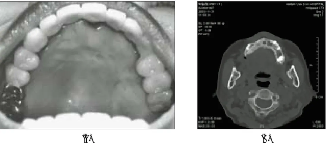
2002년 1월 17일 55세의 여자환자가 구개점막의 종창을 주소로 본원에 전원되어 방문하였다(Fig. 1). 발병 시기는 20년 전이었으 며 개인치과의원에서 치근단 낭종으로 진단 받았었다. 그곳에서 병소의 흡인을 여러 번 시행했으며 환자는 흡인이 시행됐을 때 종창이 감소되었으나 완전히 가라앉지는 않았으며 여러 번 다시 종창이 발생하였다고 진술하였다. 건전한 구개점막으로 싸여진 종물은 구강 내에서 뭉툭하게 튀어나온 양상을 보여줬다(Fig. 1).
병소의 촉진시 동통은 호소하지 않았고, 어떤 부위에서는 파동 성(fluctuation)이 감지되었다. 컴퓨터단층촬영을 시행한 결과 거 대 연조직 종물이 구개에서 관찰되었고, 비구개관이 넓어져있는 것을 관찰하였다(Fig. 2). 골 흡수나 변위는 관찰되지 않았다. 임
Abstract (J. Kor. Oral Maxillofac. Surg. 2004;30:56-59)절치신경 기원의 신경 섬유종: 증례보고와 면역조직화학적 연구
전효상∙손대일∙김성곤∙김미자∙박혜림**∙이동근*∙조병욱∙조남성***∙박영주***
한림대학교 성심병원 치과학교실 구강악안면외과 한림대학교 성심병원 치과학교실 교정과*
한림대학교 성심병원 병리과**, 한림대학교 강남성심병원 구강악안면외과***
조 병 욱
경기도 안양시 동안구 평촌동 896
한림대학교 성심병원 치과학교실 구강악안면외과 Byoung-Ouck Cho
Department of OMFS, Sacred Heart Hospital, Hallym University
#896, Pyungchon-Dong, Dongan-Gu, Anyang-city, Kyungki-do, 431-070, Korea Tel : 82-31-380-3873 Fax : 82-31-380-1900
E-mail : epker@chollian.net
SOLITARY NEUROFIBROMA OF THE INCISIVE NERVE: A CASE REPORT AND IMMUNOHISTOCHEMICAL STUDY
Hyo-Sang Jeon, Dai-Il Son, Seong-Gon Kim, Mi-Ja Kim, Hye-Rim Park
**
, Dong-Geun Lee*
, Byoung-Ouck Cho, Nam-Sung Cho***
, Young-Joo Park***
Dept. of Oral & Maxillofacial Surgery, Sacred heart hospital, Hallym University Dept. of Orthodontics, Sacred heart hospital, Hallym University*
Dept. of Pathology, Sacred heart hospital, Hallym University**
Dept. of Oral & Maxillofacial Surgery, Kangnam sacred heart hospital, Hallym University***
The neurofibroma in oral cavity is typically associated with neurofibromatosis. The solitary neurofibroma is commonly observed in skin. It is relatively rare in oral cavity and usually observed in the tongue, buccal mucosa, and vestibule. The rare types of solitary neu- rofibromas have been reported as a case report and they were in the inferior alveolar nerve, infratemporal fossa, maxilla, and palatal ginviva. In our hospital, the presented case was the first case as reported as solitary neurofibroma in the oral cavity. The prognosis after excision and the review of literatures were presented.
Key words: Neurofibroma, Neurofibromatosis, Oral cavity
절치신경 기원의 신경 섬유종: 증례보고와 면역조직화학적 연구
57 상적으로는 치성기원의 낭종 또는 양성종양으로 잠정진단 내려
졌다. 구내절개를 통해 수술을 시행하였는데, 이때 종물 내부로 함입되어있는 절치신경을 관찰할 수 있었고, 절치신경관 내에 있는 절치신경은 보존하였다. 하방의 구개골은 비교적 건전한 상태를 보였으며 술후 병소가 제거된 부위는 바셀린 거즈와 splint로 보호하였다.
술후 치료는 통상의 방법으로 시행하였으며, splint는 적출한 부위의 자극을 방지하기 위하여 술 후 4주 동안 장착 되었다. 병 소의 정확한 진단을 위하여 조직병리학적 검사를 시행하여 신경 섬유종으로 확진하였다. 술전 방사선 사진과 병소의 위치로 보 아 절치신경 기원의 신경섬유종일 가능성이 높았으나 신경관 내 에 위치하고 있는 절치신경의 절제가 기술적으로 어렵고 병소가 20년동안 서서히 자라왔으며 현재 환자의 나이가 55세임을 감안 하여 추가적인 처치는 유보하고 계속 추적조사하기로 하였다.
현미경 사진 상에서 병소는 미세한 결합조직으로 둘러싸인 물 결모양의 핵으로 이루어진 방추형 세포가 관찰되었다(Fig. 3). 신
경섬유종증에대한 평가를 위해 전신적인 검사를 시행하였으나, 여타의 비정상적 사항이나 친족과의 연관성은 관찰되지 않았다.
다음의 방법으로 면역조직화학적 염색을 시행하였다. 인체에 서 기원한 S100, CD-34, CD-56의 단백질의 아미노기 말단을 map-
Fig. 1. Intra oral view of the lesion. There was no ulceration in the overlying mucosa.
Fig. 2. Coronal CT showing soft tissue lesion on the palate and enlarged nasopalatine canal.
Fig. 3. Photomicrograph showing spindle-shaped cells with wavy nuclei in a delicate connective tissue matrix (hematoxylin-eosin stain, original magnif- ication x200).
(A) (B) (C)
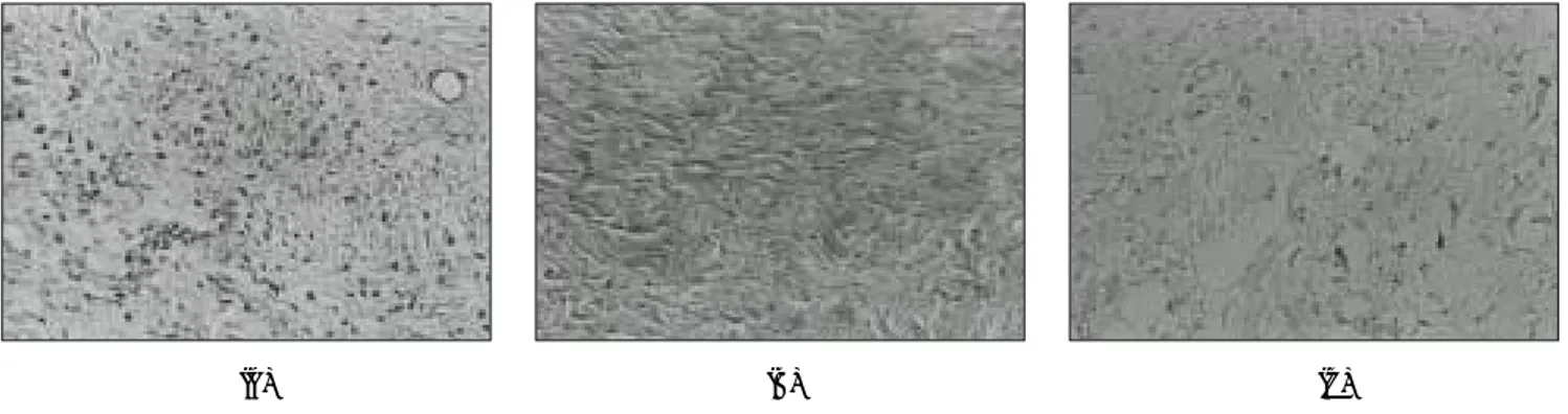
Fig. 4. Immunohistochemical results. A. The lesion showed moderate S-100 immunoreactivity. B. The lesion showed CD-34 positive immunoreactivity. C. The lesion showed negative CD-56 immunoreactivity (original magnification x200)
대구외지 2004;30:56-59
58
ping한 단백질을 쥐에 주입하여 얻은 단클론 항체를 실험에 이용 하였는데, S100, CD-34는 Ventna사(Tuscon, AZ, USA)에서 CD-56은 ZYmed( Sanfrancisco, LA, USA)에서 구입하였다. 4㎛ 두께의 절편 을 제작하여 면역조직화학적 염색을 Universal LSAB+kitds (DAKO, Glostrup, Denmark) 으로 시행하였고, 이후 과정은 제조 자의 지침서를 따랐다. 면역조직화학염색이 끝난 시편은 Mayer hematoxylin으로 counterstaining 하였다. 본 병소는 S-100과 CD-34 에 양성임을 보여주었다 (Fig. 4a, b). 그러나 CD-56에 대하여는 음 성임을 보여주었다 (Fig. 4c).
본 환자의 23개월간의 추적조사 결과 비구개관은 여전히 넓어 진 상태로 있었으나 특별한 재발 소견은 보이지 않았으며 구개 점막은 잘 치유된 상태로 유지되었다.(Fig. 5a. b)
Ⅲ. 고 찰
본 병소의 경우 조직학적으로 명확한 진단이 내려지기 전에는 감별진단으로 neurofibroma를 생각하지 못하였는 데 그 이유는 구개에서 발생하는 solitary neurofibroma가 거의 보고된 바 없기 때문이었다. 병소가 치조골과 근접하여 위치하고 있어 치성 농 양이나 낭종을 의심하게 하였지만 방사선 사진 상에 병소와 치 근과의 교통은 관찰되지 않았고 병소도 골막 상의 결합조직 내 에 있어 치성병소와는 구별되었다. 염증이나 궤양이없는 건전한 점막은 그 병소가 치성병소의 상피적 특징을 갖지 않음을 뒷받 침해준다.
병소의 위치를 감안하면 타액선 종양과의 감별진단이 필요한 데, 소타액선의 종양은 모든 타액선 종양의 25%를 차지하며 종 종 단단하며 무증상적, 그리고 단독병소의 성향을 나타낸다
9). 가 장 흔한 타액선 종양인 다형성 선종(pleomorphic adenoma)은 모 든 소타액선 종양 중 50%를 차지하며 무통적이며, 느린 성장과 단단한 종물의 특성을 나타냄으로써 감별진단으로 고려되어야 한다
10). 그리고 좀더 중요한 사실은 pleomorphic adenoma 와 관련
된 소 타액선의 가장 흔한 병발 위치는 구개라는 점이다
11). 비록 드물 지라도 타액선 조직을 제외하고 점막하 종괴(sub- mucosal lump)가 일어날 수 있는 조직의 종류에는 신경조직, 결 합조직 그리고 림프조직 등이 있다. 궤양이 없으며 가철식 치과 보철물과 관련이 없다는 점에서 외상성 섬유종(traumatic fibro- ma)에대한 가능성을 배제할 수 있다. Granular cell tumor와 지방 종(lipoma)과 같은 다른 결합조직 병소들은 명확한 진단을 위해 거론될 수 있다. 그러나 granular cell tumor의 현미경적 주 특징인 빈번한 위상피성 증식(pseudoepithelimatous hyperplasia)와 lipoma 의 임상특징인 yellowish submucosal mass는 관찰되지 않았다
9).
고려할 만한 또 다른 병소로 임파종(lymphoma)을 생각할 수 있는데, 구강 내에서는 non-Hodgkin's lymphoma가 Hodgkin's lymphoma보다 발생에 있어서 더욱 빈번하고 전신에 걸쳐서 넓 게 재발되며 구강 내에서는 구개가 두 번째로 흔한 병발 장소이 다. 경구개의 임파성 증식 병소라면 bonafide lymphoma를 고려 해 볼 수 있다
9).
또 다른 감별진단 가능한 병소로는 타액선의 악성종양이 있다.
비록 건전한 구강점막과 느린 성장의 특징으로 양성종양 또는 낮은 악성도의 악성종양으로 진단 할 수 있으나, 악성종양도 느 리게 성장할 수 있다는 것을 간과하여서는 안 된다.
Adenoid cystic carcinoma는 비교적 드문 느린 성장을 하는 malignant tumor이고, 재발성 경향이 있으며 주로 타액선에서 발 생하지만, 구개또한 3번째로 빈발하는 장소이다
12). 특히 본 병소 의 재발적 특징은 어느 정도 까지는 adenoid cystic carcinoma를 연상할 수 있다. 점액상피암종(mucoepidermoid carcinoma)는 소 타액선에선 꽤 흔하고 등급에 따라 느린 성장과 궤양을 동반하 지 않을 수도 있다. 또한 polymorphous low-grade adenocarcinoma 가 주로 노년층에서 소타액선에 가장 흔하게 나타난다
10). Poly- morphous low-grade adenocarcinoma는 단단하고, 거상된 양상으 로 궤양이 없는 침윤성 종창으로 나타난다. Acinic cell carcinoma 는 꽤 드문 악성 타액선 신생물이며, 느리게 성장하는 종물로써
(A) (B)
Fig. 5. Follow up Image 23 months later A. The soft tissue lesion showed well healing appearance. B. Coronal CT showing still enlarged nasopalatine canal
절치신경 기원의 신경 섬유종: 증례보고와 면역조직화학적 연구
59 궤양은 거의 발생하지 않는다
13).
조직학적으로 고려할 만한 또 다른 병소는 단독 섬유성 종양 (solitary fibrous tumor) 이며, 이것은 localized fibrous mesothelioma 또는 submesothelial fibroma 라고 알려져 있다. 비록 그 종양은 늑 막외에서나 복막외에서 기원할 수 있는 드문 방추 세포종양 (spindle cell neoplasm)으로 두경부 영역에서는 드물게 발생한다
14).
부드럽고 물결치는 모양의 핵이 주로 보여지는 병소는 신경초 종(schwannoma)나 신경섬유종(neurofibroma)과 같은 신경돌기와 비슷하다. 그러나 일반적으로는 조직 형태학적 특징에서 신경섬 유종과는 다르므로 이것이 감별진단에 도움을 준다.
면역조직화학적 염색은 neural tumor의 특징에 대한 기준을 정 하는 데에 유용하게 사용되어져 왔다
15). 예를 들자면, CD34 면역 반응은 단독 섬유성 종양에서 지속적으로 나타난다
16). 그러나 neurofibroma는 solitary fibrous tumor와는 달리 S-100에 대해 강한 양성을 보이나 neurofibroma에서 CD34에 양성인 것은 어느 정도 범위 내에서 다양성을 보인다
15). 그리고 이러한 특징은 진단에 유용하게 도움을 준다. 신경세포의 부착분자(N-CAM)인 CD56은 악성 신경초종(malignant schwannoma)에서 강하고 지속적인 면 역반응을 자아내는 또 다른 신경 항원 표시자(neural antigenic marker)이고 양성 신경초종(benign schwwanoma)에서 유용한 염 색이다
17).
모든 신경기원의 양성종양은 S-100에 대해서 면역양성 이고 이 번 증례에서 S-100 단백질에 대해 중등도의 면역반응을 나타냈 던 것은 조직학적 진단과 종양의 양성적 특성을 뒷받침 해준다.
그러므로 이번 증례에서 나타났던 S-100, CD-34 양성 그리고 CD- 56 음성은 총괄적으로 neurofibroma를 암시하는 것이었다. 이전 연구에 따르면 benign nerve sheath tumor의 대략 45%가량이 두경 부 영역에 위치하며 이중 10% 미만에서 구강 내에 위치하였다
18). Neurofibroma의 증례에서 성비가 2:1로 여성이 좀더 많았으며, 대 부분의 환자들은 20대와 30대였다
19).
신경 섬유종에대한 치료법으로는 대개 외과적 절제술을 선호 하며 술 후 예후에 있어서 신경초종과 신경섬유종의 감별은 매 우 중요하다. 왜냐하면 신경초종의 악성변이는 극도로 드물고
20)반면에 신경섬유종의 악성변이는 신경섬유종증 환자의 5-16%에 서 발생한다고 보고되었기 때문이다. 하지만, solitary neurofibro- ma의 악성변이는 아직 보고된 바 없다
21). 따라서 이번 증례에 대 해서는 세심한 추적조사가 요구된다.
참고문헌
1. Regezi JA, Sciubba JJ: Oral pathology, Clinical pathologic correla- tions. Philadelphia, PA, WB Sounders, 1999, p204.
2. Enzinger FM, Weiss SW: Soft tissue tumors. St. Louis, MO, Mosby, 1995, p843.
3. Apostolidis C, Anterriotis D, Rapidis AD, Angelopoulos AP: Solitary intraosseous neurofibroma of the inferior alveolar nerve: report of a case. J Oral Maxillofac Surg 2001;59:232-5.
4. Ueda M, Suzuki H, Kaneda T: Solitary intraosseous neurofibroma of the mandible: report of a case. Nagoya J Med Sci 1993;55:97-101.
5. Papageorge MB, Doku HC, Lis R: Solitary neurofibroma of the mandible and infratemporal fossa in a young child. Report of a case. Oral Surg Oral Med Oral Pathol 1992;73:407-11.
6. Skouteris CA, Sotereanos GC: Solitary neurofibroma of the maxilla:
report of a case. J Oral Maxillofac Surg 1988;46:701-5.
7. Shimoyama T, Kato T, Nasu D, Kaneko T, Horie N, Ide F: Solitary neurofibroma of the oral mucosa: a previously undescribed variant of neurofibroma. J Oral Sci 2002;44:59-63.
8. Nagahata S, Takagi S, Nishijima K. Neurofibroma of the tongue: a case report. Acta Med Okayama 1983;37:269-72.
9. Albert MM, Michael JW, Louis KR: Rapidly enlarging lesion of the upper lip. J Oral Maxillofac Surg 2000;58:883-7.
10. Waldron CA: Mixed tumor (pleomorphic adenoma) and myoepithe- lioma. In: Ellis GL, Auclair PL, Gnepp DR, editors. Surgical patholo- gy of the salivary glands. Philadelphia, PA, WB Saunders, l991, p 177.
11. Steven M, Joseph D, Danielle S, et al: Slowly expanding palatal mass. J Oral Maxillofac Surg 2001;59:655-9.
12. Ellis GL, Auclair PL. Malignant epithelial tumors. In: Rosai J, editor.
Tumors of the salivary glands. Atlas of tumor pathology. 3rd series fascicle 17. Washington DC, WA, Armed Forces Institute of Pathology, 1996, p155.
13. Waldron CA, El-Mofty SK, Gnepp DR: Tumors of the intraoral minor salivary glands: a demographic and histologic study of 426 cases.
Oral Surg Oral Med Oral Pathol 1988;66:323-33.
14. Gangopadhyay K,Taibah K, Manohar MB, et al : Solitary fibrous tumor of the parapharyngeal space: a case report and review of the literature. Ear Nose Throat J 1996;75:681-4.
15. Evanthia C, Stavros IP, Nusi PD, et al: Benign neural tumors of the oral cavity: A comparative immunohistochemical study. Oral Surg Oral Med Oral Pathol Oral Radiol Endod 1999;84:381-90.
16. Souichi I, Mitsuhiro N, Fumihiro Y, et al: Solitary fibrous tumor of the buccal mucosa: report of a case with immunohistochemical studies. Osomopore, 1999;88:461-5.
17. Miettinen M, Cupo W: Neural cell adhesion molecule distribution in soft tissue tumors. Hum Pathol 1993;24:62-6.
18. Das Gupta TK, Brasfield RD, Strong EW, et al: Benign solitary schwannomas (neurilemomas). Cancer 1969;24:355-66.
19. Leopoldo EL, Marc HH, Mia C N-P, et al: Recurrent myxoma of the zygoma: A case report. J Oral Maxillofac Surg, 2002;60:704-8.
20. Carstens PH, Schrodt GR: Malignant transformation of a benign encapsulated neurilemoma. Am J Clin Pathol 1969;51:144-9.
21. Muraki Y, Tateishi A, Tominaga K, Fukuda J, Haneji T, Lwata Y:
Malignant peripheral nerve sheath tumour in the maxilla associated with von Reclinghausen's disease. Oral Dis 1999;5:250-2.