한국컴퓨터정보학회 하계학술대회 논문집 제29권 제2호 (2021. 7)
701
● 요 약 ●
The purpose of the study was to identify fiber changes induced by electrical stimulation of a certain neural substrate in the rat brain. In the stimulation group, the peripheral nerve was injured and the brain area associated to inhibit sensory information was electrically stimulated. There were sham and sham stimulation groups as controls. Then high-field diffusion tensor imaging (DTI) was acquired. 35 features were taken from the DTI measures from 7 different brain pathways. To compare the efficacy of the classification for 3 animal groups, the linear regression analysis (LDA) and the machine learning technique (MLP) were applied. It was found that the testing accuracy by MLP was about 77%, but that of accuracy by LDA was much higher than MLP. In conclusion, machine learning algorithm could be used to identify and predict the changes of the brain white matter in some situations. The limits of this study will be discussed.
키워드: machine learning (기계학습), multi-layer perceptron (다층퍼셉트론),
linear discriminant analysis (선형판별분석), rat (흰쥐), diffusion tensor image (확산텐서영상)
신경 손상과 전기 뇌 자극에 의한 흰쥐의 뇌 섬유 경로 변화에 대한 기계학습 판별
손진훈O, 음영지**, 정재준**, 차명훈*, 이배환(교신저자)*
O연세대학교 의과대학, 생리학 교실,
*연세대학교 의과대학, 생리학 교실,
**한국기초과학지원연구원, 연구장비운영부
e-mail: [email protected]O, {youngjieum, cheong}@kbsi.re.kr**, {mhcha, bhlee}@yuhs.ac*
Classification of Fiber Tracts Changed by Nerve Injury and Electrical Brain Stimulation Using Machine Learning
Algorithm in the Rat Brain
Jin-Hun SohnO, Young-Ji Eum**, Chaejoon Cheong**, Myeounghoon Cha*, Bae Hwan Lee(Corresponding Author)*
ODepartment of Physiology, Yonsei University College of Medicine,
*Department of Physiology, Yonsei University College of Medicine,
**Center for Research Equipment, Korea Basic Science Institute
I. Introduction
Diffusion tensor imaging (DTI) is a non-invasive method for visualizing white matter fiber tracts in the brain. It is possible to observe fiber changes due to nerve development or degeneration through white matter fiber tracking or tractography [1]. However, there are few studies methods of discrimination using DTI. As a pilot study, the study attempted to establish the way of classifying DTI data using machine learning. For this, the study was to identify fiber changes induced by electrical stimulation of a certain neural substrate in the rat brain with multi-layer perceptron (MLP).
II. Method
1. Subjects
Sprague–Dawley rats were used in this study. Groups were divided into three groups; 1) sham peripheral nerve in with sham stimulation (sham), 2) the peripheral nerve injury with sham stimulation (sham stimulation), and 3) peripheral nerve injury with a specific brain region stimulation (stimulation).
한국컴퓨터정보학회 하계학술대회 논문집 제29권 제2호 (2021. 7)
702 2. Imaging acquisition
DTI data were acquired on a 9.4 T high-field MRI scanner developed for imaging of small animals (Bruker Biospin, Ettlingen, Germany). The following parameter was used: TR/TE
= 12,500/0.512 msec, in-plane resolution = 0.134 mm, 30 directions, b-value = 3,063.4 s/mm2, diffusion time/encoding duration = 20/4 msec. The data were reconstructed in the Waxholm Space atlas of the Sprague Dawley rat brain using q-space diffeomorphic reconstruction [2] to obtain the spin distribution function [3].
3. Feature Selection and Classification
Thirty-five features were extracted from 5 DTI measures in each of 7 pathways of interest, were extracted and used as feature vectors. Two classifiers, the linear discriminant analysis (LDA) and the multi-layer perceptron (MLP), were applied to classify the brain pathways among 3 groups.
III. Results
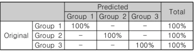
Results of LDA showed 100.0% classification of 3 groups (Table 1).
Predicted
Total Group 1 Group 2 Group 3 Original
Group 1 100% - - 100%
Group 2 - 100% - 100%
Group 3 - - 100% 100%
Table 1. Confusion matrix for classification using LDA
For applying the MLP, the data was randomly assigned into 70% subjects as the training set and 30% the testing set. The classification accuracy of testing was as high as was 77.8%
(Table 2).
Set Observed Predicted
Group 1 Group 2 Group 3 % Correct Training
Group 1 100% - - 100%
Group 2 - 100% - 100%
Group 3 - - 100% 100%
Testing
Group 1 100% - 100%
Group 2 75% 50% 75%
Group 3 25% 50% 50%
The average accuracy of testing set: 77.8%
Table 2. Confusion matrix for classification using MLP
IV. Conclusions
Results showed that features of fibers obtained by DTI on rat brain could be useful for classifying changes of nerve fibers induced by nerve injury and brain stimulation with MLP. But LDA seemed to be more effective in discriminating nerve changes.
We will try to find the difference between the two methods in the future. Also, we will seek out more useful features from 35 ones to get better classification.
We suggest that DTI is a useful technique to find out the changes of nerve fibers in the brain. The machine learning would be also useful method for identifying changes of brain fibers.
ACKNOWLEDGEMENT
This study was supported by the National Research Foundation of Korea (NRF) grants funded by the Korean government (MSIT) (2020R1A2C3008481).
REFERENCES
[1] R. Watts, C. Liston, S. Niogi, and A. M. Uluğ, "Fiber tracking using magnetic resonance diffusion tensor imaging and its applications to human brain development," Mental retardation and developmental disabilities research reviews, vol. 9, no. 3, pp. 168-177, 2003.
[2] F.-C. Yeh and W.-Y. I. Tseng, "NTU-90: a high angular resolution brain atlas constructed by q-space diffeomorphic reconstruction," Neuroimage, vol. 58, no. 1, pp. 91-99, 2011.
[3] F.-C. Yeh, V. J. Wedeen, and W.-Y. I. Tseng,
"Generalized q-sampling imaging," IEEE transactions on medical imaging, vol. 29, no. 9, pp. 1626-1635, 2010.