INTRODUCTION
Obstructive cholestasis is defined as a mechanical blockage in the bile ducts, which prevents bile from flowing into the duode- num, leading to accumulation of potentially toxic biliary com- pounds in the liver and bile ducts.1 Toxicity constituents, such as
bile acids and bilirubin, can injure hepatocytes and bile duct cells. Long-term cholestasis may develop into fibrosis, cirrhosis, and even carcinoma. The spontaneous anticholestatic defense mechanisms of hepatocytes comprise adaptive induction of ba- solateral alternative export pumps, which are important cana- licular bile transporters. Interestingly, expression levels of he- patic membrane transporters and bile acid metabolic enzymes have been shown to be significantly altered in cholestasis.2-4 For example, as an ATP-dependent organic anion transporter be- longing to the ABC transporter superfamily, multidrug resis- tance-associated protein 4 (MRP4/ABCC4) deficiency has been found to worsen liver injury and decrease plasma bile acid levels in bile duct-ligated mice.5-7 MRP4 is widely expressed in many human and rodent tissues, such as liver, kidney, intestine, brain, and breast. Published studies have shown that MRP4 expression is substantially increased in the livers of mice with disruption of bile acid nuclear receptor, accompany by increased levels of
TNFα Induces Multidrug Resistance-Associated
Protein 4 Expression through p38-E2F1-Nrf2 Signaling in Obstructive Cholestasis
Wei Lian, Xiaocong Liu, and Wensheng Chen
Department of Gastroenterology, Southwest Hospital, Army Medical University (Third Military Medical University), Chongqing, China.
Purpose: To explore the molecular mechanism of the upregulation of multidrug resistance-associated protein 4 (MRP4) in cholestasis.
Materials and Methods: The mRNA and protein levels of MRP4 in liver samples from cholestatic patients were determined by quantitative real-time PCR and Western blot. In human hepatoma HepG2 cells, electrophoretic mobility shift assay (EMSA) was used to determine the affinity of nuclear factor-E2-related factor (Nrf2) binding to MRP4 promoter. Dual-luciferase reporter assay was used to detect the binding of tumor necrosis factor α (TNFα) to the promotor of E2F1. The bile duct ligation mouse models were established using male C57BL/6 mice.
Results: The mRNA and protein levels of MRP4 were significantly increased in cholestatic patients. TNFα treatment induced the expression of MRP4 and Nrf2 and enhanced cell nuclear extract binding activity to MRP4 promoter, as demonstrated by EMSA. Nrf2 knockdown reduced MRP4 mRNA levels in both HepG2 and Hep-3B cells. In addition, TNFα increased Rb phosphorylation and ex- pression of MRP4 and Nrf2 and activated E2F1 and phosphorylated p38 in HepG2 and Hep-3B cells. These effects were markedly in- hibited by pretreatment with E2F1 siRNA. Dual-luciferase reporter assay validated that TNFα induces the transcription of E2F1. Fur- thermore, the expression of MRP4, Nrf2, E2F1, and p-p38 proteins was improved with treatment of TNFα in a mouse model of cholestasis. E2F1 siRNA lentivirus or SB 203580 (p38 inhibitor) inhibited these positive effects.
Conclusion: Our findings indicated that TNFα induces hepatic MRP4 expression through activation of the p38-E2F1-Nrf2 signal- ing pathway in human obstructive cholestasis.
Key Words: Obstructive cholestasis, multidrug resistance-associated protein 4, Nrf-2, TNFα
pISSN: 0513-5796 · eISSN: 1976-2437
Received: March 22, 2019 Revised: July 24, 2019 Accepted: August 14, 2019
Corresponding author: Wensheng Chen, PhD, Department of Gastroenterology, Southwest Hospital, Army Medical University (Third Military Medical University), No. 30, Gaotanyan Main Street, Shapingba District, Chongqing, 400038, China.
Tel: 86-023-68754124, Fax: 86-023-65415803, E-mail: [email protected]
•The authors have no potential conflicts of interest to disclose.
© Copyright: Yonsei University College of Medicine 2019
This is an Open Access article distributed under the terms of the Creative Com- mons Attribution Non-Commercial License (https://creativecommons.org/licenses/
by-nc/4.0) which permits unrestricted non-commercial use, distribution, and repro- duction in any medium, provided the original work is properly cited.
Yonsei Med J 2019 Nov;60(11):1045-1053 https://doi.org/10.3349/ymj.2019.60.11.1045
serum and hepatocellular bile acids, and that MRP4 can be fur- ther upregulated by cholic acid feeding, consistent with a role in bile acid homeostasis.8 Thus, hepatic MRP4 levels are normally low in rodents, but elevated in obstructive cholestasis, similar to elevated multidrug resistance-associated protein 3 (MRP3/
ABCC3) expression in obstructive cholestasis.9,10
The transcriptional regulation of hepatic organic anion trans- porters comprises a complex interacting network of ligand-ac- tivated nuclear receptors, including farnesoid X-activated recep- tor, pregnane X receptor (PXR), constitutive androstane receptor (CAR), retinoid X receptor, nuclear factor-E2-related factor (Nrf2), liver receptor homolog-1 (LRH-1), peroxisome proliferator-ac- tivated receptor-α, and others. These nuclear factors play essen- tial roles in the transcriptional regulation of hepatobiliary trans- port systems and have been reported to be involved in bile acid metabolism in animal models of cholestasis and cell lines.11,12 In addition, Nrf2 has been reported to participate in the transcrip- tional regulation of drug transporter genes, MRPs.13 The mech- anisms by which proinflammatory cytokines modulate trans- porter gene expression have been intensively studied over the past decade. Recent reports indicate that the proposed mecha- nism by which tumor necrosis factor α (TNFα) upregulates MRP3 is via induction of the liver receptor LRH-1.14 Thus, TNFα may be the factor that mediates bile acid-dependent induction because it upregulates LRH-1 expression in vitro.
In a previous study, we found that the upregulation of hepatic MRP3/ABCC3 expression in human obstructive cholestasis is likely triggered by TNFα and mediated by the activation of SP1.15 However, whether TNFα signaling plays a role in Nrf2-stimulat- ed MRP4/ABCC4 expression is still unknown. Decades of study have shown that several intracellular stress signals, including mi- togen-activated protein kinases, extracellular signal-regulated kinase (ERK), and c-Jun-N-terminal kinase (JNK), can be stim- ulated by lipopolysaccharides in hepatic Kupffer cells.16 In this study, we investigated whether p38 MAPK signaling is involved in the TNFα-mediated regulation of MRP4. To this end, we as- sessed MRP4/ABCC4 expression and plasma levels of TNFα in the livers of patients with obstructive cholestasis by quantita- tive real-time PCR (qRT-PCR), Western blot, and ELISA. The ex- pression of MRP4, Nrf2, E2F1, p-p38 MAPK and p-Rb proteins was detected by Western blot analysis. The binding activity of Nrf2 to the MRP4/ABCC4 promoter was analyzed by electropho- retic mobility shift assay (EMSA). In this report, we described the association of increased hepatic MRP4/ABCC4 expression with elevated TNFα levels and enhanced Nrf2 expression, show- ing that p38-Rb-E2F1 signaling is involved in this progression.
MATERIALS AND METHODS
Patients and samples
All liver samples were collected from patients at Southwest Hos- pital (Chongqing, China) with informed consent from all partic-
ipants, and this study was approved by the Southwest Hospital Institutional Ethics Review Board and carried out in accordance with the Declaration of Helsinki of the World Medical Associa- tion. Control liver samples were also obtained during resection of liver metastases without cholestasis (n=14; three rectal me- tastases, four colorectal metastases, seven colonic metastases).
Cholestatic liver samples (n=14) were surgically resected from patients with obstructive cholestasis caused by biliary stones originating from the intrahepatic bile duct and common bile duct. Surgery was carried on these patients out within 3 days of admission due to severe symptoms of biliary obstruction and jaundice. Neither ursodeoxycholic acid (UDCA) nor other pre- operative therapy was administered. Isolated liver samples were immediately cut into small pieces and stored in liquid nitrogen.
The clinical characteristics of the patients are listed in Table 1.
Gender was not a factor involved in altering adaptive response gene expression in cholestatic patients.
Cell culture and treatment
Human hepatoma HepG2 cells and Hep-3B cells and human embryonic kidney HEK293T cells (ATCC, Manassas, VA, USA) were maintained at 37˚C in DMEM (Sigma Chemical Co., St.
Louis, MO, USA) containing 10% FBS, 1% L-glutamine and 1%
streptomycin (Invitrogen, San Diego, CA, USA). Before chemical treatment, cells were serum-starved overnight and then treat- ed with the indicated dose of chemicals for designated times.
For p38-Rb-E2F1 signaling inhibition experiments, HepG2 or Hep-3B cells were transfected with E2F1 siRNA (GenePharma, Shanghai, China) for 48 h prior to the addition of TNFα. For elim- inating reactive oxygen species (ROS), cells were pre-incubat- ed with 1 mM N-acetyl-L-cysteine (NAC, Beyotime Biotechnol- ogy, Shanghai, China) for 30 min before being exposed to TNFα.
Cell transfection
The siRNA constructs used in this study were all purchased from GenePharma, including a negative control siRNA (neg. siRNA), Nrf2 siRNA, and E2F1 siRNA. Transfection of HepG2 cells was performed using Lipofectamine® RNAiMAX Transfection Re- agent (Invitrogen, Carlsbad, CA, USA) following the manufac- turer’s protocol. Cells were transfected with 100 nM siRNA and were cultured for 48 h, after which the following experiments were performed.
Animals and treatments
Male C57BL/6 mice (8–9 weeks, weighing 21–25 g) were ob- tained from the experimental animal center of the Field Sur- gery Research Institute (Daping Hospital, Chongqing, China) and underwent bile duct ligation (BDL) or sham-operation, as previously described.17 The p38 inhibitor SB 203580 (Sigma Chemical Co.) was dissolved in 3% DMSO. Mice in the experi- mental group were pretreated with 30 mL of SB 203580 (100 μM) or 100 μL of E2F1 siRNA lentivirus. The recombinant lentivirus of E2F1 siRNA was prepared and titered to 108 TU/mL. TNFα was
intravenously administered at various time periods as indicated.
ELISA
ELISA kits for human and mouse TNFα, IL-6, and IL-1β detec- tion were all purchased from Beyotime Biotechnology. Plasma levels of TNFα, IL-6, and IL-1β in cholestatic patients or in mouse models of cholestasis were assayed using the corresponding ELISA kit according to the manufacturer’s instructions.
Liver function
At the end of the treatment period, blood samples were collect- ed from the orbital veins of mice under anesthesia. After 2 h of coagulation at room temperature, blood samples were centri- fuged at 5000 rpm for 15 min The serum was collected for the de- tection of alanine aminotransferase (ALT) and aspartate trans-
aminase (AST) levels using an automatic biochemical analyzer (AU5800 Series, Beckman Coulter, Brea, CA, USA).
Detection of reactive oxygen species
Intracellular ROS levels were determined using Reactive Oxy- gen Species Assay Kits (Nanjing Jiancheng Bioengineering In- stitute, Nanjing, China). Briefly, HepG2 cells and Hep-3B cells were cultured in DMEM in the presence of 50 ng/mL of TNFα.
After 12 h, cells were digested with 0.25% trypsin and were then collected into centrifuge tubes. The collected cells were rinsed twice with 10 mM fresh phosphate buffer saline, re-suspended in serum-free medium containing 10 μM 2’,7’-dichlorofluorescin diacetate (DCFH-DA), and then incubated in the dark for 30 min at 37°C. At the end of incubation, cells were washed three times with serum-free medium to fully remove DCFH-DA that Table 1. Clinical Features of Patients
No. Gender Age (yr) TBIL (mg/dL) TBA (μmol/L) ALT (IU/L) AST (IU/L) ALP (IU/L)
Control (n=14)
1 Female 44 0.2 12.4 24.5 20.6 92.1
2 Male 47 0.3 15.6 20.3 29.5 88.6
3 Female 54 0.2 9.3 58.7 62.3 130.4
4 Female 57 0.1 17.1 62.5 65.9 114.2
5 Male 47 0.2 12.9 13.6 34.1 94.5
6 Male 50 0.3 14.6 23.8 29.6 93.4
7 Female 51 0.4 10.3 20.2 27.4 37.7
8 Male 53 0.1 9.1 67.3 48.9 127.2
9 Female 48 0.2 17.6 64.4 44.2 98.7
10 Female 51 0.2 9.4 56.6 45.9 47.9
11 Male 53 0.2 14.7 20.5 17.4 44.2
12 Male 55 0.3 9.6 15.6 28.5 37.3
13 Female 44 0.3 14.9 57.1 37.4 73.3
14 Female 47 0.2 8.8 59.0 31.2 36.8
Mean±SD 50.0±4.0 0.2±0.1 12.6±3.2 40.3±21.6 37.4±14.5 79.7±33.7
Obstructive (n=14)
1 Male 58 4.0 221.6 143.5 325.6 445.3
2 Male 59 2.6 139.3 176.2 374.1 493.9
3 Female 49 4.2 148.2 353.3 561.8 239.1
4 Male 60 4.5 122.4 119.9 251.6 308.7
5 Female 62 3.2 240.5 159.8 287.3 276.5
6 Female 59 3.1 143.9 177.2 296.3 456.4
7 Male 45 4.5 152.7 265.6 403.6 522.8
8 Female 57 3.0 214.5 146.4 247.5 318.9
9 Female 49 4.7 219.1 237.1 316.9 426.3
10 Male 52 3.1 154.5 121.6 268.2 338.3
11 Male 48 5.0 229.3 169.9 306.7 256.8
12 Male 56 3.0 238.6 251.1 413.4 370.8
13 Female 44 2.9 213.5 302.9 389.1 561.5
14 Female 58 3.3 152.4 137.8 231.0 344.6
Mean±SD 54.0±6.0 3.7±0.8 185.0±43.1 197.3±72.5 333.8±88.4 382.9±102.3
p value (vs. control) 0.052 <0.001 <0.001 <0.001 <0.001 <0.001
TBIL, total bilirubin; TBA, total bile salts; ALT, alanine aminotransferase; AST, aspartate transaminase; ALP, alkaline phosphatase.
did not enter cells. The intensity of ROS fluorescence was mea- sured by flow cytometry (BD Biosciences, San Diego, CA, USA) at the wavelength of 488 nm.
RNA extraction and qRT-PCR
Samples of liver tissue frozen (100 mg) in liquid nitrogen were ground and subsequently used for the extraction of total RNA with TRIzol reagent (Invitrogen). Total RNA was reverse tran- scribed into cDNA using a Revert AidTM first strand cDNA syn- thesis kit (MBI Fermentas Inc., Ontario, Canada). qRT-PCR us- ing a SYBR® premix Ex TaqTM II kit (Takara Biotechnology, Tokyo, Japan) was performed in a Bio-Rad CFX96 real-time system ma- chine (Bio-Rad, Hercules, CA, USA) to determine the mRNA lev- els of specific genes, whose primers are listed in Table 2. The am- plification efficiency of target genes was considered using CFX manager 2.0 (Bio-Rad) for data analysis. GAPDH was used as a reference for normalizing data.
Western blot analysis
Total membranes and nuclear extracts were prepared as de- scribed previously,18 and protein concentrations were determined using a Bradford kit (Thermo Scientific, Rockford, IL, USA). The dilutions of primary antibodies were MRP4, 1:1300; Nrf2, 1:1000;
p-p38 MAPK, 1:2000; E2F1, 1:2000; and p-Rb, 1:2500 (Cell Signal- ing, Danvers, MA, USA). GAPDH and SH-PTP1 (Santa Cruz Bio- technology, Santa Cruz, CA, USA) were used as loading referenc- es for data analysis.
Electrophoretic mobility shift assay
Oligos containing the Nrf2 response elements in the human MRP4/ABCC4 promoter or its mutant forms, as previously re- ported,18 were labeled with γ-32P-ATP (10 mCi/mL). EMSA was performed using nuclear extracts from HepG2 cells as described previously.19
Dual-luciferase reporter assay
cDNA fragments containing the predicted TNFα binding se- quences in E2F1 were amplified and subcloned into the pGL3 luciferase promoter-reporter vector (Promega, Madison, WI, USA). As a control, the pGL3-mutE2F1 plasmids were also con- structed using cDNA fragments containing corresponding mu- tated nucleotides for TNFα. HEK-293T cells were seeded into six-well plates at 1×10⁵ cells per well, transfected with 0.1 μg of pGL3-E2F1 or pGL3-mutE2F1 vectors in the presence of pre- TNFα or pre-DMSO negative controls using Lipofectamine 2000 (Invitrogen, Carlsbad, CA, USA), and incubated for 48 h. Cells were harvested and luciferase activities were measured using
the dual-luciferase reporter assay kit (Promega) according to the operating instructions.
Statistical analysis
All data are expressed as the mean±SD of at least three determi- nations and were analyzed using one-way ANOVA and subse- quent Tukey’s multiple comparison test for comparison of two groups. A difference was accepted as significant if p<0.05.
RESULTS
Hepatic MRP4 expression and plasma TNFα are ele- vated in patients with obstructive cholestasis
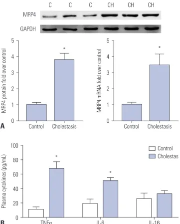
In this study, Western blot analysis showed that the expression of MRP4 in liver samples of patients with obstructive cholesta- sis was significantly increased (3.8-fold) when compared with control liver tissues samples. qRT-PCR assay revealed that the expression of MRP4 mRNA was increased 3.4-fold in liver sam- ples of patients with obstructive cholestasis (Fig. 1A). ELISA kit
Fig. 1. Expression of MRP4 and plasma levels of TNFα were increased in liver samples of patients with obstructive cholestasis. (A) The expres- sion of MRP4 protein and mRNA was detected by Western blot and qRT-PCR analysis. Representative blots and corresponding densitome- try are shown (n=14). (B) Plasma levels of TNFα, IL-6, and IL-1β in patients with obstructive cholestasis were detected using ELISA kits (n=14).
*p<0.05, vs. controls. C, control liver tissues samples; CH, cholestasis liver samples, MRP4, multidrug resistance-associated protein 4; Nrf2, nuclear factor-E2-related factor; TNFα, tumor necrosis factor α.
100 80 60 40 20
0 TNFα IL-6 IL-1β
Plasma cytokines (pg/mL)
Control Cholestas
*
*
B 5 4 3 2 1 0
5 4 3 2 1
Control Cholestasis 0 Control Cholestasis
MRP4 protein fold over control MRP4 mRNA fold over control
* *
MRP4
C C C CH CH CH
GAPDH
A
Table 2. Sense and Antisense Primers Used for qRT-PCR
Gene Sense primer (5’→3’) Antisense primer (5’→3’) MRP4 (ABCC4) TGCCTTTGGGTCCCGATTC TGGTGGTGGGCGTTTCTGAT
Nrf2 TCTATGTGCCTCCAAAGG CTCAGCATGATGGACTTGGA
GAPDH ACCACAGTCCATGCCATCAC TCCACCACCCTGTTGCTGTA
was used to evaluate the plasma levels of cytokines. Compared to the control group, the levels of TNFα and IL-6 were signifi- cantly increased in cholestatic patients, whereas levels of IL-1β were not significantly altered (Fig. 1B).
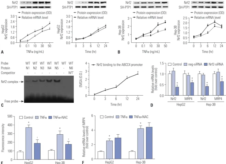
TNFα increases Nrf2 expression and DNA binding to MRP4 promoter
Here, we determined the regulatory mechanism of the promot- er of the second rescue transporter MRP4 and related receptors in response to TNFα in obstructive cholestasis. Our results indi- cated that TNFα increases Nrf2 mRNA and nuclear protein ex- pression in a dose and time-dependent manner in both HepG2 (Fig. 2A) and Hep-3B cells (Fig. 2B). Furthermore, EMSA dem- onstrated that TNFα increases binding capacity to MRP4 pro- moter in a time-dependent manner when using the Nrf2 re- sponse element in nuclear extracts from HepG2 cells (Fig. 2C).
To confirm that Nrf2 is critical to inducing MRP4 expression, siR- NA against Nrf2 was transfected into HepG2 cells or Hep-3B cells in the presence of TNFα treatment. As shown in Fig. 2D, the tran- scription of MRP4 was significantly inhibited by Nrf2-siRNA transfection. These results confirm that TNFα treatment induces Nrf2 expression and enhances Nrf2 binding to MRP4 promoter.
Since it has been reported that cholestasis can induce ROS production in hepatocytes and that TNFα increases ROS levels, we investigated whether ROS is an initial trigger that activates MRP4 under the stimulation of TNFα. NAC, a ROS scavenger, was used to inhibit ROS production in this study. We demon- strated that ROS levels were significantly elevated in both HepG2 cells and Hep-3B cells after TNFα stimulation and were posi- tively correlated with mRNA levels of MRP4 (Fig. 2E and F). NAC pre-treatment significantly inhibited ROS production, but did not change the mRNA levels of MRP4, compared to the TNFα
Fig. 2. TNFα-induced Nrf2 expression and binding activity to Nrf2 response element in MRP4 promoter. The influence of TNFα on Nrf2 expression in HepG2 cells (A) and Hep-3B cells (B) was detected by qRT-PCR and Western blot analysis. (C) HepG2 cells were serum-starved overnight before TNFα treatment. Nrf2 binding activity to the Nrf2 response element in MRP4/ABCC4 promoter after TNFα treatment was detected by EMSA analysis. (D) HepG2 cells and Hep-3B cells were transfected with Nrf2 siRNA upon treatment with 50 ng/mL of TNFα. Transcription of Nrf2 and MRP4 in HepG2 and Hep-3B cells was detected by qRT-PCR. (E) ROS levels in cells were determined using Reactive Oxygen Species Assay kits. NAC was used to eliminate ROS. The intensity of ROS fluorescence was measured by flow cytometry at 488 nm. (F) The mRNA levels of MRP4 were detected by qRT-PCR. *p<0.05, vs. controls;
†p<0.05, vs. TNFα. MRP4, multidrug resistance-associated protein 4; Nrf2, nuclear factor-E2-related factor; TNFα, tumor necrosis factor α; ROS, reactive oxygen species.
Nrf2 SH-PTP1
Nrf2 SH-PTP1 3.0
2.4 1.8 1.2 0.6
0.0 0 0.1 10 TNFα (ng/mL) Protein expression (OD) Relative mRNA level
30 50 HepG2 Nrf2 expression
3.0 2.4 1.8 1.2 0.6
0.0 0 3 6
Time (hr) Protein expression (OD) Relative mRNA level
12 24 HepG2 Nrf2 expression
A
Nrf2 SH-PTP1
Nrf2 SH-PTP1
3 2 1
0 0 0.1 10 TNFα (ng/mL) Protein expression (OD) Relative mRNA level
30 50 Hep-3B Nrf2 expression
2.5 2.0 1.5 1.0 0.5
0.0 0 3 6
Time (hr) Protein expression (OD) Relative mRNA level
12 24 Hep-3B Nrf2 expression
B
4 3 2 1
0 0 3 6
Time (hr)
Nrf2 binding to the ABCC4 promoter
12 24
EMSA (O.D.)
Probe Protein Competitor Nrf2 complex
Free probe
WT N2
- WT N3
- WT N4
- WT
N5 -
WT - -
WT N6 WT’
WT N1
-
C
1.5
1.0
0.5
0.0 Nrf2 MRP4 Nrf2 MRP4
Relative mRNA levels (fold over control)
Control neg-siRNA Nrf2-siRNA
HepG2 Hep-3B
D
*
*
*
*
500 400 300 200 100 0
6
4
2
HepG2 Hep-3B 0 HepG2 Hep-3B
Fluorescence intensity Relative mRNA levels of MRP4 (fold over control)
Control TNFα TNFα+NAC Control TNFα TNFα+NAC
E F
† †
*
*
*
*
group (Fig. 2E and F). These data implied that ROS is not an es- sential mediator of MRP4 expression stimulated by TNFα.
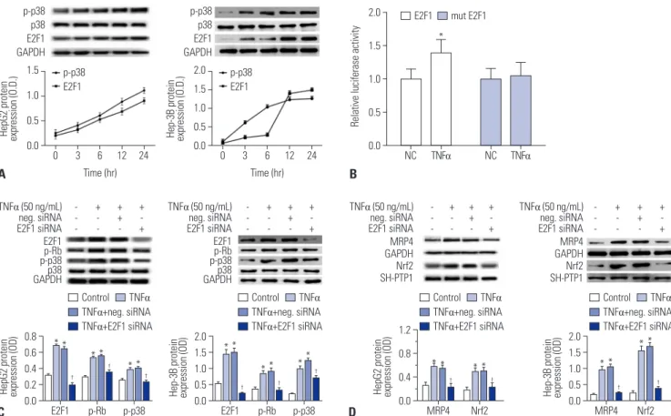
TNFα mediates MRP4/Nrf2 interactions via p38-Rb- E2F1 activation
To investigate whether p38 MAPK and E2F1 are part of the TNFα- enhanced MRP4/Nrf2 signaling pathway, we treated HepG2 cells and Hep-3B cells with 50 ng/mL of TNFα for 12 h. The re- sults showed that phosphorylated p38 and E2F1 were activated in a time-dependent manner of TNFα treatment in both HepG2 cells and Hep-3B cells (Fig. 3A). The results of luciferase report- er gene assays showed that pre-TNFα significantly induces lu- ciferase activity in pGL3-E2F1-3’-UTR-transfected cells, where- as luciferase activity is not affected by pre-TNFα in the pGL3- mutated E2F1-3’-UTR transfected group in comparison with pre- TNFα-negative control treatment (Fig. 3B). When HepG2 or Hep-3B cells were pretreated with E2F1 siRNA, TNFα-induced E2F1 activation and phosphorylation of Rb and p38 were abol- ished, whereas reductions in Nrf2 and MRP4 protein levels were observed in these cells (Fig. 3C and D).
TNFα injection increases MRP4/Nrf2 via p38-Rb-E2F1 pathway in mouse models of cholestasis
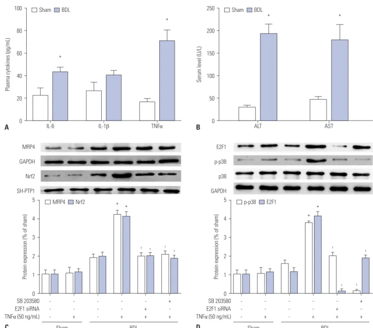
Expression of MRP4 protein in patients with obstructive cho- lestasis has been observed. In addition, we determined the ex- pression of Nrf2 and MRP4 in BDL mice. As in patients with cho- lestasis, TNFα levels in mouse models of cholestasis were markedly upregulated, compared with levels of IL-6 and IL-1β (Fig. 4A).
Serum levels of ALT and AST in BDL mice were also significant- ly elevated (Fig. 4B). The expression of MRP4 and Nrf2 proteins in Western blot analysis was induced in BDL mice when com- pared with sham rats. Moreover, after injection with 50 ng/mL of TNFα, the expressions of MRP4 and Nrf2 were significantly upregulated in BDL mice (Fig. 4C). In addition, p-p38 and E2F1 expression was significantly elevated after injection with TNFα (Fig. 4D). Similar to results in patients with obstructive cholesta- sis, these data confirmed that TNFα participates in the regula- tion of MRP4/Nrf2 expression and that E2F1 may be involved in this regulatory mechanism. The expression of these proteins was decreased by pre-treatment with either E2F1 siRNA lentivirus or SB 203580.
Fig. 3. The p38-Rb-E2F1 pathway mediates TNFα-induced MRP4 expression. (A) The influence of TNFα on p-p38 and E2F1 in HepG2 cells and Hep-3B cells was detected by Western blot analysis. (B) The interaction between E2F1 3’-UTR and TNFα was detected by luciferase activity assay. Wild-type or mutated E2F1 3’-UTR containing binding sites for TNFα was co-transfected with pre-TNFα or pre-DMSO negative control in HEK293T cells. (C and D) The expression of p-p38, p38, E2F1, p-Rb, MRP4, and Nrf2 protein levels in both HepG2 cells and Hep-3B cells was detected by Western blot and their corre- sponding densitometry values after TNFα and siRNA treatment were quantified. *p<0.05, vs. controls; †p<0.05, vs. TNFα. MRP4, multidrug resistance-as- sociated protein 4; Nrf2, nuclear factor-E2-related factor; TNFα, tumor necrosis factor α.
C D
2.0 1.5 1.0 0.5 0.0 2.0
1.5 1.0 0.5 0.0 0.8
0.6 0.4 0.2 0.0
1.2 0.8 0.4
0.0 MRP4
E2F1
E2F1 MRP4 Nrf2 Nrf2
-- - --
- --
-
-- -
TNFα (50 ng/mL) neg. siRNA E2F1 siRNA TNFα (50 ng/mL)
neg. siRNA E2F1 siRNA TNFα (50 ng/mL)
neg. siRNA E2F1 siRNA
TNFα (50 ng/mL) neg. siRNA E2F1 siRNA
+- - +-
- +-
-
+- -
++ - ++
- ++
-
++ -
+- + +-
+ +-
+
+- +
MRP4 GAPDH SH-PTP1Nrf2 E2F1p-Rb
p-p38 GAPDHp38 E2F1p-Rb
p-p38 GAPDHp38
MRP4 GAPDH SH-PTP1Nrf2
Hep-3B protein expression (OD) Hep-3B protein expression (OD)
HepG2 protein expression (OD) HepG2 protein expression (OD)
Control TNFα TNFα+neg. siRNA TNFα+E2F1 siRNA Control TNFα
TNFα+neg. siRNA TNFα+E2F1 siRNA Control TNFα
TNFα+neg. siRNA TNFα+E2F1 siRNA
Control TNFα TNFα+neg. siRNA TNFα+E2F1 siRNA
*
* *
*
*
* *
* *
*
*
*
†
†
†
p-Rb p-Rb
*
** *
†
†
p-p38 p-p38
* **
* †
† † † †
p-p38 p38 E2F1 GAPDH
p-p38 p38 E2F1 GAPDH 1.5
1.0 0.5 0.0
2.0 1.5 1.0 0.5
0 3 6 0.0 0 3 6
Time (hr) Time (hr)
p-p38 E2F1
p-p38 E2F1
12 24 12 24
HepG2 protein expression (O.D.) Hep-3B protein expression (O.D.)
A
E2F1 mut E2F1
* 2.0
1.5
1.0
0.5
0.0 NC TNFα NC TNFα
Relative luciferase activity
B
DISCUSSION
Cholestasis is a clinical syndrome characterized by systemic and intrahepatic retention of excessive toxic bile acids that causes liver injury, which may result either from a functional defect in bile formation at the level of the hepatocyte or from an impair- ment in bile secretion and flow at the bile duct level.10 Both con- genital and acquired pathogenic factors can cause bile acid se- cretion disorders and homeostatic imbalance, and eventually develop into cholestasis. However, treatment methods available for cholestasis and the current knowledge of related molecular mechanisms are seriously lacking. Recent research has shown that reduced expression and function of transport systems may
underlie the pathogenesis of cholestasis.
In recent years, transcription factors have been proven to play crucial roles in bile acid detoxification and output in cholestasis, such as multidrug resistance-associated protein (MRP), bile salt export pump, and the basolateral Na+-dependent bile acid up- take system (NTCP).15,20 The elevated expression of MRP4/ABCC4 has been previously reported in the liver tissues of cholestasis rodent models and some patients with primary biliary cirrhosis and obstructive cholestasis.21 Upregulation of MRP4 protects against the accumulation of toxic bile acids during cholestasis by facilitating their efflux into the blood for ultimate renal excre- tion.22 In this report, we found that protein and mRNA expression of MRP4/ABCC4 and TNFα levels were significantly increased 100
80
60
40
20
0
250
200
150
100
50
IL-1β 0 AST
IL-6 ALT
*
* *
*
TNFα
Plasma cytokines (pg/mL) Serum level (U/L)
Sham BDL Sham BDL
A B
5
4
3
2
1
0
5
4
3
2
1
0 -
- -
- - -
Sham BDL Sham BDL
- - +
- - + -
- -
- - - -
- +
- - + -
+ +
- + + +
- +
+ - +
*
*
* *
† † †
†
†
†
† †
Protein expression (% of sham) Protein expression (% of sham)
MRP4 Nrf2 p-p38 E2F1
C D
SB 203580 E2F1 siRNA TNFα (50 ng/mL) MRP4 GAPDH Nrf2 SH-PTP1
E2F1 p-p38 p38 GAPDH
SB 203580 E2F1 siRNA TNFα (50 ng/mL)
Fig. 4. The p38-Rb-E2F1 pathway mediates TNFα-induced MRP4/ABCC4 expression in mouse models of cholestasis. (A) Plasma levels of TNFα, IL-6, and IL-1β in BDL mice were detected by ELISA after BDL operation. (B) Serum levels of ALT and AST in mice were detected using an automatic biochemical meter. (C and D) Representative Western blots for MRP4, Nrf2, p-p38, and E2F1 and their corresponding densitometry values after E2F1 siRNA lentiviral, SB 203580 pretreatment and TNFα injection (% of control group). *p<0.05, vs. controls. †p<0.05, vs. TNFα. MRP4, multidrug resistance-associated protein 4;
Nrf2, nuclear factor-E2-related factor; TNFα, tumor necrosis factor α; ALT, alanine aminotransferase; AST, aspartate transaminase; BDL, bile duct ligation.
both in obstructive cholestatic patients and in mouse models of cholestasis. However, whether TNFα induction of MRP4/ABCC4 may also be mediated through an Nrf2 pathway needs further study. Thus, we explored the role of TNFα in hepatic MRP4 ex- pression and the expression of related nuclear receptors, Nrf2, in the HepG2 cells, which may have major clinical and therapeu- tic implications.
TNFα is a pleiotropic cytokine that mediates inflammatory, proliferative, cytostatic, and cytotoxic effects in a variety of cell types. TNFα has been thought to be crucial for liver injury and subsequent liver fibrosis.23 Interestingly, in contrast to its nega- tive regulatory effects, TNFα plays a protective role against liver injury.24 The current study showed that proinflammatory cy- tokines (IL-1β and TNFα) regulate ABCG2 and PXR expression, as well as NF-κB activity, in some breast cancer and normal cells.25 Moreover, recent studies have shown that Nrf2 activators, such as UDCA and oltipraz, induce MRP efflux transporters in rodent liver.26 Our data demonstrated that TNFα significantly increases the expression of Nrf2 and enhances Nrf2 binding to the MRP4 promoter in HepG2 cells in a time- and dose-dependent manner.
Our previous study indicated that upregulation of hepatic MRP3/ABCC3 expression in human obstructive cholestasis is likely triggered by TNFα and mediated by activation of the JNK/
SAPK signaling pathway.15 Major members of the SAPK family include ERK, JNK, and p38 MAPK. To investigate whether p38 MAPK was part of the mechanism of TNFα-enhanced MRP4/
Nrf2, we pretreated BDL mice with the p38 inhibitor SB 203580.
Blocking p38 activity with SB 203580 had no effect on JNK, ERK, or several other protein kinases. The Rb/E2F1 pathway, which is a vital regulator of G1-S phase transition, has been shown to act as a converging point for multiple signaling events.27 In Jurkat cells, Rb/E2F1 is modulated by p38 in response to Fas activation.28 Our results showed that TNFα increases p-p38 and E2F1 expres- sion. However, both E2F1 siRNA and SB 203580 blocked TNFα- induced Rb phosphorylation, induction of expression of the transcription factor Nrf2, and MRP4/ABCC4, p-p38, and E2F1 expression in HepG2 cells. In the present report, we demonstrat- ed that, indeed, Rb/E2F1 is activated by TNFα-activated p-p38.
Otherwise, Rb/E2F1 can modulate upstream p38 signals. Ac- cordingly, we speculated that TNFα-induced hepatic MRP4/
ABCC4 expression via activation of the p38-Rb-E2F1 signaling pathway is accompanied by an increase in Nrf2 expression and functions in human obstructive cholestasis.
In severe cholestasis, liver inflammation may increase TNFα expression and result in upregulation of MRP4/ABCC4 expres- sion. Hepatic MRP4/ABCC4 expression does not change, be- cause TNFα levels may be normal in the early stages of cholesta- sis. Our findings indicated that Nrf2 plays a role in TNFα-mediated induction of MRP4/ABCC4 expression. Nrf2 has also been im- plicated in the upregulation of MRP3 in mouse liver and may theoretically be activated in obstructive cholestasis via the ac- cumulation of hydrophobic bile acids. The mechanism of MRP4 and 3 induction has not yet been fully elucidated. Hypothetical
mechanisms include upregulation via nuclear factors, such as Nrf2, which have been shown to be activated by biliary com- pounds. In a previous study, we demonstrated that PXR and CAR protein levels were also significantly increased in obstructive cholestasis.18 Whyte-Allman, et al.29 reported that MRP4 mRNA and protein expression was significantly upregulated in an in vitro cell line model of the blood-testis barrier after exposure to PXR or CAR ligands. Meanwhile, PXR or CAR knockdown at- tenuated the expression of MRP4.29 This positive correlation be- tween MRP4 and PXR/CAR suggested that PXR/CAR might also be involved in the regulation of TNFα-induced expression of MRP4. Nevertheless, the relative contribution of each tran- scription factor remains to be determined.
This study mainly focused on the mechanisms of the adaptive response of TNFα-upregulated expression of MRP4/ABCC4 in obstructive cholestatic patients. Nevertheless, we also noted al- terations in the expression of other genes (MDR3/ABCB3, NTCP/
SLC10A1) involved in bile salt transport and synthesis in obstruc- tive cholestatic patients. These changes in gene expression were consistent with previous reports and appeared to contribute to the overall adaptive protective responses in cholestatic liver in- jury.9,30 A sketch map was provided in Fig. 5 to illustrate the path- way of TNFα-induced expression of MRP4.
In summary, the current study demonstrated that the upregu- lation of MRP4/ABCC4 in obstructive cholestasis is positively associated with serum levels of TNFα. We also showed that TNFα induces MRP4/ABCC4 expression via p38-Rb-E2F1 activation with a concomitant enhancement of Nrf2 expression in human hepatocytes. These findings suggest that the proinflammatory cytokine TNFα plays a cytoprotective role in cholestatic liver injury.
AUTHOR CONTRIBUTIONS
Conceptualization: Wensheng Chen. Data curation: Wei Lian. Formal analysis: Wei Lian and Xiaocong Liu. Methodology: Wei Lian and Xiaocong Liu. Project administration: Wensheng Chen. Resources:
Wei Lian and Xiaocong Liu. Software: Xiaocong Liu. Supervision: Wen- sheng Chen. Validation: Wensheng Chen. Visualization: Wei Lian.
Writing—original draft: Wei Lian. Writing—review & editing: Wensh- eng Chen, Wei Lian, and Xiaocong Liu.
ORCID iDs
Wei Lian https://orcid.org/0000-0001-7006-4315 Xiaocong Liu https://orcid.org/0000-0003-1760-8162 Wensheng Chen https://orcid.org/0000-0002-8496-7870
TNF-α p38/Rb E2F1 Nrf2 MRP4
Fig. 5. Pathway of TNFα-induced expression of MRP4. MRP4, multidrug resistance-associated protein 4; Nrf2, nuclear factor-E2-related factor;
TNFα, tumor necrosis factor α.
REFERENCES
1. Miszczuk GS, Banales JM, Zucchetti AE, Pisani GB, Boaglio AC, Saez E, et al. Adaptive downregulation of Cl-/HCO3- exchange activity in rat hepatocytes under experimental obstructive cho- lestasis. PLoS One 2019;14:e0212215.
2. Wagner M, Trauner M. Transcriptional regulation of hepatobili- ary transport systems in health and disease: implications for a ra- tionale approach to the treatment of intrahepatic cholestasis. Ann Hepatol 2005;4:77-99.
3. Zollner G, Marschall HU, Wagner M, Trauner M. Role of nuclear receptors in the adaptive response to bile acids and cholestasis:
pathogenetic and therapeutic considerations. Mol Pharm 2006;3:
231-51.
4. Wagner M, Zollner G, Trauner M. New molecular insights into the mechanisms of cholestasis. J Hepatol 2009;51:565-80.
5. Schuetz JD, Connelly MC, Sun D, Paibir SG, Flynn PM, Srinivas RV, et al. MRP4: a previously unidentified factor in resistance to nucleoside-based antiviral drugs. Nat Med 1999;5:1048-51.
6. Borst P, Elferink RO. Mammalian ABC transporters in health and disease. Annu Rev Biochem 2002;71:537-92.
7. Wielinga PR, van der Heijden I, Reid G, Beijnen JH, Wijnholds J, Borst P. Characterization of the MRP4- and MRP5-mediated trans- port of cyclic nucleotides from intact cells. J Biol Chem 2003;278:
17664-71.
8. Schuetz EG, Strom S, Yasuda K, Lecureur V, Assem M, Brimer C, et al. Disrupted bile acid homeostasis reveals an unexpected in- teraction among nuclear hormone receptors, transporters, and cytochrome P450. J Biol Chem 2001;276:39411-8.
9. Wagner M, Fickert P, Zollner G, Fuchsbichler A, Silbert D, Tsyb- rovskyy O, et al. Role of farnesoid X receptor in determining he- patic ABC transporter expression and liver injury in bile duct-li- gated mice. Gastroenterology 2003;125:825-38.
10. Donner MG, Schumacher S, Warskulat U, Heinemann J, Häussinger D. Obstructive cholestasis induces TNF-alpha- and IL-1 -mediat- ed periportal downregulation of Bsep and zonal regulation of Ntcp, Oatp1a4, and Oatp1b2. Am J Physiol Gastrointest Liver Physiol 2007;293:G1134-46.
11. Stedman CA, Liddle C, Coulter SA, Sonoda J, Alvarez JG, Moore DD, et al. Nuclear receptors constitutive androstane receptor and pregnane X receptor ameliorate cholestatic liver injury. Proc Natl Acad Sci U S A 2005;102:2063-8.
12. Petrick JS, Klaassen CD. Importance of hepatic induction of con- stitutive androstane receptor and other transcription factors that regulate xenobiotic metabolism and transport. Drug Metab Dis- pos 2007;35:1806-15.
13. Chen P, Zeng H, Wang Y, Fan X, Xu C, Deng R, et al. Low dose of oleanolic acid protects against lithocholic acid-induced cholesta- sis in mice: potential involvement of nuclear factor-E2-related fac- tor 2-mediated upregulation of multidrug resistance-associated proteins. Drug Metab Dispos 2014;42:844-52.
14. Bohan A, Chen WS, Denson LA, Held MA, Boyer JL. Tumor ne- crosis factor alpha-dependent up-regulation of Lrh-1 and Mrp3 (Abcc3) reduces liver injury in obstructive cholestasis. J Biol Chem 2003;278:36688-98.
15. Chai J, He Y, Cai SY, Jiang Z, Wang H, Li Q, et al. Elevated hepatic multidrug resistance-associated protein 3/ATP-binding cassette subfamily C 3 expression in human obstructive cholestasis is me- diated through tumor necrosis factor alpha and c-Jun NH2-termi- nal kinase/stress-activated protein kinase-signaling pathway.
Hepatology 2012;55:1485-94.
16. Zhang FX, Kirschning CJ, Mancinelli R, Xu XP, Jin Y, Faure E, et al.
Bacterial lipopolysaccharide activates nuclear factor-kappaB through interleukin-1 signaling mediators in cultured human der- mal endothelial cells and mononuclear phagocytes. J Biol Chem 1999;274:7611-4.
17. Lee J, Azzaroli F, Wang L, Soroka CJ, Gigliozzi A, Setchell KD, et al.
Adaptive regulation of bile salt transporters in kidney and liver in obstructive cholestasis in the rat. Gastroenterology 2001;121:
1473-84.
18. Chai J, Luo D, Wu X, Wang H, He Y, Li Q, et al. Changes of organic anion transporter MRP4 and related nuclear receptors in human obstructive cholestasis. J Gastrointest Surg 2011;15:996-1004.
19. Renga B, Migliorati M, Mencarelli A, Cipriani S, D’Amore C, Dis- trutti E, et al. Farnesoid X receptor suppresses constitutive andro- stane receptor activity at the multidrug resistance protein-4 pro- moter. Biochim Biophys Acta 2011;1809:157-65.
20. Chai J, Cai SY, Liu X, Lian W, Chen S, Zhang L, et al. Canalicular membrane MRP2/ABCC2 internalization is determined by Ezrin Thr567 phosphorylation in human obstructive cholestasis. J Hep- atol 2015;63:1440-8.
21. Denk GU, Soroka CJ, Takeyama Y, Chen WS, Schuetz JD, Boyer JL.
Multidrug resistance-associated protein 4 is up-regulated in liver but down-regulated in kidney in obstructive cholestasis in the rat.
J Hepatol 2004;40:585-91.
22. Geier A, Wagner M, Dietrich CG, Trauner M. Principles of hepatic organic anion transporter regulation during cholestasis, inflam- mation and liver regeneration. Biochim Biophys Acta 2007;1773:
283-308.
23. Osawa Y, Hoshi M, Yasuda I, Saibara T, Moriwaki H, Kozawa O.
Tumor necrosis factor-α promotes cholestasis-induced liver fi- brosis in the mouse through tissue inhibitor of metalloproteinase-1 production in hepatic stellate cells. PLoS One 2013;8:e65251.
24. Nagaki M, Naiki T, Brenner DA, Osawa Y, Imose M, Hayashi H, et al. Tumor necrosis factor alpha prevents tumor necrosis factor re- ceptor-mediated mouse hepatocyte apoptosis, but not fas-medi- ated apoptosis: role of nuclear factor-kappaB. Hepatology 2000;
32:1272-9.
25. Malekshah OM, Lage H, Bahrami AR, Afshari JT, Behravan J. PXR and NF-κB correlate with the inducing effects of IL-1β and TNF-α on ABCG2 expression in breast cancer cell lines. Eur J Pharm Sci 2012;47:474-80.
26. Eba S, Hoshikawa Y, Moriguchi T, Mitsuishi Y, Satoh H, Ishida K, et al. The nuclear factor erythroid 2-related factor 2 activator oltip- raz attenuates chronic hypoxia-induced cardiopulmonary altera- tions in mice. Am J Respir Cell Mol Biol 2013;49:324-33.
27. Trimarchi JM, Lees JA. Sibling rivalry in the E2F family. Nat Rev Mol Cell Biol 2002;3:11-20.
28. Wang S, Nath N, Minden A, Chellappan S. Regulation of Rb and E2F by signal transduction cascades: divergent effects of JNK1 and p38 kinases. EMBO J 1999;18:1559-70.
29. Whyte-Allman SK, Hoque MT, Jenabian MA, Routy JP, Bendayan R. Xenobiotic nuclear receptors pregnane X receptor and consti- tutive androstane receptor regulate antiretroviral drug efflux trans- porters at the blood-testis barrier. J Pharmacol Exp Ther 2017;363:
324-35.
30. Soroka CJ, Lee JM, Azzaroli F, Boyer JL. Cellular localization and up-regulation of multidrug resistance-associated protein 3 in he- patocytes and cholangiocytes during obstructive cholestasis in rat liver. Hepatology 2001;33:783-91.