INTRODUCTION
Clinicians frequently encounter infiltrative renal masses in their practices. These lesions lack a sharp border of demarcation with
the normal parenchyma, showing ill-defined zones of transition between the lesion and normal parenchyma.1 The masses rep- resent a number of pathologies, such as infiltrative renal cell car- cinoma (RCC), transitional cell carcinoma (TCC), metastatic cancer, medullary carcinoma, renal sarcoma, lymphoma, and inflammatory diseases.2 However, due to radiologic similarities among these conditions, multi-detector computed tomography (MDCT) is not helpful in distinguishing between lesion types.
Distinguishing infiltrative RCC from TCC is a particularly critical process, because of differences in surgical treatment: nephrec- tomy is carried out in cases of RCC, while nephroureterectomy with lymphadenectomy is usually performed in TCC.1,3,4
RCC is the most common type of kidney parenchymal can- cer, accounting for 85% to 90% of cases.4 About 6% of RCCs manifest as infiltrative lesions.1 Meanwhile, TCC of the renal
Preoperative Lymphocyte-Monocyte Ratio Ameliorates the Accuracy of Differential Diagnosis in
Non-Metastatic Infiltrative Renal Masses
Jang Hee Han, Young Eun Yoon, Sook Young Kim, Young In Cho, Koon Ho Rha, Young Deuk Choi, and Woong Kyu Han
Department of Urology, Urological Science Institute, Yonsei University College of Medicine, Seoul, Korea.
Purpose: Distinguishing infiltrative renal cell carcinoma (RCC) from transitional cell carcinoma (TCC) is a challenging issue due to their radiologic similarities. We evaluated systemic inflammatory biomarkers as parameters for distinguishing tumor types.
Materials and Methods: A computerized search of medical records from November 2005 to October 2015 identified 116 patients with infiltrative renal masses who were difficult to diagnose confirmatively in radiological study. We investigated the diagnostic efficacy among these patients with their preoperative absolute neutrophil counts (ANC), absolute lymphocyte counts (ALC), ab- solute monocyte counts (AMC), neutrophil-lymphocyte ratio (NLR), and lymphocyte-monocyte ratio (LMR).
Results: The infiltrative RCC group demonstrated significantly lower ALC {1449/μL (1140–1896), median [interquartile range (IQR)]} than the TCC group [1860/μL (1433–2342), p=0.016]. LMR [median (IQR)] also was lower in the infiltrative RCC group [2.98 (2.32–4.14) vs. TCC group 4.10 (2.86–6.09); p=0.011]. In subgroup analysis, non-metastatic infiltrative RCC showed lower ALC and LMR and higher NLR than non-metastatic TCC. Within non-metastatic infiltrative renal masses, multivariate logistic re- gression analysis revealed that younger patient age and lower LMR were associated with infiltrative RCC [odds ratios (OR) 0.874, p=0.024 and OR 0.461, p=0.048, respectively]. Receiver operating characteristic curve analysis showed that younger age and lower LMR were highly predictive of non-metastatic RCC (area under the curve=0.919, p<0.001).
Conclusion: Age and LMR were significantly different between patients with infiltrative renal mass. These are potential markers for distinguishing between infiltrative RCC and TCC without metastasis.
Key Words: Infiltration, renal cell carcinoma, transitional cell carcinoma, lymphocyte, monocyte
pISSN: 0513-5796 · eISSN: 1976-2437
Received: July 20, 2016 Revised: September 18, 2016 Accepted: October 15, 2016
Corresponding author: Dr. Woong Kyu Han, Department of Urology, Urological Science Institute, Yonsei University College of Medicine, 50-1 Yonsei-ro, Seodae- mun-gu, Seoul 03722, Korea.
Tel: 82-2-2228-2310, Fax: 82-2-312-2538, E-mail: [email protected]
•The authors have no financial conflicts of interest.
© Copyright: Yonsei University College of Medicine 2017
This is an Open Access article distributed under the terms of the Creative Com- mons Attribution Non-Commercial License (http://creativecommons.org/licenses/
by-nc/3.0) which permits unrestricted non-commercial use, distribution, and repro- duction in any medium, provided the original work is properly cited.
Yonsei Med J 2017 Mar;58(2):388-394 https://doi.org/10.3349/ymj.2017.58.2.388
pelvis usually displays an infiltrative growth pattern.2 Both infil- trative RCC and TCC show up as poorly marginated areas of di- minished enhancement in MDCT.1 A few studies have tried to use CT to differentiate between the two cancers. Raza, et al.2 re- ported six representative CT findings that could be used to dis- tinguish centrally located RCC from intrarenal TCC. Bata, et al.3 reported an additional parameter using attenuation ratios in different phases. Nevertheless, Li, et al.5 refuted these two re- ports, reporting that imaging findings of hypovascular RCC are indistinguishable from TCC, and that the clinical application of CT in this area is still not acceptable for confirmative diagnoses.
Since RCC is an immunologic cancer,6 our study focused on the altered immunology that is characteristic of RCC pathogen- esis. Several serum biomarkers and hematological indices rep- resentative of inflammatory response, including C reactive pro- tein, fibrinogen, absolute monocyte count (AMC), absolute neutrophil count (ANC),7 absolute lymphocyte count (ALC),6,8 lymphocyte-monocyte ratio (LMR),9,10 neutrophil-lymphocyte ratio (NLR), and platelet-lymphocyte ratio,11-19 have been inves- tigated as biomarkers to predict prognosis, oncologic outcomes, and treatment responses in RCC patients.20-22 Herein, we hypoth- esized that systemic inflammatory biomarkers could play a major role in distinguishing infiltrative RCC from TCC and at- tempted to investigate their accuracy in differential diagnosis of patients with infiltrative renal masses.
MATERIALS AND METHODS
Good clinical practice protocols
The study was performed in accordance with applicable laws and regulations, good clinical practices, and ethical principles as described in the Declaration of Helsinki. Severance Hospital Institutional Review Board approved this study protocol (Ap- proval number: 4-2016-0021).
Study design
We retrospectively reviewed a database of RCC and TCC pa- tients whose diagnoses were not confirmed in preoperative MDCT imaging. A total of 117 patients from November 2005 to October 2015 with typically infiltrative renal masses were fur- ther assessed. They were confirmatively diagnosed by either surgical resection or percutaneous needle biopsy. From this initial cohort, 25 patients were excluded due to inability to un- dergo pathological diagnosis for personal reason or incomplete preoperative blood test or other pathologic findings, such as 1) acute pyelonephritis (n=5), 2) spindle cell sarcoma (n=3), 3) other primary cancer metastasis (n=3), and 4) oncocytoma (n=1). Patients with relevant comorbidities affecting systemic inflammatory response markers, such as chronic liver disease, immunosuppression, cytotoxic medication, hemato-oncologi- cal disease, autoimmune disease, or acute infection status, were also excluded (n=4). Finally, 88 patients with pathologi-
cally proven RCC or TCC that showed an infiltrative growth pattern in MDCT were included in our study (Fig. 1).
Study variables and measurements of systemic inflammation
Blood samples were collected in calcium ethylenediaminetet- raacetic acid tubes, and an auto-analyzer (XN-9000-Hematolo- gy Analyzer, Sysmex, IL, USA) was used to evaluate ANC, ALC, AMC, NLR, and LMR as systemic inflammatory biomarkers.
Blood counts were measured within 1 month prior to surgery.
Equipment and scanning
As described in detail by Keskin, et al.,11 all of the patients were examined using a 64-multidetector CT scanner (Siemens SO- MATOM Sensation 64, Erlangen, Germany) set for 0.6-mm col- limation, 3-mm slice thickness, 3-mm increments, 100 kV, 135 mAs, pitch of 0.9, and a gantry rotation time of 0.33 s. A scout image was obtained first, followed by an unenhanced phase, corticomedullary phase (25 seconds after contrast injection), nephrographic phase (60 seconds after contrast injection), and excretory phase (5 minutes after contrast injection), sequentially.
All patients received 100 mL of nonionic contrast medium (Ul- travist 300; Bayer Schering Pharma, Berlin, Germany) with a flow rate of 5 mm/sec.
Statistical analysis
Kolmogorov-Smirnov and Shapiro-Wilk normality tests were used to verify the normal distribution of continuous variables.
The differences between two groups for normally distributed variables were tested using an independent Student’s t-test, while the Mann-Whitney U test was used to compare non-parametri- cally distributed variables. Normally distributed variables are expressed as median±SD, and non-parametrically distributed variables are expressed as median (IQR). The differences be- tween the categorical variables were determined by a χ2-test.
Univariate and multivariate logistic regression analyses were performed to determine correlations with age, sex, tumor size, and systemic inflammatory biomarkers. In a multivariate mod- el, significant variables in univariate analysis were taken into account. Odds ratios estimated from the logistic analyses are reported as relative risks with corresponding 95% confidence intervals (CIs).
Receiver operating characteristic (ROC) curve analysis was subsequently performed using the multivariable model. The area under the curve (AUC) was calculated. All statistical analy- ses were performed using SPSS software version 20.0 (SPSS Inc., Chicago, IL, USA). All p-values <0.05 were considered sta- tistically significant, and all statistical tests were two-sided.
RESULTS
A total of 63 patients with infiltrative RCC and 25 patients with
TCC were enrolled in our study. RCC patients were significantly younger than TCC patients (55.4±13.6 years vs. 69.7±10.2 years, p<0.001). Tumor size tended to be larger in the RCC group, with a mean size of 7.6±4.1 cm, compared to 6.1±3.1 cm for the TCC group; however, the difference was not statistically significant.
Fewer than half of the patients in both groups showed nodal metastasis (Table 1). Likewise, there were more non-metastatic diseases than metastatic diseases. Infiltrative RCC patients tended to have high Fuhrman grades with clear cell dominant features.
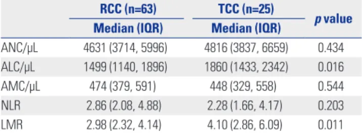
ANC, AMC, and NLR did not statistically differ between the two groups (Table 2). ALC was significantly lower for the RCC group than the TCC group [1499/μL (1140, 1896) vs. 1860/μL (1433, 2342), p=0.016]. Likewise, LMR was significantly lower in the RCC group than in the TCC group [2.98 (2.32, 4.14) vs. 4.10 (2.86, 6.09), p=0.011].
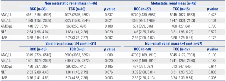
In subgroup analyses, ALC, NLR, and LMR significantly dif- fered between the two groups in N0M0 renal masses (Table 3).
In particular, ALC and LMR were useful markers for distin- guishing small RCC and TCC tumors (≤ 4 cm) (p-values=0.020 and 0.003, respectively). Conversely, no inflammatory bio- marker was useful in distinguishing metastatic RCC from meta- static TCC or in classifying a renal mass larger than 4 cm.
When we performed logistic regression within the non-met- astatic group, young age at diagnosis, low ALC, and low LMR were significantly associated with RCC instead of TCC in uni- variate analysis (Table 4). In multivariate analysis, age at diag- nosis and LMR were the only two variables that were statistical- ly significant differentiators of RCC and TCC, with young age and low LMR correlating with higher probability of RCC than TCC. The odds ratio (95% CI) for age at diagnosis was 0.874 (0.778–0.982; p=0.024); for LMR it was 0.461 (0.214–0.994;
p=0.048). We subsequently performed ROC curve analysis with probabilities extracted from the logistic regression, and the AUC was 0.919 (p<0.001), indicating strong correlations be- tween RCC and younger age or LMR (date not shown). For the
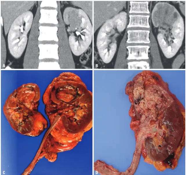
Fig. 1. (A and B) Pathologically proven infiltrative renal cell carcinoma and transitional cell carcinoma in preoperative multi-detector computed tomogra- phy. (C and D) Cross sections of the pathologic specimens of each tumor after surgical resection.
A B
C D
small renal mass (≤4 cm) group, however, multivariate logistic regression analysis revealed no significant correlations with any parameters (Supplementary Table 1, only online).
DISCUSSION
About 6% of RCC cases involve infiltration of surrounding tis- sues, appearing irregular and with ill-defined margins on MDCT.
For this reason, in suspected RCC cases, MDCT is technically limited for excluding other infiltrative diseases and defining the correct diagnosis. Our study demonstrated the clinical useful- ness of inflammatory biomarkers in distinguishing infiltrative RCC from TCC, which are the two most common infiltrative re- nal diseases. The usefulness of these biomarkers is already es- tablished in many other fields. These markers are widely used as prognostic factors not only for acute and chronic infections, but also for cancerous and non-cancerous inflammatory dis- eases. Because inflammatory biomarker evaluation is relatively cheap and blood samples are easy to access, these parameters
are important to consider as diagnostic tools.
Cancer has recently been considered to be as a disease of chronic inflammation. This classification is based on the patho- logical presence of inflammatory cells and its related media- tors, such as cytokines in tumor tissues. Furthermore, accord- ing to Mantovani, et al.,23 tissue remodeling and angiogenesis processes in cancers are similar to those seen in chronic in- flammatory responses and tissue repair. Additionally, Man- tovani, et al.24 demonstrated that immature myeloid cells play a key role in both chronic infection and tumor microenviron- ment.
RCC is one of the most well-known cancers associated with a pathogenesis that includes altered immune activity. Several studies demonstrate poorer prognosis in RCC patients with higher monocyte and neutrophils levels and lower lymphocyte counts.7 According to Frankenberger, et al.,25 growing RCC tu- mors influence the activity of effector lymphocytes and modu- late the composition of immune infiltrates. As the disease pro- gresses, peripheral circulation are also affected as an increase in the number of circulating myeloid cells and regulatory T cells.
There are several reports that neutrophils and macrophages play major roles in RCC tumor progression. The level of circu- lating monocytes can reflect the formation or presence of tu- mor-associated macrophages. Many macrophage-released sol- uble factors directly stimulate the growth of tumor cells and promote tumor cell migration and metastasis.9 Donskov, et al.7 reported that among macrophage and neutrophil products, ROS may not only induce genomic instability, but also damage antitumor immune effector cells. Another study demonstrated that co-cultivation of tumor cells with macrophages leads to enhanced invasiveness of the malignant cells by TNF-α- dependent MMP induction in the macrophages.24 Given these laboratory findings, clinical researchers have focused on the role of inflammatory biomarkers, especially monocyte and lymphocyte counts, in peripheral circulation as oncologic out- come predictors or prognostic factors in RCC patients. Based on these above findings, the LMR has been assessed as a good candidate inflammatory biomarker for RCC. Several studies fo- cused on the value of preoperative and postoperative LMR as an important prognostic factor in metastatic and non-metastat- Table 1. Characteristics of Patients with Infiltrating Renal Cell Carcino-
mas (RCC) and RCC-Mimicking Transitional Cell Carcinomas (TCC)
RCC TCC p value
Number of patients enrolled 63 25
Age at diagnosis* 55.4±13.6 69.7±10.2 <0.001
Sex ratio (M:F)† 47:16 13:12 0.040
Tumor size (cm)* 7.6±4.1 6.1±3.1 0.105
Necrosis (%)‡ 7 (11.1) 1 (4.0) 0.431
Sarcomatoid change (%)‡ 11 (17.5) 3 (12.0) 0.749 Stage
pT stage, n (%)
pT1 14 (23.3) 1 (4.0)
pT2 4 (6.7) 0 (0)
pT3 34 (56.7) 15 (60.0)
pT4 8 (13.3) 9 (36.0)
pN stage, n (%)† 0.278
pNx/N0 43 (68.3) 14 (56.0)
pN+ 20 (31.7) 11 (44.0)
M stage, n (%)† 0.655
M0 41 (65.1) 15 (60.0)
M1 22 (34.9) 10 (40.0)
Fuhrman grade, n (%)
G1–2 7 (15.6)
G3–4 38 (84.4)
Histologic grade
Low grade 0 (0.0)
High grade 22 (100.0)
Histology, n (%)
Clear cell 36 (57.1)
Non-clear cell 27 (42.9)
M, male; F, female.
*Independent t-test, †χ2-test, ‡Fisher’s exact test.
Table 2. Inflammatory Biomarker Measurements in RCC and TCC RCC (n=63) TCC (n=25)
p value Median (IQR) Median (IQR)
ANC/µL 4631 (3714, 5996) 4816 (3837, 6659) 0.434 ALC/µL 1499 (1140, 1896) 1860 (1433, 2342) 0.016
AMC/µL 474 (379, 591) 448 (329, 558) 0.544
NLR 2.86 (2.08, 4.88) 2.28 (1.66, 4.17) 0.203 LMR 2.98 (2.32, 4.14) 4.10 (2.86, 6.09) 0.011 RCC, renal cell carcinoma; TCC, transitional cell carcinoma; ANC, absolute neutrophil count; ALC, absolute lymphocyte count; AMC, absolute monocyte count; NLR, neutrophil-lymphocyte ratio; LMR, lymphocyte-monocyte ratio.
All variables examined by Mann-Whitney test.
ic RCC patients.9,26-29
Additionally, there are several reports of a predictive role for LMR in upper urinary tract TCC. Elevated preoperative LMR also has prognostic value in non-metastatic upper urinary tract TCC.30 Furthermore, the presence of neutrophilia with relative lymphocytopenia predicts a worse oncological prognosis in pa- tients with localized upper urinary tract TCC.31-33
As a novel perspective, we focused on the possibility that in- flammatory biomarkers could distinguish RCC from other infil- trative renal masses, especially TCC that mimics RCC on diag- nostic images. We hypothesized that infiltrative RCC and TCC might demonstrate different degrees of biomarker changes due to differences in tumor biology. Of the 63 pathologically-proven RCC patients in our study, more than half (35/63, 55.6%) re- quired renal biopsy (20/63, 31.7%) or diagnostic ureterorenos- copy (15/63, 23.8%). Eight (12.7%) cases of pathologically-proven RCC underwent nephroureterectomy instead of nephrectomy, due to relatively low suspicion of RCC in preoperative MDCT.
Likewise, of the 25 pathologically-proven TCC patients, 15 (60%) underwent renal biopsy or diagnostic ureterorenoscopy
preoperatively. Six patients (24%) underwent nephrectomy in- stead of nephroureterectomy, and consequently required sub- sequent remnant-ureterectomy surgery. Overall, about 16% of infiltrative renal masses in our cohort were misdiagnosed pre- operatively, and underwent inappropriate surgery. Adding cy- tology results also did not help. Among 25 pathologically-prov- en TCC patients, cytology was performed in 16 cases. Only 2 cases out of 16 (12.5%) showed positive results for TCC.
Previous studies report that as RCC tumor burden grows, the values of NLR and LMR increase and decrease, respectively. We performed subgroup analysis to see if the biomarker parame- ters could be diagnostically relevant in cases with relatively lower tumor burden. We especially focused on the clinical power of inflammatory biomarkers as tools for diagnosis in sur- gical candidates without metastasis. In the non-metastatic group, we identified ALC, NLR, and LMR as potential biomark- ers that distinguished infiltrative RCC from TCC. Logistic re- gression analysis revealed that LMR is the only significant use- ful inflammatory biomarker for distinguishing RCC from TCC.
In the ROC curve analysis, the AUC was 0.817 (p-value=0.002),
Table 3. Inflammatory Biomarker Associations with Pathological Parameters in Subgroups of Infiltrative RCC and TCC
Non metastatic renal mass (n=46) Metastatic renal mass (n=42)
RCC (n=36) TCC (n=10) p value RCC (n=27) TCC (n=15) p value
ANC/µL 4161 (3154, 4925) 4076 (2845, 4697) 0.537 5770 (4430, 6584) 5945 (4627, 8663) 0.423
ALC/µL 1599 (1153, 2008) 2227 (1550, 2544) 0.021 1335 (981, 1766) 1747 (1331, 2153) 0.101
AMC/µL 448 (351, 578) 368 (256, 497) 0.108 501 (399, 616) 480 (427, 641) 0.783
NLR 2.54 (1.96, 4.04) 1.66 (1.41, 2.38) 0.020 4.6 (2.35, 7.05) 3.31 (1.96, 6.23) 0.572
LMR 3.69 (2.54, 4.43) 5.78 (3.79, 7.57) 0.002 2.55 (2.05, 4.07) 3.90 (2.29, 5.47) 0.176
Small renal mass (≤4 cm) (n=21) Non-small renal mass (>4 cm) (n=67)
RCC (n=13) TCC (n=8) p value RCC (n=50) TCC (n=17) p value
ANC/µL 3819 (2724, 6510) 3900 (3063, 5392) 1.000 4790 (1169, 1910) 5945 (4172, 7993) 0.103
ALC/µL 1501 (1079, 2022) 2166 (1795, 2372) 0.020 1489 (1169, 1910) 1741 (1259, 2380) 0.185
AMC/µL 430 (337, 595) 396 (256, 445) 0.185 487 (381, 597) 513 (347, 645) 0.614
NLR 2.53 (2.00, 4.46) 1.81 (1.43, 2.79) 0.076 3.02 (2.08, 5.01) 3.31 (1.93, 5.86) 0.885
LMR 3.76 (2.41, 4.83) 5.74 (4.66, 7.86) 0.003 2.82 (2.26, 4.13) 3.74 (2.39, 5.51) 0.306
RCC, renal cell carcinoma; TCC, transitional cell carcinoma; ANC, absolute neutrophil count; ALC, absolute lymphocyte count; AMC, absolute monocyte count;
NLR, neutrophil-lymphocyte ratio; LMR, lymphocyte-monocyte ratio.
Table 4. Univariate and Multivariate Analysis of Correlations between Demographic and Blood Count Variables with Non-Metastatic Infiltrative Renal Cell Carcinoma
Variables Univariate analysis Multivariate analysis
OR (95% CI) p value OR (95% CI) p value
Age at diagnosis 0.85 (0.768–0.950) 0.004 0.87 (0.778–0.982) 0.024
Size (cm) 1.10 (0.870–0.139) 0.444
Male 1.14 (0.201–6.494) 0.880
ANC/µL 1.00 (1.000–1.001) 0.400
AMC/µL 1.00 (0.999–1.010) 0.125
ALC/µL 0.998 (0.997–1.000) 0.019 1.00 (0.998–1.003) 0.728
NLR 2.32 (0.944–5.718) 0.067
LMR 0.44 (0.249–0.775) 0.005 0.46 (0.214–0.994) 0.048
OR, odds ratio; CI, confidence interval; ANC, absolute neutrophil count; AMC, absolute monocyte count; ALC, absolute lymphocyte count; NLR, neutrophil-lym- phocyte ratio; LMR, lymphocyte-monocyte ratio.
and the cut off value was 5.250 (data not shown). This value is slightly higher than the already mentioned values ranging from 3.0–5.2 as an independent prognostic factor. Furthermore, in patients with small renal masses less than 4 cm, we confirmed that ALC and LMR could be helpful in distinguishing RCC from TCC by Mann-Whitney U tests (Table 3), although multivariate regression showed no statistically significant biomarkers. With the help of these biomarkers, we may avoid choosing the wrong surgical method in planning resections; moreover, we may be able to recommend additional preoperative renal biopsies or diagnostic ureterorenoscopies in cases of mismatch between imaging results and biomarker results. Nevertheless, the in- flammatory biomarkers were less effective in distinguishing in- filtrative large (>4 cm) or metastatic cancers. We think this re- sult originated from the common biologic characteristics of metastatic solid cancers. According to the results of a meta-anal- ysis performed by Teng, et al.,34 increased pretreatment LMR is correlated with significantly favorable outcomes in patients with general solid tumors. Although the exact mechanism of the prognostic value of LMR in solid tumor is poorly under- stood, we could infer that the effect of cancer-related inflam- mation could be maximized for general advanced solid cancers with metastasis, obscuring the role of inflammatory biomarkers in distinguishing cancer types.
This study focused primarily on non-metastatic infiltrative renal masses. For patients with lesions of this nature, we typi- cally consider surgical resection regardless of the exact pathol- ogy. By simple calculations based on a basic blood workup, urologists can gain insight into the type of infiltrative lesion for designing surgical plans in preoperative settings.
The current study has several limitations. First, although we attempted to control for possible confounding factors that could lead to biased results, this was a retrospective study with a relatively small number of patients. Another potential limita- tion is that we did not compare serum LMR with the extent of inflammatory cell infiltration within and surrounding tumor tissue. Such histological correlation should be considered in fu- ture analyses. However, even considering these limitations, we believe our study outlines the clinically benefits of the noted biomarkers in distinguishing infiltrative RCC from TCC.
In conclusion, inflammatory biomarkers could be helpful in distinguishing infiltrative renal masses. Younger age and lower LMR are more likely to imply infiltrative RCC rather than TCC.
Using these inflammatory biomarkers in conjunction with ra- diological imaging, non-metastatic infiltrative RCC could be di- agnosed with greater certainty.
REFERENCES
1. Pickhardt PJ, Lonergan GJ, Davis CJ Jr, Kashitani N, Wagner BJ.
From the archives of the AFIP. Infiltrative renal lesions: radiologic- pathologic correlation. Armed Forces Institute of Pathology. Ra- diographics 2000;20:215-43.
2. Raza SA, Sohaib SA, Sahdev A, Bharwani N, Heenan S, Verma H,
et al. Centrally infiltrating renal masses on CT: differentiating in- trarenal transitional cell carcinoma from centrally located renal cell carcinoma. AJR Am J Roentgenol 2012;198:846-53.
3. Bata P, Tarnoki DL, Tarnoki AD, Novak PK, Gyebnar J, Kekesi D, et al. Transitional cell and clear cell renal carcinoma: differentiation of distinct histological types with multiphase CT. Acta Radiol 2014;
55:1112-9.
4. Sheir KZ, El-Azab M, Mosbah A, El-Baz M, Shaaban AA. Differen- tiation of renal cell carcinoma subtypes by multislice computer- ized tomography. J Urol 2005;174:451-5.
5. Li Y, Ding YU, Chen D, Yu Z, Gui Y, Yang S, et al. Renal cell carci- noma growing into the renal pelvis and mimicking transitional cell carcinoma: a case report and literature review. Oncol Lett 2015;
9:1869-72.
6. Saroha S, Uzzo RG, Plimack ER, Ruth K, Al-Saleem T. Lymphope- nia is an independent predictor of inferior outcome in clear cell renal carcinoma. J Urol 2013;189:454-61.
7. Donskov F, Hokland M, Marcussen N, Torp Madsen HH, von der Maase H. Monocytes and neutrophils as ‘bad guys’ for the out- come of interleukin-2 with and without histamine in metastatic renal cell carcinoma--results from a randomised phase II trial. Br J Cancer 2006;94:218-26.
8. Mehrazin R, Uzzo RG, Kutikov A, Ruth K, Tomaszewski JJ, Dulaimi E, et al. Lymphopenia is an independent predictor of inferior out- come in papillary renal cell carcinoma. Urol Oncol 2015;33:388.
e19-25.
9. Hutterer GC, Stoeckigt C, Stojakovic T, Jesche J, Eberhard K, Pum- mer K, et al. Low preoperative lymphocyte-monocyte ratio (LMR) represents a potentially poor prognostic factor in nonmetastatic clear cell renal cell carcinoma. Urol Oncol 2014;32:1041-8.
10. Park YH, Ku JH, Kwak C, Kim HH. Post-treatment neutrophil-to- lymphocyte ratio in predicting prognosis in patients with meta- static clear cell renal cell carcinoma receiving sunitinib as first line therapy. Springerplus 2014;3:243.
11. Keskin S, Keskin Z, Taskapu HH, Kalkan H, Kaynar M, Poyraz N, et al. Prognostic value of preoperative neutrophil-to-lymphocyte and platelet-to-lymphocyte ratios, and multiphasic renal tomog- raphy findings in histological subtypes of renal cell carcinoma.
BMC Urol 2014;14:95.
12. Hu K, Lou L, Ye J, Zhang S. Prognostic role of the neutrophil-lym- phocyte ratio in renal cell carcinoma: a meta-analysis. BMJ Open 2015;5:e006404.
13. Wen RM, Zhang YJ, Ma S, Xu YL, Chen YS, Li HL, et al. Preopera- tive neutrophil to lymphocyte ratio as a prognostic factor in pa- tients with non-metastatic renal cell carcinoma. Asian Pac J Cancer Prev 2015;16:3703-8.
14. Pichler M, Hutterer GC, Stoeckigt C, Chromecki TF, Stojakovic T, Golbeck S, et al. Validation of the pre-treatment neutrophil-lym- phocyte ratio as a prognostic factor in a large European cohort of renal cell carcinoma patients. Br J Cancer 2013;108:901-7.
15. Santoni M, De Giorgi U, Iacovelli R, Conti A, Burattini L, Rossi L, et al. Pre-treatment neutrophil-to-lymphocyte ratio may be asso- ciated with the outcome in patients treated with everolimus for metastatic renal cell carcinoma. Br J Cancer 2013;109:1755-9.
16. Ohno Y, Nakashima J, Ohori M, Hatano T, Tachibana M. Pretreat- ment neutrophil-to-lymphocyte ratio as an independent predic- tor of recurrence in patients with nonmetastatic renal cell carci- noma. J Urol 2010;184:873-8.
17. Ohno Y, Nakashima J, Ohori M, Gondo T, Hatano T, Tachibana M.
Followup of neutrophil-to-lymphocyte ratio and recurrence of clear cell renal cell carcinoma. J Urol 2012;187:411-7.
18. de Martino M, Pantuck AJ, Hofbauer S, Waldert M, Shariat SF, Belldegrun AS, et al. Prognostic impact of preoperative neutro-
phil-to-lymphocyte ratio in localized nonclear cell renal cell car- cinoma. J Urol 2013;190:1999-2004.
19. Viers BR, Houston Thompson R, Boorjian SA, Lohse CM, Leibov- ich BC, Tollefson MK. Preoperative neutrophil-lymphocyte ratio predicts death among patients with localized clear cell renal car- cinoma undergoing nephrectomy. Urol Oncol 2014;32:1277-84.
20. Riemann D, Hase S, Fischer K, Seliger B. Granulocyte-to-dendrit- ic cell-ratio as marker for the immune monitoring in patients with renal cell carcinoma. Clin Transl Med 2014;3:13.
21. Fox P, Hudson M, Brown C, Lord S, Gebski V, De Souza P, et al.
Markers of systemic inflammation predict survival in patients with advanced renal cell cancer. Br J Cancer 2013;109:147-53.
22. Lucca I, de Martino M, Hofbauer SL, Zamani N, Shariat SF, Klatte T. Comparison of the prognostic value of pretreatment measure- ments of systemic inflammatory response in patients undergoing curative resection of clear cell renal cell carcinoma. World J Urol 2015;33:2045-52.
23. Mantovani A, Allavena P, Sica A, Balkwill F. Cancer-related in- flammation. Nature 2008;454:436-44.
24. Mantovani A, Schioppa T, Porta C, Allavena P, Sica A. Role of tu- mor-associated macrophages in tumor progression and invasion.
Cancer Metastasis Rev 2006;25:315-22.
25. Frankenberger B, Noessner E, Schendel DJ. Immune suppression in renal cell carcinoma. Semin Cancer Biol 2007;17:330-43.
26. Chang Y, An H, Xu L, Zhu Y, Yang Y, Lin Z, et al. Systemic inflam- mation score predicts postoperative prognosis of patients with clear-cell renal cell carcinoma. Br J Cancer 2015;113:626-33.
27. Chang Y, Fu Q, Xu L, Zhou L, Liu Z, Yang Y, et al. Prognostic value of preoperative lymphocyte to monocyte ratio in patients with nonmetastatic clear cell renal cell carcinoma. Tumour Biol 2016;37:
4613-20.
28. Gu L, Ma X, Wang L, Li H, Chen L, Li X, et al. Prognostic value of a systemic inflammatory response index in metastatic renal cell carcinoma and construction of a predictive model. Oncotarget 2016 Jul 16 [Epub]. http://dx.doi.org/10.18632/oncotarget.10626.
29. Xia WK, Wu X, Yu TH, Wu Y, Yao XJ, Hu H. Prognostic significance of lymphocyte-to-monocyte ratio and CRP in patients with non- metastatic clear cell renal cell carcinoma: a retrospective multi- center analysis. Onco Targets Ther 2016;9:2759-67.
30. Hutterer GC, Sobolev N, Ehrlich GC, Gutschi T, Stojakovic T, Mannweiler S, et al. Pretreatment lymphocyte-monocyte ratio as a potential prognostic factor in a cohort of patients with upper tract urothelial carcinoma. J Clin Pathol 2015;68:351-5.
31. Luo HL, Chen YT, Chuang YC, Cheng YT, Lee WC, Kang CH, et al.
Subclassification of upper urinary tract urothelial carcinoma by the neutrophil-to-lymphocyte ratio (NLR) improves prediction of oncological outcome. BJU Int 2014;113:E144-9.
32. Dalpiaz O, Ehrlich GC, Mannweiler S, Hernández JM, Gerger A, Stojakovic T, et al. Validation of pretreatment neutrophil-lympho- cyte ratio as a prognostic factor in a European cohort of patients with upper tract urothelial carcinoma. BJU Int 2014;114:334-9.
33. Dalpiaz O, Pichler M, Mannweiler S, Martín Hernández JM, Stoja- kovic T, Pummer K, et al. Validation of the pretreatment derived neutrophil-lymphocyte ratio as a prognostic factor in a European cohort of patients with upper tract urothelial carcinoma. Br J Can- cer 2014;110:2531-6.
34. Teng JJ, Zhang J, Zhang TY, Zhang S, Li BS. Prognostic value of pe- ripheral blood lymphocyte-to-monocyte ratio in patients with solid tumors: a meta-analysis. Onco Targets Ther 2016;9:37-47.