Yonsei Med J http://www.eymj.org Volume 52 Number 1 January 2011 192
Case Report
DOI 10.3349/ymj.2011.52.1.192pISSN: 0513-5796, eISSN: 1976-2437 Yonsei Med J 52(1):192-195, 2011
Three Cases of Manifesting Female Carriers in Patients with Duchenne Muscular Dystrophy
Tae-Jin Song,
1Kyung-A Lee,
2Seong-Woong Kang,
3Hanna Cho,
1and Young-Chul Choi
1Departments of 1Neurology and 2Laboratory Medicine, Brain Korea 21 Project for Medical Science, Yonsei University College of Medicine, Seoul;
3Department of Rehabilitation Medicine and Rehabilitation Institute of Muscular Disease, Yonsei University College of Medicine, Seoul, Korea.
Received: March 2, 2009 Revised: May 1, 2009 Accepted: May 15, 2009
Corresponding author: Dr. Young-Chul Choi, Department of Neurology,
Yonsei University College of Medicine, Gangnam Severance Hospital, 712 Eonju-ro, Gangnam-gu, Seoul 135-720, Korea.
Tel: 82-2-2019-3323, Fax: 82-2-3462-5904 E-mail: [email protected]
∙ The authors have no financial conflicts of interest.
© Copyright:
Yonsei University College of Medicine 2011 This is an Open Access article distributed under the terms of the Creative Commons Attribution Non- Commercial License (http://creativecommons.org/
licenses/by-nc/3.0) which permits unrestricted non- commercial use, distribution, and reproduction in any medium, provided the original work is properly cited.
Duchenne muscular dystrophy usually affects males. However, females are also affected in rare instances. Approximately 8% of female Duchenne muscular dys- trophy (DMD) carriers are manifesting carriers and have muscle weakness to some extent. We investigated the clinical features of 3 female patients with dystro- phinopathy diagnosed by clinical, pathological, and genetic studies at our neuro- muscular disease clinic. The onset age of manifesting symptoms varied (8-28 years). Muscle weakness grade varied as follows: patient 1 showed asymmetrical bilateral proximal upper and lower extremities weakness, patient 2 showed asym- metrical bilateral upper extremities weakness similar to scapulohumoral muscular dystrophy, and patient 3 had only bilateral asymmetric proximal lower extremities weakness. Two patients had familial histories of DMD (their sons were diagnosed with DMD), but the 1 remaining patient had no familial history of DMD. The se- rum creatine kinase level was elevated in all patients, but it was not correlated with muscular weakness. An electromyography study showed findings of myopathy in all patients. One patient was diagnosed with a DMD carrier by a muscle biopsy with an immunohistochemical stain (dystrophin). The remaining 2 patients with familial history of DMD were diagnosed by multiplex ligation-dependent probe amplification (MLPA). There were inconsistent clinical features in the female car- riers. An immunohistochemical analysis of dystrophin could be useful for female carrier patients. Also, multiplex ligation-dependent probe amplification is essential for the diagnosis of a manifesting female carrier DMD in female myopathic pa- tients because conventional multiplex PCR could not detect the duplication and is less accurate compared to MLPA.
Key Words: Dystrophinopathy, female carrier, multiplex ligation-dependent probe amplification
INTRODUCTION
Two-thirds of mothers of affected males are thought to be Duchenne muscular dystro- phy (DMD) gene carriers and approximately 8% of female DMD carriers have mus- cle weakness to some extent and are designated as manifesting DMD carriers.1-3 We investigated the clinical features of 3 female myopathic carrier patients with DMD.
Manifesting Female Carriers in Patients with Duchenne Muscular Dystrophy
Yonsei Med J http://www.eymj.org Volume 52 Number 1 January 2011 193
ic patterns on a dystrophin immunohistochemical stain from a muscle biopsy (Fig. 1). The other immunohistochemical stain for detecting limb girdle muscular dystrophy and con- genital myopathy revealed no abnormality. Accordingly, she was diagnosed as a DMD carrier.
Case 2
A 32 year-old woman was admitted with complaints of pro- gressive limb weakness that started 9 years prior. She had 2 sons who were diagnosed by exon duplication (exon 52, 53, 56-61) with DMD. On a neurological examination, she showed intellectual disability (IQ = 70) and her bilateral up- per extremities, especially scapulohumoral lesion, showed muscular weakness (right arm abductor, elevator, adductor G4°/left arm abductor, elevator, adductor G4+). The power of other muscles was within a relatively normal range.
Gower signs and bilateral calf pseudohypertrophy were not observed. Serum CK was mildly elevated (CK: 3708 U/L).
Cardiac echocardiography showed normal findings, but ECG findings revealed the R/S > 1 in Vl, 2 leads, and deep Q wave in I, V4-6 leads. An electromyography study re- vealed spontaneous activity (fibrillation and positive sharp waves) and short duration polyphasic potentials in volun- tary contraction. A conventional multiplex polymerase chain reaction (PCR) had negative findings, but a multiplex liga- tion-dependent probe amplification (MLPA) revealed exon duplication at exon 52, 53, 56-61.
Case 3
A 34 year-old woman was admitted with complaints of progressive weakness in both legs that started 6 years prior.
She had 2 sons, who were diagnosed by exon deletion (exon 43, 44 and 45) with DMD. On a neurological examination, she showed intellectual disability (IQ = 78) and her bilater- al lower extremities showed muscular weakness (right hip flexor, hip extensor, knee flexor, knee extensor G3°/left hip flexor, hip extensor, knee flexor, knee extensor G4-). The power of the other muscles was within a relatively normal range. Gower signs and bilateral calf pseudohypertrophy were not observed. Serum CK was mildly elevated (CK:
1289 U/L). Electrocardiography and cardiac echocardiogra- phy showed no abnormal findings. An electromyography study revealed spontaneous activity (fibrillation and posi- tive sharp waves) and short duration polyphasic potentials in voluntary contraction. A conventional multiplex PCR had negative findings but a MLPA revealed exon deletion at 43, 44 and 45.
CASE REPORT
Case 1
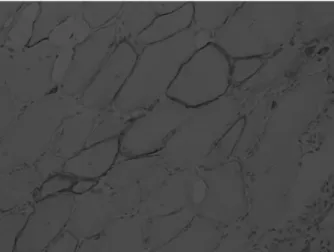
A 9 year-old girl was admitted with complaints of slow, progressive proximal limb weakness that started 1 year pri- or. Until the age of 8, she had grown up without abnormali- ty, developed a normal walking course, and normal cogni- tive function. There was no familial history of neuromus- cular disease, including her elder brother. Upon neurological examination, she showed normal intelligence and no crani- al nerve abnormality. However, her bilateral limb-girdle muscular power decreased (right arm abductor, arm eleva- tor, arm adductor, forearm flexor, forearm extensor G4+/left arm abductor, arm elevator, arm adductor, forearm flexor, forearm extensor G4°/right hip flexor, hip extensor, knee flexor, knee extensor G4°/left hip flexor, hip extensor, knee flexor, knee extensor G4+). Gower signs and bilateral calf muscle pseudohypertrophy were also observed. Liver en- zymes, serum CK, lactate dehydrogenase, and aldolase were elevated (AST: 97 IU/L; ALT: 225 IU/L; and CK: 456 1 U/L, CK-MB 225 U/L, LDH 1714 IU/L, and Aldolase 16.4 U/mL). The patient also showed normal findings for viral hepatitis markers and arterial blood gas analysis. Elec- trocardiography and cardiac echocardiography showed no abnormal findings. An electromyography study revealed spontaneous activity (positive sharp waves) and short dura- tion polyphasic potentials in voluntary contraction. We car- ried out a muscle biopsy on the left vastus lateralis muscle because limb-girdle muscular dystrophy was suspected as the patient was female and did not have a familial history of DMD. The expression of dystrophin protein showed mosa-
Fig. 1. Skeletal muscle, left vastus laterals: increased size variability and degenerating fibers were revealed. Dystrophin immunostaining revealed a mosaic distribution of positive and negative fibers on C-terminals.
Tae-Jin Song, et al.
Yonsei Med J http://www.eymj.org Volume 52 Number 1 January 2011 194
the deletions in male patients, but duplications are not iden- tified in DMD.10-12 However, the MLPA method has turned out to be reliable and accurate for identifying duplications and deletions in DMD.13,14 The MLPA technique could in- crease mutation pick-up rate by 33% rather than PCR.14 Moreover, the technique could confidently identify carrier individuals.14 In our cases, exon duplication detected in case 2, on the other hand, exon deletion also detected in case 3 but the PCR could not detect genetic abnormality. These re- sults support that MLPA is essential for diagnosis of mani- festing female carrier DMD in female myopathic patients because conventional multiplex PCR could not detect the duplication and is less accurate compared to MLPA.
REFERENCES
1. Walcher T, Kunze M, Steinbach P, Sperfeld AD, Burgstahler C, Hombach V, et al. Cardiac involvement in a female carrier of Duchenne muscular dystrophy. Int J Cardiol 2010;138:302-5.
2. Ceulemans BP, Storm K, Reyniers E Jr, Callewaert L, Martin JJ.
Muscle pain as the only presenting symptom in a girl with dystro- phinopathy. Pediatr Neurol 2008;38:64-6.
3. Hoffman EP, Arahata K, Minetti C, Bonilla E, Rowland LP. Dys- trophinopathy in isolated cases of myopathy in females. Neurolo- gy 1992;42:967-75.
4. Hoogerwaard EM, Bakker E, Ippel PF, Oosterwijk JC, Majoor- Krakauer DF, Leschot NJ, et al. Signs and symptoms of Duchenne muscular dystrophy and Becker muscular dystrophy among carri- ers in The Netherlands: a cohort study. Lancet 1999;353:2116-9.
5. Griggs RC, Mendell JR, Brooke MH, Fenichel GM, Miller JP, Province M, et al. Clinical investigation in Duchenne dystrophy:
V. Use of creatine kinase and pyruvate kinase in carrier detection.
Muscle Nerve 1985;8:60-7.
6. Politano L, Nigro V, Nigro G, Petretta VR, Passamano L, Pappar- ella S, et al. Development of cardiomyopathy in female carriers of Duchenne and Becker muscular dystrophies. JAMA 1996;275:
1335-8.
7. Lukasik E. Electrocardiographic studies in female carriers of Duchenne muscular dystrophy. J Neurol 1975;209:279-85.
8. Hoffman EP, Brown RH Jr, Kunkel LM. Dystrophin: the protein product of the Duchenne muscular dystrophy locus. Cell 1987;51:
919-28.
9. Arahata K, Ishihara T, Kamakura K, Tsukahara T, Ishiura S, Baba C, et al. Mosaic expression of dystrophin in symptomatic carriers of Duchenne’s muscular dystrophy. N Engl J Med 1989;320:138- 10. Chamberlain JS, Gibbs RA, Ranier JE, Nguyen PN, Caskey CT. 42.
Deletion screening of the Duchenne muscular dystrophy locus via multiplex DNA amplification. Nucleic Acids Res 1988;16:11141- 11. Beggs AH, Koenig M, Boyce FM, Kunkel LM. Detection of 98% 56.
of DMD/BMD gene deletions by polymerase chain reaction. Hum Genet 1990;86:45-8.
12. Kunkel LM, Snyder JR, Beggs AH, Boyce FM, Feener CA.
Searching for dystrophin gene deletions in patients with atypical
DISCUSSION
The clinical features of female carriers in this study were inconsistent. In Duchenne and Becker dystrophinopathic carriers, muscular weakness is predominantly asymmetric (81.8%). 41% of dystrophinopathic carriers have weakness limited to the upper extremities, 23% of carriers have weak- ness of the lower extremities, and 36% of carriers have muscular weakness of the upper and lower extremities.4 In this study, the patients revealed asymmetrical muscular weakness in different lesion.
Serum CK levels are elevated in approximately 45-70%
of carriers and the measurement of serum CK is the most commonly used method for detecting carriers.5 In our cas- es, serum CK levels were elevated in all patients. But, al- though muscular weakness was more severe in the third case, the serum CK level was lowest. The CK level is usu- ally due to the progressive involvement of muscle function.
Further study regarding the correlation between the serum CK level and muscular weakness is requested in female carriers. In DMD carriers, incidences of cardiac involve- ment progresses with age. Researchers reported latent and clinical cardiac involvement in 55% of carriers under the age of 16 and in 90% of carriers after the age of 16, espe- cially dilated cardiomyopathy.6 Abnormal ECG patterns were reported (R/S > 1 in Vl, 2: deep Q wave in I, V4-6) in 6.6-16.44% of female carriers of DMD.7 In this case, ECG findings of the second case were compatible with the previ- ous report. However, ECG and echocardiographs of the other patients showed whole normal findings.
The dystrophin is localized on the cytoplasmic surface of the plasma membrane of skeletal and cardiac muscle cells.8 The mosaic distribution of dystrophin-positive and -nega- tive fibers in the skeletal and cardiac muscles of biopsy is the characteristic finding of a DMD carrier.9 The surface membranes of muscles in DMD patients do not react with anti-dystrophin antiserum, while those from carriers show a mosaic pattern.9 In the first case, we could diagnosis the DMD manifesting carrier because the patient’s biopsy re- vealed a typical mosaic pattern in immunohistochemical stains. Therefore, the dystrophin immunohistochemistry could be useful to diagnosis what type of myopathy is pres- ent and whether mosaicism is present in female myopathic patients.
The multiplex PCR have traditionally been used in a clin- ical diagnostic set up and allow a detection of 90-95% of
Manifesting Female Carriers in Patients with Duchenne Muscular Dystrophy
Yonsei Med J http://www.eymj.org Volume 52 Number 1 January 2011 195 Res 2002;30:e57.
14. Schwartz M, Dunø M. Multiplex ligation-dependent probe ampli- fication is superior for detecting deletions/duplications in Duch- enne muscular dystrophy. Clin Genet 2005;67:189-91.
presentations. In: Lindsten J, Petterson U, editors. Etiology of Hu- man Diseases at the DNA Level. New York: Raven Press; 1991.
p.51-60.
13. Schouten JP, McElgunn CJ, Waaijer R, Zwijnenburg D, Diepvens F, Pals G. Relative quantification of 40 nucleic acid sequences by multiplex ligation-dependent probe amplification. Nucleic Acids